Designer Genes
2027 season
On this page(17 sections)
Designer Genes is a written, quantitative genetics event for Division C. Teams work through problems in classical (Mendelian) genetics, population and evolutionary genetics, and molecular biology. The 2027 rules say outright that questions will lean on process skills: quantitative reasoning, calculations, reading experimental results and drawing conclusions from evidence. Expect fewer pure-vocabulary items and more "here is a data set, what does it show and why." Points are awarded for reasoning as well as answers, so always show the setup. Teams that lose points usually know the material but skip a step, mix up two similar terms or misread which strand a question is about.
This guide covers the whole event. The sections below follow the six topic areas of the 2027 rules in order, then cover strategy, pitfalls and how tournaments write the test.
Event format
| Rule | Detail |
|---|---|
| Team | Up to 2 students |
| Time | About 50 minutes |
| Format | A written test, which may be run as stations |
| Calculators | Up to two Class II calculators per team |
| Notes | One 8.5 × 11 in sheet, both sides, any form and any source. It may be laminated or put in a sheet protector. Attached labels and extra sheets (even inside one protector) are not allowed |
| Other | Writing utensils |
| Scoring | High score wins. Points for accuracy, quality of supporting reasoning and proper scientific method. Preselected questions break ties |
| Division | Division C |
The notes rule is broad: formulas, a codon table, epistasis ratios, a χ² critical-value row, the steps of each technique and a worked template for each calculation type all fit. Keep it to what you will actually use, since searching a crowded sheet costs more than it saves.
What the rules cover
The 2027 rules split the event into six topic areas, paraphrased below. Items marked State or National are not part of Invitational or Regional tests. The rules call the topic list exhaustive and say no single topic should be overemphasized, so spread your preparation.
| Area | Every level (Invitational and Regional) | State and National adds |
|---|---|---|
| Mendelian genetics | Mendel's laws and the patterns that break them (linkage, incomplete dominance and codominance, complementation); mono-, di- and trihybrid Punnett squares; probability rules applied to data; reading and building pedigrees; epistasis | Mapping genes from recombination data: two genes at State, three genes at National |
| Mitosis and meiosis | Stages and key structures of both divisions and how they compare; effects of nondisjunction; reading karyotypes for abnormalities | Somatic recombination: V(D)J recombination and immunoglobulin class switching |
| Population and evolutionary genetics | Hardy–Weinberg assumptions, calculations and violations; drift, founder effects, bottlenecks, migration and relative fitness; additive alleles and the number of genes behind a trait; gene duplication with homologs, orthologs and paralogs; reading phylogenies and the steps of building one from sequences | Heritability: broad-sense, narrow-sense and realized |
| Molecular biology of DNA | Nucleotide structure, strand direction and base pairing; replication from pre-replication complex to termination in bacteria, archaea and eukaryotes; polymerase fidelity and proofreading; plasmids, chromatin and chromosome organization; damage from UV, ionizing radiation and intercalators and its repair (NER, NHEJ, HDR) in bacteria and eukaryotes; mutation classes and protein effects | The end-replication problem and telomere synthesis |
| Eukaryotic gene expression and regulation | The central dogma; transcription stages and RNA polymerase; cis and trans regulatory elements including epigenetics and riboswitches, and yeast two-hybrid screening; RNA maturation (capping, polyadenylation, splicing); translation stages and its regulation | Mobile genetic elements: retroviruses, retrotransposons and DNA transposons, and the enzymes that move them |
| Technology and techniques | PCR and its applications (Sanger sequencing, error-prone PCR, RT-qPCR); Illumina vs Nanopore sequencing; molecular cloning (plasmids, restriction enzymes, PCR, Gibson and Golden Gate assembly, stable lines and transient transfection); knockout, knockdown and complementation logic for necessity and sufficiency | How knockdowns and knockouts are made (RNAi, homologous recombination including Cre-Lox, CRISPR); ChIP-seq, Hi-C and RNA-seq (single-cell vs bulk) |
Strategy
Splitting the work
Two students means two parallel streams of calculations. A workable split is one partner on inheritance (Mendelian ratios, pedigrees, probability, mapping, Hardy–Weinberg, heritability) and the other on molecules and techniques (replication, repair, expression, cloning, sequencing). Both read the whole test first and flag questions that need the other person, then check each other's arithmetic. Agree on notation up front (RF as a percentage or in cM, how you write genotypes) so a single answer never mixes conventions.
Time and the answer sheet
With 50 minutes, answer the short items first, then the multi-step calculations and data questions. For every calculation, write the setup on one line before the number: the cross, the table, or the formula with its variables defined. For every experiment question, give a claim, the evidence from the data and a mechanism, and say what control or follow-up experiment would test it. Because the rules reward supporting reasoning and proper scientific method, a short answer that names the control and the mechanism beats a long one that restates the question.
Mendelian genetics and pedigrees
Mendel's first law, segregation, says that each gamete carries only one allele of each gene, so the two alleles in a diploid parent separate during gamete formation. The second law, independent assortment, says that genes on different chromosomes sort independently. Both laws describe what happens in meiosis I, which is why the cell-division and Mendelian material are tested together. Violations happen when genes are linked (on the same chromosome and close enough that they usually travel together), when alleles are incompletely or co-dominant, or when two genes complement each other to make a new phenotype.
The baseline dihybrid ratio is 9:3:3:1 from a cross of two heterozygotes at two unlinked loci. A monohybrid cross of two heterozygotes gives 3:1, and a testcross of a heterozygote with a homozygous recessive gives 1:1. Punnett squares for three genes (trihybrid crosses) have 64 boxes, so the per-gene product method is faster: treat each gene as its own monohybrid cross, then multiply. For AaBbCc × AaBbCc, the chance of the A_B_C_ phenotype is ¾ × ¾ × ¾ = 27/64, and the chance of aabbcc is (¼)³ = 1/64. The full phenotypic ratio with complete dominance and independent assortment is 27:9:9:9:3:3:3:1.
Probability
Most calculations use two rules. The product rule applies to independent events joined by "and": multiply. The sum rule applies to mutually exclusive outcomes joined by "or": add. Write out the connecting word before you choose the rule.
Testing data against a ratio
Because the 2027 rules emphasize evaluating experimental results, expect a cross with real counts and the question "do these data fit the expected ratio?" The standard tool is the chi-square goodness-of-fit test.
Pedigrees
A pedigree problem asks you to decide the mode of inheritance from which individuals are affected and how the trait passes between sexes and generations. Work through the pattern systematically rather than guessing from one generation.
| Mode | What the pedigree shows |
|---|---|
| Autosomal dominant | Affected individuals in every generation; an affected parent passes the trait to about half of children; both sexes equally affected |
| Autosomal recessive | Often skips generations; affected children of unaffected carrier parents; both sexes equally affected |
| X-linked recessive | Many more affected males; an affected son usually has a carrier mother; an affected father passes the allele to all daughters and no sons |
| X-linked dominant | An affected father passes the trait to all daughters and none of his sons; an affected mother passes it to about half of all children |
| Y-linked | Every son of an affected male is affected; no daughters are affected |
| Mitochondrial | Passed from mother to all children; an affected father passes it to none of his children |
A pedigree construction question hands you a case study and asks you to draw it. Use squares for males, circles for females, filled symbols for affected individuals and a horizontal line for a mating; half-filled symbols mark carriers if the key says so.
Complementation and epistasis
A complementation test asks whether two recessive mutants with the same phenotype carry mutations in the same gene. Cross the two homozygous mutants. If the offspring are normal, the mutations are in different genes, because each parent supplies a working copy of the gene the other lacks. If the offspring are still mutant, the mutations are in the same gene (they fail to complement).
Epistasis happens when one gene masks or modifies another. Start from the 9:3:3:1 baseline, merge the classes the interaction makes identical, and name the resulting ratio.
| Ratio | Interaction type | What it means |
|---|---|---|
| 9:7 | Complementary genes | A homozygous recessive at either locus blocks the pathway |
| 9:3:4 | Recessive epistasis | Homozygous recessive at one locus masks the other locus |
| 12:3:1 | Dominant epistasis | A dominant allele at one locus masks the other locus |
| 15:1 | Duplicate dominant genes | Any dominant allele at either locus gives the same phenotype |
| 13:3 | Dominant suppression | A dominant allele at one locus suppresses the other's dominant phenotype |
Linkage and mapping (State and National)
Genes on the same chromosome are linked. During prophase I, homologs exchange segments by crossing over, and crossovers between two linked genes create recombinant gametes. The recombination frequency (RF) is the proportion of recombinant progeny. Genes farther apart are more likely to be separated, so RF rises with distance up to a cap of 50%, the value for unlinked genes. Two-gene mapping is a State task and three-gene mapping a National task.
In three-point mapping the most common classes are parental and the rarest are double crossovers. The middle gene is the one whose allele differs between the double-crossover and parental classes. Each adjacent distance is (single crossovers in that interval + double crossovers) / total, because a double crossover counts in both intervals.
Mitosis, meiosis and nondisjunction
Mitosis produces two genetically identical diploid cells. Meiosis produces four haploid gametes that differ because of crossing over in prophase I and random orientation of homolog pairs at metaphase I.
| Feature | Mitosis | Meiosis |
|---|---|---|
| Divisions | One | Two (I and II) |
| Daughter cells | Two, diploid | Four, haploid |
| Homologs pair | No | Yes, in prophase I (synapsis) |
| Crossing over | No | Yes, in prophase I |
| What separates at anaphase | Sister chromatids | Homologs at anaphase I; sister chromatids at anaphase II |
| Genetic result | Identical to the parent cell | Different from each other |
Stages and key structures
| Stage | What happens | Key structures |
|---|---|---|
| Prophase | Chromatin condenses into chromosomes of two sister chromatids; centrosomes move apart | Centrosomes, condensin, cohesin |
| Prometaphase | Nuclear envelope breaks down; microtubules attach to chromosomes | Kinetochores on centromeres |
| Metaphase | Chromosomes align; the spindle assembly checkpoint confirms every kinetochore is attached | Spindle, metaphase plate |
| Anaphase | Separase cleaves cohesin; sister chromatids move to opposite poles | Shortening kinetochore microtubules |
| Telophase and cytokinesis | Nuclear envelopes re-form; cytoplasm divides (cleavage furrow in animals, cell plate in plants) | Actin–myosin contractile ring |
Meiosis I differs in two ways. Homologs pair as bivalents held together by chiasmata, and the two sister kinetochores face the same pole, so whole homologs rather than chromatids are pulled apart. Meiosis II then resembles mitosis in a haploid cell, with no DNA replication in between. Prophase I has five substages: leptotene (condensation), zygotene (synapsis begins), pachytene (synapsis complete, crossing over), diplotene (the synaptonemal complex disassembles while chiasmata hold homologs together) and diakinesis (full condensation). Homologs separating at anaphase I is the physical basis of the law of segregation.
Nondisjunction and karyotypes
Nondisjunction is a failure of chromosomes to separate. In meiosis I the homologs fail to separate, so two gametes are n + 1 and two are n − 1. In meiosis II one pair of sister chromatids fails, so one gamete is n + 1, one is n − 1 and two are normal. Counting gametes tells you which division failed.

A karyotype is written as the total count, the sex chromosomes, then any change. Trisomy 21 is 47,XX,+21 or 47,XY,+21; Klinefelter syndrome is 47,XXY; Turner syndrome is 45,X. Karyotypes also reveal structural changes such as deletions, inversions and translocations, written with the chromosome arm and band. A common example is a Robertsonian translocation, which fuses the long arms of two acrocentric chromosomes.
Somatic recombination (State and National)
B and T lymphocytes rearrange their own DNA to make an enormous variety of antigen receptors from a limited set of gene segments. In V(D)J recombination, a variable (V), diversity (D) and joining (J) segment are brought together and the DNA between them is deleted. The heavy chain and the T-cell receptor β chain use V, D and J; light chains and TCR α use only V and J. RAG1 and RAG2 recognize recombination signal sequences and cut the DNA, non-homologous end joining repairs the breaks, and TdT adds random nucleotides at the junctions.
Class switch recombination happens later in an activated B cell. The variable region (and so the antigen specificity) stays the same while the heavy-chain constant region is swapped, for example IgM to IgG, IgA or IgE. The intervening DNA is looped out and deleted, so switching is irreversible. Activation-induced cytidine deaminase (AID) starts it by deaminating cytosines in switch regions; AID also drives somatic hypermutation of the variable region.
Population and evolutionary genetics
Hardy–Weinberg equilibrium describes allele and genotype frequencies in a population that is not evolving. It assumes no mutation, random mating, no selection, no migration and a population large enough that drift is negligible.
Drift is random change in allele frequency, strongest in small populations. A bottleneck is a sharp drop in size that leaves a small sample of the original alleles. A founder effect is drift when a small group starts a new population. Migration (gene flow) makes populations more similar. Relative fitness, w, compares each genotype with the fittest, which is set to 1; the selection coefficient is s = 1 − w.
Inbreeding and assortative mating change genotype frequencies but not allele frequencies; mutation, migration, selection and drift change allele frequencies. For an X-linked recessive trait, the frequency in males equals q and in females q².
Quantitative traits and the number of genes
Continuous variation comes from several loci whose alleles each add a small amount to the trait. With n unlinked loci of equal additive effect, there are 2n + 1 phenotypic classes, and in a cross of two all-heterozygous parents each extreme phenotype appears in (¼)ⁿ of progeny. If 1/256 of an F2 matches one parental extreme, (¼)ⁿ = 1/256 gives n = 4 genes and 9 classes.
Gene duplication, homologs, orthologs and paralogs
Homologs share ancestry. Orthologs were separated by speciation (the same gene in two species). Paralogs arose by duplication within one genome. Duplication is a major source of new genes: after it, one copy keeps the original job while the other can drift, gain a new function (neofunctionalization), split the old functions with its partner (subfunctionalization) or decay into a pseudogene. The globin family is the classic example: α- and β-globin are paralogs, and human β-globin and mouse β-globin are orthologs.
Phylogenetic trees
Tips are current taxa and internal nodes are inferred common ancestors. A monophyletic group (clade) contains an ancestor and all its descendants; a paraphyletic group leaves some descendants out; a polyphyletic group does not include its members' common ancestor. An outgroup roots the tree and fixes the direction of time. Building a tree from sequences follows clear steps: collect homologous sequences, align them, choose a model of sequence change, infer the tree (distance methods such as neighbor-joining, or character methods such as maximum parsimony and maximum likelihood), then assess support with bootstrap resampling.
Which DNA you sample matters. Nuclear genes come from both parents and recombine. Mitochondrial and chloroplast (organellar) genomes are usually inherited from one parent and do not recombine, so they trace a single lineage; organellar genes also sit on bacterial branches, the evidence for endosymbiosis. Extrachromosomal elements such as plasmids can move between species by horizontal transfer, so their trees may disagree with the species tree.
Heritability (State and National)
Heritability is the fraction of phenotypic variance due to genetic differences. V_P = V_G + V_E, and V_G = V_A + V_D + V_I (additive, dominance and interaction variance). Only additive variance responds predictably to selection.
DNA structure and organization
DNA is a double helix of two antiparallel strands. Each nucleotide is a deoxyribose sugar, a phosphate and one of four bases. The backbone is joined by covalent phosphodiester bonds; the bases pair by hydrogen bonds, A with T (two) and G with C (three). Chargaff's rules follow: in double-stranded DNA, A = T and G = C, so a genome that is 30% A is 30% T and 20% each G and C. GC-rich DNA melts at a higher temperature. Each strand has a 5′ phosphate end and a 3′ hydroxyl end, and polymerases add only to the 3′ end. RNA differs by its ribose (an extra 2′ hydroxyl) and uracil in place of thymine. The common form in cells is right-handed B-DNA, about 10.5 bp per turn.
Bacteria usually carry one circular chromosome plus small circular plasmids that replicate independently. Archaea also have circular chromosomes, and many wrap DNA around histone-like proteins. Eukaryotic DNA is wrapped about 1.7 times around a histone octamer (H2A, H2B, H3, H4) to form nucleosomes, which pack into chromatin. Euchromatin is loosely packed, gene-rich and acetylated; heterochromatin is dense, often DNA-methylated and mostly silent (centromeres and telomeres are constitutive heterochromatin).
DNA replication in the three domains
Replication is semiconservative (the Meselson–Stahl experiment): each daughter helix keeps one old strand. It starts at origins and runs in both directions. At each fork, helicase unwinds, single-strand binding proteins hold the strands apart, topoisomerase relieves supercoiling ahead, primase lays down RNA primers, and a sliding clamp keeps the polymerase on the template. The leading strand is made continuously; the lagging strand is made in Okazaki fragments, whose primers are removed and gaps sealed by DNA ligase.

The 2027 rules ask for this in detail for bacteria, archaea and eukaryotes. Archaea are the key comparison: their replication proteins resemble eukaryotic ones, even though their chromosomes are circular like bacterial ones.
| Step or role | Bacteria (E. coli) | Archaea | Eukaryotes |
|---|---|---|---|
| Origins | One (oriC) | One to several (Sulfolobus has three) | Thousands per genome |
| Initiator / licensing | DnaA binds oriC | Orc1/Cdc6 proteins | ORC + Cdc6 + Cdt1 load MCM2-7 in G1 (pre-replication complex) |
| Replicative helicase | DnaB (loaded by DnaC), moves 5′→3′ on the lagging template | MCM homohexamer with GINS, moves 3′→5′ | CMG (Cdc45–MCM–GINS), moves 3′→5′ on the leading template |
| Primase | DnaG | PriS/PriL | Pol α–primase |
| Main polymerases | Pol III holoenzyme (both strands) | Family B and family D polymerases | Pol ε (leading), Pol δ (lagging) |
| Clamp / loader | β clamp / γ complex | PCNA / RFC | PCNA / RFC |
| Primer removal | Pol I (5′→3′ exonuclease) | RNase H, FEN1 | RNase H, FEN1 |
| Termination | Forks meet at Ter sites bound by Tus; Topo IV unlinks daughters | Forks meet; topoisomerase separates circles | Forks converge; CMG unloaded; Topo II decatenates; telomeres finish ends |
The eukaryotic licensing step is why DNA is copied only once per cycle. MCM double hexamers are loaded onto origins in G1 while CDK activity is low, but they are inactive. In S phase, CDK and DDK kinases recruit Cdc45 and GINS to form the active CMG helicase. High CDK activity then blocks new loading until the next G1, so no origin fires twice.
Fidelity and proofreading
Polymerases make few errors for three reasons. The active site fits a correct Watson–Crick pair far better than a mismatch (base selection, about 1 error in 10⁴–10⁵). A 3′→5′ exonuclease removes a mispaired nucleotide before the chain continues (proofreading, roughly another 100-fold). Mismatch repair after replication catches most of the rest, recognizing the new strand (by hemimethylation in E. coli, via MutS, MutL and MutH). The combined rate is about 1 error per 10⁹–10¹⁰ bases.
The end-replication problem and telomeres (State and National)
A linear chromosome cannot be fully copied. When the last RNA primer at the end of the lagging strand is removed, nothing can fill its gap, because polymerases only extend an existing 3′ end. Each round leaves a 3′ overhang and a shorter daughter. Telomeres solve this: they are tandem repeats (TTAGGG in vertebrates) capped by the shelterin protein complex, which folds the overhang back into a protective T-loop so the end is not mistaken for a double-strand break.
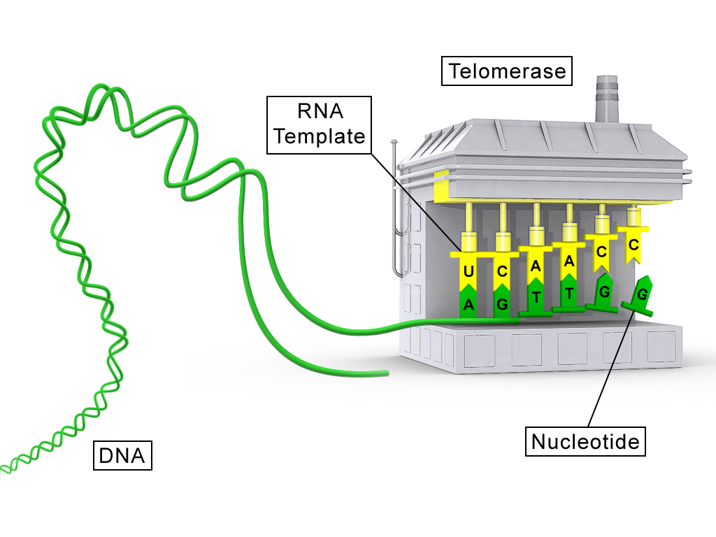
Telomerase is a reverse transcriptase (TERT) carrying its own RNA template (TERC). It base-pairs the RNA to the 3′ overhang, adds one repeat, translocates and repeats. Pol α–primase then fills in the complementary strand. Germ cells, stem cells and most cancers express telomerase; most somatic cells do not, so their telomeres shorten until they stop dividing (replicative senescence). Bacteria and most archaea avoid the problem with circular chromosomes.
DNA damage and repair
| Agent | Main lesion | Main fix |
|---|---|---|
| UVB (280–315 nm) | Absorbed directly by DNA: cyclobutane pyrimidine dimers and 6-4 photoproducts that kink the helix | Nucleotide excision repair |
| UVA (315–400 nm) | Mostly indirect: reactive oxygen species oxidize bases (8-oxoguanine); fewer dimers | Dimers by NER; oxidative lesions by other pathways |
| Ionizing radiation (X-rays, γ-rays) | Hydroxyl radicals and direct hits break the backbone; double-strand breaks are the dangerous lesion | NHEJ or HDR |
| Intercalating agents (ethidium bromide, acridines, doxorubicin) | Flat molecules slip between base pairs, stretch the helix and cause insertions or deletions during replication; some trap topoisomerase II, creating breaks | Frameshift mutations if unrepaired |
Nucleotide excision repair (NER) removes bulky, helix-distorting lesions by cutting out a short stretch of the damaged strand and resynthesizing it from the intact strand. In bacteria, UvrA and UvrB find the lesion, UvrC cuts on both sides (a 12–13 nucleotide piece), UvrD helicase removes it, and Pol I and ligase fill in. Eukaryotes do the same job with more proteins: XPC (or a stalled RNA polymerase, in transcription-coupled repair) detects damage, the TFIIH helicases open the DNA, and XPG and XPF–ERCC1 cut out about 24–32 nucleotides. Defects cause xeroderma pigmentosum, extreme UV sensitivity.
Double-strand breaks have two fates. Non-homologous end joining (NHEJ) ligates the ends directly. In eukaryotes, Ku70/80 binds the ends, DNA-PKcs and Artemis trim them, and XRCC4–ligase IV seals them. It works in any cell-cycle phase but often adds or deletes a few bases. Many bacteria (such as mycobacteria) have a minimal NHEJ of Ku plus ligase D; E. coli lacks it. Homology-directed repair (HDR) copies an intact homologous template, usually the sister chromatid, so it is accurate but limited to S and G2. The ends are resected to 3′ single-stranded tails (RecBCD in bacteria; MRN and CtIP in eukaryotes). A recombinase filament (RecA; Rad51, loaded by BRCA2) invades the homolog, polymerase copies across the gap, and Holliday junctions are resolved (RuvABC in bacteria).
Mutations
Substitutions swap one base: a transition swaps purine for purine or pyrimidine for pyrimidine, and a transversion swaps between classes. Insertions and deletions add or remove bases. Chromosomal rearrangements move large segments: deletions, duplications, inversions (pericentric if they include the centromere, paracentric if not) and translocations.
| Mutation | Effect on the protein |
|---|---|
| Silent | Codon changes but the amino acid does not (the code is redundant) |
| Missense | One amino acid changes; effect ranges from none to severe |
| Nonsense | A premature stop codon truncates the protein |
| Frameshift | Insertion or deletion not a multiple of three shifts every downstream codon |
| In-frame indel | Whole codons added or removed; frame preserved |
Eukaryotic gene expression and regulation
The central dogma describes information flowing from DNA to RNA to protein. In eukaryotes, transcription and RNA processing happen in the nucleus and translation in the cytoplasm, so every step offers a control point that bacteria lack.
Transcription
Eukaryotes use three nuclear RNA polymerases: Pol I makes most rRNA, Pol II makes mRNA (and most small nuclear and micro RNAs), and Pol III makes tRNA and 5S rRNA. Pol II cannot find a promoter alone. Initiation: TFIID, through its TATA-binding protein, binds the core promoter (often a TATA box about 25–30 bp upstream of the start site), then TFIIB, Pol II with TFIIF, TFIIE and TFIIH assemble into the preinitiation complex, with the Mediator complex relaying signals from activators. TFIIH unwinds the DNA and phosphorylates serine 5 of Pol II's C-terminal domain (CTD), releasing the polymerase. Elongation: Pol II reads the template strand 3′→5′ and builds RNA 5′→3′; serine 2 phosphorylation of the CTD recruits splicing and polyadenylation factors. Termination: after Pol II passes the polyadenylation signal, the RNA is cleaved, and an exonuclease (Xrn2) degrades the trailing RNA and knocks the polymerase off (the torpedo model).
The RNA matches the coding (non-template) strand with U for T. If a question gives the template strand, the mRNA is its reverse complement. Write the 5′ and 3′ labels before you transcribe.
RNA maturation

All three processing steps happen while the RNA is still being made, coordinated by the Pol II CTD.
5′ capping comes first, after about 20–30 nucleotides. A 7-methylguanosine is attached to the first nucleotide by an unusual 5′–5′ triphosphate bridge. The cap protects the mRNA from 5′ exonucleases, helps export, and is recognized by eIF4E to start translation.
Polyadenylation sets the 3′ end. CPSF recognizes the AAUAAA signal and CstF binds a GU-rich stretch downstream; the RNA is cut between them, about 10–30 nucleotides past AAUAAA, and poly(A) polymerase adds roughly 200 adenines without a template. Poly(A)-binding protein coats the tail, which stabilizes the mRNA and helps translation. Tail shortening (deadenylation) is usually the first step of mRNA decay.
Splicing removes introns. Most introns begin with GU at the 5′ splice site, end with AG at the 3′ splice site, and contain a branch-point A upstream. The spliceosome (snRNPs U1, U2, U4, U5 and U6 plus many proteins) does it in two transesterifications: the branch-point A's 2′ OH attacks the 5′ splice site, forming a lariat; then the freed 3′ OH of the upstream exon attacks the 3′ splice site, joining the exons and releasing the lariat. U1 pairs with the 5′ splice site and U2 with the branch point. Alternative splicing (exon skipping, alternative 5′ or 3′ sites, mutually exclusive exons, intron retention) lets one gene make several proteins, which is why humans have about 20,000 protein-coding genes but far more distinct proteins.
Regulatory elements
Cis-regulatory elements are DNA (or RNA) sequences that act only on the molecule they sit on. The core promoter sits at the start site; proximal promoter elements (CAAT and GC boxes) lie within a few hundred base pairs upstream. Enhancers can lie thousands of base pairs away, upstream, downstream or in introns, and work in either orientation, because the DNA loops (helped by Mediator and cohesin) to bring bound activators to the promoter. Silencers bind repressors, and insulators block an enhancer from acting on the wrong gene. Riboswitches are cis elements in RNA that change shape when they bind a metabolite. They are common in bacteria, but the eukaryotic examples known are thiamine pyrophosphate riboswitches in plants, fungi and algae, which control expression by changing splicing or 3′-end processing.
Trans-acting factors are diffusible molecules, mainly transcription factors. They are modular: a DNA-binding domain (zinc finger, helix-turn-helix, leucine zipper, helix-loop-helix) chooses the site, and a separate activation or repression domain recruits coactivators, Mediator or chromatin modifiers.
Epigenetic regulation changes expression without changing sequence, and the marks can be inherited through cell division. DNA methylation at CpG sites (by DNMTs) in a promoter usually silences it. Histone acetylation (by HATs, removed by HDACs) neutralizes lysine charges, loosens chromatin and favors transcription. Histone methylation depends on the site: H3K4me3 marks active promoters, while H3K9me3 and H3K27me3 mark silent chromatin. X-chromosome inactivation and genomic imprinting are classic epigenetic examples.
Yeast two-hybrid screening
The modular structure of transcription factors makes the yeast two-hybrid screen possible. The yeast activator Gal4 is split into its DNA-binding domain (BD) and activation domain (AD), which do nothing apart. The protein of interest (the bait) is fused to BD, and a library of candidate partners (the prey) is fused to AD. If bait and prey bind each other inside a yeast nucleus, BD and AD are brought together, the reconstituted activator turns on reporter genes (for example HIS3 for growth without histidine, or lacZ), and only those colonies grow or turn blue. Sequencing the prey plasmid identifies the partner, which is how transcription-factor partners and cofactors are found. Limits: interactions must happen in the nucleus, fusions can block binding, and false positives (a prey that activates alone) need a control with BD alone.
Translation
In eukaryotes, initiation starts at the cap. eIF4F (cap-binding eIF4E, scaffold eIF4G and helicase eIF4A) binds the cap, and eIF4G also binds poly(A)-binding protein, looping the mRNA into a closed circle. The 43S complex (small 40S subunit with eIF2–GTP carrying the initiator Met-tRNA) joins and scans 5′→3′ to the first AUG in a good Kozak context (a purine at −3 and G at +4). GTP hydrolysis releases the factors and the 60S subunit joins. Elongation: eEF1A delivers each charged tRNA to the A site, the large-subunit rRNA catalyzes peptide bond formation (the ribosome is a ribozyme), and eEF2 translocates the tRNAs from A to P to E. Termination: eRF1 recognizes UAA, UAG or UGA and eRF3 helps release the protein. Aminoacyl-tRNA synthetases charge each tRNA with the right amino acid beforehand; the tRNA anticodon pairs with the codon, with wobble at the third position.
Translation is regulated mostly at initiation. Stress kinases phosphorylate eIF2α, which shuts down most protein synthesis. When nutrients are scarce, mTOR is inactive and 4E-binding proteins sequester eIF4E. Specific mRNAs are controlled by elements in their untranslated regions: iron-regulatory proteins bind an iron-responsive element in the ferritin 5′ UTR and block scanning when iron is low; microRNAs guide the RISC complex to 3′ UTRs to repress translation and speed decay; upstream ORFs divert scanning ribosomes; and internal ribosome entry sites (IRES) allow cap-independent initiation, for example during viral infection.
Mobile genetic elements (State and National)
Transposable elements make up roughly 45% of the human genome. Barbara McClintock discovered them in maize (the Ac/Ds system). They are classified by how they move.
| Element | Intermediate | Enzymes | Hallmarks |
|---|---|---|---|
| DNA transposons (class II) | DNA; usually cut-and-paste | Transposase | Terminal inverted repeats; short target-site duplication flanking each insertion |
| LTR retrotransposons (class I) | RNA, copy-and-paste | Reverse transcriptase, integrase, RNase H (from pol) | Long terminal repeats; gag and pol but no env; replicate inside virus-like particles |
| Non-LTR retrotransposons: LINEs | RNA, copy-and-paste | ORF2 protein with endonuclease and reverse transcriptase | Target-primed reverse transcription: the RNA is copied directly at a nick in the target. LINE-1 is about 17% of the human genome |
| SINEs (such as Alu) | RNA | Borrow LINE-1 machinery | Short and non-autonomous; Alu is about 10% of the genome |
| Retroviruses (such as HIV) | RNA genome → DNA provirus | Reverse transcriptase, integrase, protease | LTRs plus gag, pol and env; env makes them infectious between cells |
A DNA transposon encodes a transposase that binds its inverted repeats, cuts the element out and inserts it at a staggered cut elsewhere; filling those staggered ends creates the target-site duplication. Because the copy leaves its old site, copy number rises mainly when transposition happens during replication. Retrotransposons are transcribed, and the RNA is reverse-transcribed into DNA that inserts at a new site, so the original stays put and copy number grows each time. Retroviruses follow the same route as LTR retrotransposons (reverse transcription in the cytoplasm, integrase inserting the provirus) but add an envelope gene to leave the cell. Their LTRs carry the promoter and polyadenylation signals for the provirus. Cells silence mobile elements with DNA methylation, heterochromatin and small RNAs (piRNAs in the germline), which ties this topic back to epigenetics.
Techniques
PCR
The polymerase chain reaction amplifies a target region exponentially. Denaturation at about 94–98 °C breaks the hydrogen bonds between strands. Annealing, a few degrees below the primers' melting temperature (Tm), lets two primers bind sequences flanking the target; too cool allows mispriming, too hot prevents binding. Extension at about 72 °C lets a thermostable polymerase (Taq) extend each primer 5′→3′. The reaction needs template, two primers, dNTPs, a thermostable polymerase, buffer and Mg²⁺. It needs no helicase (heat unwinds) and no ligase.

The 2027 rules name three applications.
Sanger sequencing is a one-primer extension reaction with a small fraction of fluorescent dideoxynucleotides (ddNTPs), which lack a 3′ OH and stop the chain wherever one is added. The result is a set of fragments ending at every position, separated by capillary electrophoresis and read from shortest (5′) to longest. It differs from PCR in using one primer, adding ddNTPs and making linear rather than exponential product. Read lengths are about 700–1,000 bases with very high accuracy, so it is the choice for checking a cloned insert or a single variant. Two overlapping peaks at one position on the trace mean the sample is heterozygous.
Error-prone PCR deliberately lowers fidelity (Taq, which has no proofreading, plus Mn²⁺, extra Mg²⁺ or unbalanced dNTPs) to scatter random mutations across a gene. Combined with screening, it is used for directed evolution and to find which residues matter for a function.
Reverse transcriptase–quantitative PCR (RT-qPCR) measures how much of a specific mRNA is present. Reverse transcriptase copies RNA into cDNA, then qPCR follows fluorescence each cycle. The Ct (threshold cycle) is the cycle at which signal crosses a set threshold: more starting template means a lower Ct, and each one-cycle difference is about a twofold difference.
Illumina and Nanopore sequencing
| Feature | Illumina (next-generation) | Nanopore (third-generation) |
|---|---|---|
| How it works | DNA fragments with adapters are amplified into clusters on a flow cell (bridge amplification), then read by sequencing by synthesis with reversible fluorescent terminators, one base per cycle | A motor protein ratchets a single native strand through a protein pore; the changing ionic current is decoded into bases |
| Read length | Short, about 50–300 bases (often paired-end) | Long, typically thousands to over a million bases |
| Accuracy | Very high per base (about 99.9%) | Lower per read, improving with newer chemistry |
| Amplification | Yes (cluster PCR) | Not needed; reads native DNA or RNA and can detect base modifications such as methylation |
| Best for | Counting (RNA-seq, ChIP-seq), variant calls, high throughput at low cost per base | Genome assembly across repeats, structural variants, real-time and field sequencing |
Choose the platform from the question: short, accurate reads for counting and small variants; long reads for repeats, rearrangements and full-length transcripts.
Molecular cloning
Cloning puts a gene into a vector so it can be copied and, often, expressed in a chosen organism. The major steps: (1) obtain the insert, usually by PCR from cDNA (use cDNA, not genomic DNA, for bacterial expression, since bacteria cannot splice introns); (2) assemble insert and vector; (3) transform E. coli and select on antibiotic; (4) screen colonies by colony PCR or restriction digest and confirm by Sanger sequencing; (5) move the plasmid into the expression host.
A plasmid vector needs an origin of replication, a selectable marker and a cloning site; an expression vector adds a promoter that works in the host (T7 or lac in bacteria, CMV or EF1α in mammalian cells), a ribosome-binding site or Kozak sequence, and a terminator or poly(A) signal. Restriction enzymes cut at short, usually palindromic sites (EcoRI: G^AATTC), often leaving sticky ends that pair with any end cut by the same enzyme; ligase seals them. PCR primers can add restriction sites to the insert's ends.
Two newer assembly methods join fragments without depending on sites already in the sequence:
| Method | How it works | Strengths |
|---|---|---|
| Gibson assembly | Fragments share 20–40 bp overlapping ends. In one 50 °C reaction, a 5′ exonuclease chews back to expose single-stranded 3′ overhangs, the overlaps anneal, a polymerase fills gaps and ligase seals | Seamless (no scar), any sequence, several fragments at once |
| Golden Gate cloning | Type IIS enzymes (such as BsaI) cut outside their recognition site, leaving custom 4-nt overhangs. Digestion and ligation run together; the recognition sites are lost from the correct product, so it can no longer be recut and accumulates | Many fragments in a set order in one pot, standardized parts, high efficiency |
Expression in mammalian cells can be transient or stable. In transient transfection (lipid reagents, electroporation), the plasmid stays outside the chromosomes, expression peaks within 1–3 days, and the plasmid is diluted and lost as cells divide. It is fast and good for short experiments. A stable cell line carries the gene integrated into its genome: cells are transfected (or transduced with a lentivirus), selected for weeks with a drug such as puromycin or G418, and single clones are expanded. It takes longer but gives uniform, lasting expression for long-term or large-scale work.
Knockouts, knockdowns and complementation
These experiments test what a gene does. A knockout permanently removes or disrupts the gene at the DNA level; a knockdown lowers its expression, usually at the RNA level, and is often partial and reversible. Complementation (rescue) adds a working copy back to the mutant to show the defect was caused by that gene, not an off-target change.
The logic is about necessity and sufficiency. A loss-of-function experiment (knockout or knockdown) tests necessity: if removing the gene removes the phenotype, the gene is needed for it. A gain-of-function experiment (expressing the gene where it is normally absent, or overexpressing it) tests sufficiency: if adding the gene produces the phenotype, it is enough on its own.
Making them (State and National). RNAi knocks down: a double-stranded siRNA or a vector-expressed shRNA is processed by Dicer and loaded into RISC, whose Argonaute protein uses the guide strand to cleave matching mRNA. Homologous recombination gene targeting knocks out: a vector carrying a selection marker flanked by sequences matching the gene (homology arms) replaces the gene in embryonic stem cells. Cre-Lox makes the knockout conditional.
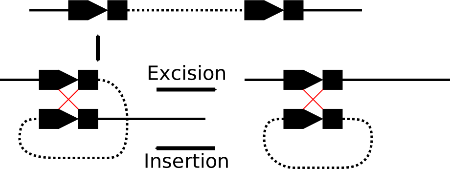
In Cre-Lox, the 34 bp loxP site (two 13 bp inverted repeats around an 8 bp asymmetric spacer) is placed around an essential exon (a "floxed" allele). The bacteriophage recombinase Cre recombines two loxP sites: same orientation excises the DNA between them, opposite orientation inverts it. Crossing a floxed mouse with one expressing Cre from a tissue-specific promoter deletes the gene only in that tissue, which avoids embryonic lethality. A tamoxifen-activated Cre (CreER) adds timing control.
CRISPR–Cas9 uses a 20-nucleotide guide RNA to direct Cas9 to a matching site next to an NGG PAM, where it makes a blunt double-strand break about 3 bp upstream of the PAM. NHEJ repair leaves small indels, and a frameshift in an early exon gives a knockout; adding a donor template lets HDR insert a precise change. A catalytically dead Cas9 fused to a repressor (CRISPRi) gives a knockdown without cutting. Limits: off-target cuts, in-frame indels that leave a working protein, and mosaicism in embryos.
Genomics assays (State and National)
| Assay | Procedure | Output and use | Main limits |
|---|---|---|---|
| ChIP-seq | Crosslink proteins to DNA with formaldehyde, shear chromatin, immunoprecipitate with an antibody to a transcription factor or histone mark, reverse crosslinks, sequence | Peaks where the protein binds; maps TF sites, enhancers, histone marks | Depends on antibody specificity; needs an input control; binding is not proof of regulation |
| Hi-C | Crosslink chromatin, cut with a restriction enzyme, mark ends with biotin, ligate nearby ends, pull down and sequence the junctions | Contact matrix showing which regions touch in 3D; reveals compartments, TADs, enhancer–promoter loops | Population average; resolution limited by sequencing depth |
| Bulk RNA-seq | Extract RNA from many cells, make cDNA, sequence | Average expression of every gene; differential expression; splice isoforms | Averages hide cell types; a change could be a shift in cell mix |
| Single-cell RNA-seq | Dissociate tissue, capture each cell (for example in droplets) with a barcode and unique molecular identifiers, then sequence | Expression per cell; clusters cell types and states, finds rare cells and trajectories | Sparse data with many missed transcripts; costly per cell; dissociation stress; loses spatial position |
Common pitfalls
How tournaments test this
The question archive holds 4,916 Designer Genes questions, 4,842 of them Division C, and the question bank holds 9,704 (9,184 Division C). About 3,090 archived questions are from the 2026 season, so recent tests dominate. The most represented tournaments are the Captains Summer Exchange (423 questions tagged 2026), the MIT Invitational (192, 2026), the Duke Invitational (160, 2021) and the Princeton Invitational (131, 2026). Older tests reflect earlier rules: the 196 archived questions that mention the lac operon, and the Sec/Tat items, come from seasons when gene expression was prokaryotic, so use them only for general transcription and translation logic.
Of the archived questions, about 47% are multiple choice, 43% free response, 5% multi-select, 3% numeric and 2% true/false. Read each multi-select option on its own. Task tags put "which of the following" at about 11%, "explain or why" at 9.7%, "calculate" at 4.3%, "compare or contrast" at 2.2% and "interpret data" at 1.2%. The tags undercount reasoning, and the 2027 rules push further toward calculations and data interpretation, so expect more of both than past tests show.
Subtopic tags show where weight has fallen: molecular biology on 2,247 archived questions, genetics 2,051, DNA 1,327, heredity 1,191, biotechnology 923 and Mendelian genetics 694. The most common correct answers are DNA ligase (33), incomplete dominance (30), autosomal recessive (29), DNA polymerase (27), autosomal dominant (24), RNA polymerase (23), epistasis (22), helicase (21) and prophase I (21); 50% and 25% are frequent numeric answers. Recurring phrasings include "what is the probability," "which stage of meiosis," "which enzyme is required," and "a cell lacking ... would be expected to." For probability, identify the rule; for stages, name the event that separates chromosomes; for enzymes, name the function first.
Test yourself
These questions come from official keys in the archive, re-solved before inclusion. Questions on topics outside the 2027 rules were left out, so there is no gene-expression block.
- 1
A pumpkin plant heterozygous for both orange pigment (orange is dominant, white is recessive) and warts (warts are dominant, no warts are recessive) is crossed with a pumpkin recessive for both traits. If the recombination frequency is 18%, how many of the 700 offspring will possess parental alleles?
Practice more Designer Genes questions →
- 1
Which model of DNA replication is generally accepted as correct?
From: University of Texas at Austin Invitational, 2019-20, Division C
Practice more Designer Genes questions →
- 1
Which components are required for a standard PCR reaction?
Practice more Designer Genes questions →
References and further reading
- Science Olympiad (soinc.org), for the 2027 rules manual and the event page.
- SciOly Wiki: Designer Genes, for event history. Use it as a pointer, not as a source for scope.
- OpenStax Biology 2e, for Mendelian genetics, meiosis, replication, eukaryotic transcription, RNA processing and translation.
- NCBI Bookshelf: Molecular Biology of the Cell and Molecular Cell Biology, for replication in the three domains, DNA repair, transposable elements and genome-editing methods.
- Addgene educational resources, for plasmid elements, Gibson and Golden Gate assembly, Cre-Lox and CRISPR.
- NHGRI Genetics Glossary, for definitions of linkage, epistasis, heritability and sequencing terms.
Official references
Sample notesheet
Download a printable, rule-compliant sample notesheet. Customize with your notes.