Heredity
2027 season
On this page(16 sections)
Heredity is a written test for teams of two about how traits pass from parents to offspring, how the information behind those traits is stored, copied, damaged and used to build proteins, and how allele frequencies change in whole populations. Almost every question comes back to four ideas: each parent passes one allele of each gene to each child, so probability does most of the work; genes sit on chromosomes, and meiosis is what sorts those chromosomes into gametes; DNA is copied, then transcribed and translated into protein, so a "what happens if" molecular question is almost always about one step in that chain; and a population's allele frequencies stay put unless something (selection, drift, migration, mutation, non-random mating) pushes them. This guide covers the whole event for Division B under the 2027 rules.
Event format
Heredity is a written test. The rules allow it to be arranged in stations, but each station is still a written question, so there is no lab, no build and no hands-on part. Here are the parameters that matter for preparation:
- Team size: up to 2 students.
- Time: about 50 minutes for the whole test.
- Calculators: each team may bring two Class II calculators.
- Writing: each team may bring writing utensils.
- Reference sheet: each team may bring one 8.5 × 11 inch sheet, with notes on either side, and the notes can come from any origin. Lamination or a protective sleeve is fine. Stuck-on labels are out, and so is more than one sheet, even if the extra sheets share a single sleeve.
- Scoring: the highest total wins, and preselected questions break ties. Points come from the quality and accuracy of answers, the quality of the supporting reasoning, and the use of proper scientific method. A correct number with no reasoning can lose points on the questions that ask you to explain.
The 2027 rules state plainly what the questions will stress: process skills. Expect quantitative reasoning, calculations, analyzing and interpreting experimental results, and drawing conclusions from evidence. In practice that means more data tables, cross results, pedigrees from case studies and "what does this experiment show" items, and fewer pure vocabulary questions.
Two things follow from this format. First, you are graded on your reasoning as well as your answer, so write the one line of logic that justifies each answer. Second, the sheet is the only reference you bring, so it has to be built for speed: ratios, equations and tables, not paragraphs.
What the rules cover
Scope below follows the 2027 Division B rules manual for Heredity (section 3). It lists six topic areas, numbered i through vi, and says the list is exhaustive, so if a topic is not on it, do not spend study time on it for this event. The rules also say no single topic should be overemphasized, and that every area expects quantitative reasoning, interpretation of experimental results and evidence-based conclusions. The table paraphrases the scope by level.
| Area (rules item) | What you must be able to do | Level |
|---|---|---|
| i. Mendelian genetics (i.1–i.4) | Explain Mendel's laws and what they imply; build mono- and dihybrid Punnett squares; predict offspring genotypes and phenotypes and their probabilities from crosses and data; read pedigrees to find the mode of inheritance (dominant or recessive, autosomal or sex-linked) and draw a pedigree from a case study | All levels |
| i. Mendelian genetics (i.5–i.6) | Describe patterns that break Mendel's laws (linkage, incomplete dominance, codominance, complementation); apply recessive epistasis to predict phenotypes | State & National |
| ii. Mitosis and meiosis (ii.1–ii.2) | Compare the stages and key structures of mitosis and meiosis; know the steps of human karyotype analysis and interpret a karyotype | All levels |
| iii. Molecular biology of DNA (iii.1–iii.4) | Explain nucleotide and backbone structure, 5′/3′ directionality and base pairing; describe replication with a focus on elongation, the fork and proofreading; describe DNA organization (plasmids, chromatin, euchromatin, heterochromatin, chromosomes); classify DNA mutations and predict their effect on the protein (frameshift, silent, missense, nonsense) | All levels |
| iii. Molecular biology of DNA (iii.5) | Explain how UVA and UVB damage DNA and how nucleotide excision repair fixes it | State & National |
| iv. Gene and protein relationship (iv.1–iv.3) | Explain the central dogma; describe initiation, elongation and termination of transcription and translation; explain how RNA polymerase and the ribosome work, the roles of mRNA and tRNA, regulation by activators and repressors, and RNA processing (splicing, capping, polyadenylation) | All levels |
| iv. Gene and protein relationship (iv.4) | Explain the function and mechanism of the lac operon | State & National |
| iv. Gene and protein relationship (iv.5) | Explain the function and mechanism of the trp operon | National only |
| v. Population and evolutionary genetics (v.1–v.2) | State the Hardy-Weinberg assumptions and what happens when they fail; do Hardy-Weinberg calculations from data; relate changing allele frequencies to genetic drift, founder effects, bottlenecks, migration and relative fitness | All levels |
| v. Population and evolutionary genetics (v.3) | Explain how additive alleles produce continuous variation, and estimate how many genes contribute to a trait | State & National |
| vi. Technology and techniques (vi.1) | Describe what happens in each step of PCR (denaturation, annealing, extension), the molecules involved, the role of temperature, the components of a reaction, and which questions PCR can answer | All levels |
| vi. Technology and techniques (vi.2) | Describe how Sanger sequencing differs from standard PCR, and which questions it can answer | State & National |
Where each rules item is taught in this guide. i: Mendelian genetics and Pedigrees and sex-linked inheritance. ii: Mitosis, meiosis and karyotypes. iii: Molecular biology of DNA and DNA replication. iv: Gene expression: transcription and translation. v: Population and evolutionary genetics. vi: Technology and techniques. The Condensed notesheet at the end collects the ratios, formulas and tables for your one allowed sheet.
What changed from last season matters for anyone reusing old notes. Population and evolutionary genetics is a whole new area (Hardy-Weinberg, drift, founder effects, bottlenecks, migration, relative fitness, and polygenic traits at State and Nationals). The trp operon is new at Nationals. Classifying mutations is now an every-level skill, and UV damage with excision repair takes the State & National slot. Base-pairing rules, proofreading at the replication fork and RNA maturation (splicing, capping, polyadenylation) are now named explicitly, while Chargaff's rules are no longer a separate item (they still follow from base pairing). Old Heredity tests therefore have very few population-genetics questions (about 50 of 3,318 archive items), so practice those calculations from the worked examples below and from Division C tests (Designer Genes and older Heredity tests), which have covered Hardy-Weinberg.
Strategy
Split the work. With two students, the most efficient split is by skill, not by topic. One partner takes the calculations: crosses, probability, pedigrees, recombination, Hardy-Weinberg, selection and PCR arithmetic. The other takes the explanation and molecular questions: meiosis, replication, repair, transcription, translation, operons and techniques. Both partners should skim every question once so that neither is surprised by a multi-part station. When one partner finishes a calculation, they should hand it to the other to check the reasoning line.
Build the sheet around what you will be asked to compute. The sheet is the only reference you bring, so spend it on the things you would otherwise have to derive under time pressure:
- The ratio table for crosses (3:1, 1:1, 9:3:3:1, and, for State and Nationals, 1:2:1 for incomplete dominance, 9:3:4 and 9:7).
- The product and sum rules, and the conditional probability formula.
- The pedigree symbols and the four basic inheritance patterns, each with one sentence of identifying clues.
- The Hardy-Weinberg equations, the five assumptions, and the one-line formulas for migration and selection against a recessive.
- For State and Nationals, the polygenic shortcuts: (1/4)ⁿ for an extreme class and 2n + 1 phenotype classes.
- The codon table (it fits in a 4 × 16 grid) and the one-letter amino-acid codes that are not the first letter (R, K, N, D, E, Q, F, W, Y).
- The chi-square formula with the critical values for 1–5 degrees of freedom, and the binomial formula.
- An enzyme → job table for replication and gene expression, and a karyotype-syndrome table.
- Short-form stages of mitosis and meiosis, with the event that separates each pair of partners (homologs in meiosis I, sister chromatids in mitosis and meiosis II).
- A lac versus trp operon comparison, PCR cycle temperatures and the Sanger difference.
Affixed labels are prohibited, so everything must be on the sheet itself. Put the most-used entries at the top. The Condensed notesheet section is a starting layout you can print and then personalize.
Manage time in stations. Work the quick items first: definitions, true/false, identify-the-structure. Then do the calculations, which are the most likely to take several minutes. Save the explanation questions for last, but leave time for them, because they carry reasoning points. If a calculation stalls, write down the setup (the genotypes, the frequencies and the operation you would apply) so that you can still earn method points.
Show your reasoning in one line. For example: "Two unaffected parents have an affected son, so the trait is recessive. Every affected individual is male and no affected father has an affected son, so X-linked recessive is most likely." That line is what earns the reasoning points.
Mendelian genetics
Mendel's work is usually summarized as three ideas: dominance (a heterozygote shows the dominant phenotype) and two laws. The law of segregation says that the two alleles for each gene separate when gametes form, so each gamete carries one allele per gene. The law of independent assortment says that alleles of genes on different chromosomes sort independently when gametes form. Independent assortment is why a dihybrid cross produces four phenotype classes in a 9:3:3:1 ratio. Both laws describe what happens to unlinked genes in meiosis, and their main implication is that inheritance is predictable with probability. Exceptions to them are tested at State and Nationals (see below).
Important vocabulary: a gene is a segment of DNA that codes for a functional product (usually a protein, sometimes an RNA) that influences a trait; its position on a chromosome is its locus; an allele is one version of that gene. A genotype is the set of alleles an organism carries (such as Aa). A phenotype is the observable trait (such as "purple flowers"). An organism with two different alleles is heterozygous (a hybrid); with two copies of the same allele, homozygous. Dominant alleles are expressed when at least one copy is present, and recessive alleles are expressed only when two copies are present, in a diploid organism. The wild type is the most common form in natural populations. A bacterium or other haploid organism has only one allele of each gene, so a recessive allele shows directly in its phenotype.
Mendel's experiments
Gregor Mendel, an Augustinian friar in Brno, crossed garden peas (Pisum sativum) for about eight years and published his results in 1866. The work was ignored until 1900, when Hugo de Vries, Carl Correns and Erich von Tschermak independently rediscovered it. Around 1902–1903, Walter Sutton and Theodor Boveri pointed out that chromosomes behave in meiosis exactly as Mendel's "factors" do: this is the chromosome theory of inheritance.
Peas were a good choice: they grow quickly and produce many seeds; they normally self-pollinate, so true-breeding lines were available, but Mendel could also cross-pollinate them by removing a flower's stamens and dusting its stigma with pollen from another plant; and he picked seven characters that came in two clear-cut forms with no intermediates. He counted every offspring and analyzed the numbers, which was new for his time.
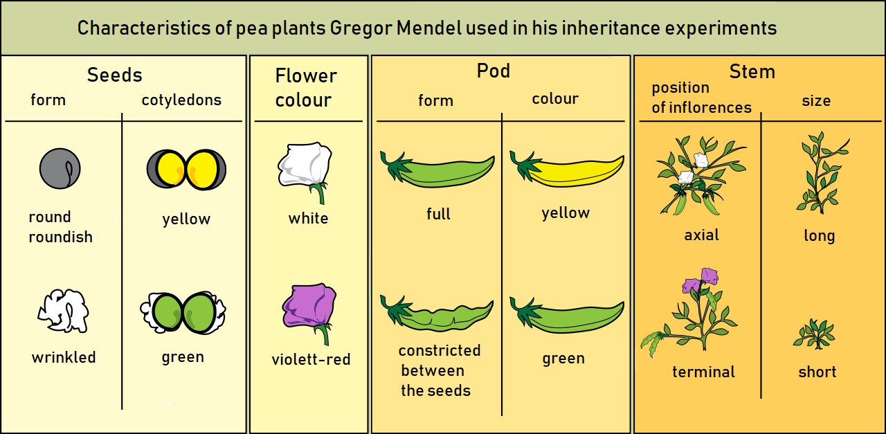
His crosses have a standard notation. The P (parental) generation is two true-breeding plants (homozygous, so they produce only offspring like themselves when selfed) that differ in a character, for example round × wrinkled seeds. Their offspring are the F₁ (first filial) generation, all hybrids. Selfing (or intercrossing) the F₁ gives the F₂ generation. In every monohybrid cross Mendel's F₁ all showed one form (which he called dominant), and the other form (recessive) reappeared in about a quarter of the F₂: for seed shape, 5,474 round to 1,850 wrinkled, a ratio of 2.96 : 1. The recessive form had not been blended away, only hidden, which is the evidence for particulate inheritance and for segregation. A reciprocal cross swaps which parent carries which trait (female round × male wrinkled, then female wrinkled × male round); identical results show that inheritance does not depend on which parent supplied the trait, ruling out sex linkage (in animals) and maternal/cytoplasmic inheritance.
Punnett squares
A Punnett square lists the gametes from each parent along the top and side, then fills each box with the combination of one allele from each parent. Each box is an equally likely offspring, so the fraction of boxes with a trait is the probability of that trait. The diagram is named after Reginald Punnett.

For a monohybrid cross between two heterozygotes (Aa × Aa), the genotype ratio is 1 AA : 2 Aa : 1 aa, and the phenotype ratio is 3 dominant : 1 recessive when the alleles show complete dominance. A test cross crosses an organism of unknown genotype with a homozygous recessive (aa). If the offspring are 1:1 dominant to recessive, the unknown parent is heterozygous. If every offspring shows the dominant trait, the unknown parent is most likely homozygous dominant. The bigger the all-dominant sample, the stronger that conclusion: with 10 dominant offspring and no recessives, the chance that a heterozygote would produce that result is (1/2)¹⁰ ≈ 0.001. (Test crosses are used in plants and animals; in humans, pedigree analysis takes their place.)
Among the dominant F₂ of an Aa × Aa cross, 2/3 are heterozygous and 1/3 homozygous, because two of the three dominant boxes are Aa. This 2/3 shows up constantly in pedigree problems.
The common crosses, for one gene with complete dominance:
| Cross | Genotypes of offspring | Phenotypes of offspring |
|---|---|---|
| AA × aa | all Aa | all dominant |
| Aa × Aa | 1 AA : 2 Aa : 1 aa | 3 dominant : 1 recessive |
| Aa × aa (test cross) | 1 Aa : 1 aa | 1 dominant : 1 recessive |
| AA × Aa | 1 AA : 1 Aa | all dominant |
| aa × aa | all aa | all recessive |
Because the 2027 rules stress experimental data, expect crosses where you are given offspring counts and must work backward. Real counts never match a ratio exactly: 152 purple to 48 white is close to 3:1 (150:50), so the parents were most likely both heterozygous. Say "consistent with" rather than "proves", and name the ratio you compared against. The chi-square test below tells you whether a deviation is small enough to be chance.
Probability rules
Probability is how the event turns genetics into arithmetic. Two rules cover nearly every cross:
The branch (forked-line) method. For three or more genes, a Punnett square gets huge (64 boxes for a trihybrid). Instead, solve each gene separately and multiply. For AaBbCc × AaBbCc, P(A_B_C_) = (3/4)³ = 27/64 and P(aabbcc) = (1/4)³ = 1/64. The full phenotype ratio is 27 : 9 : 9 : 9 : 3 : 3 : 3 : 1. The same logic handles unequal parents: for AaBbCc × aaBbCC, P(aaB_C_) = 1/2 × 3/4 × 1 = 3/8.
Binomial probability. When a question asks for a specific number of children of a given type in a family of a given size, in any birth order, you must count every possible order. Each particular order has probability , and there are orders.
Chi-square test for cross data
Observed offspring counts never match a predicted ratio exactly. The chi-square (χ²) goodness-of-fit test asks whether the difference is small enough to be due to chance. The null hypothesis is that the data fit the predicted ratio (for example, that the genes assort independently with 9:3:3:1).
Compare your χ² with the critical value for your degrees of freedom:
| Degrees of freedom | 1 | 2 | 3 | 4 | 5 |
|---|---|---|---|---|---|
| Critical χ² at p = 0.05 | 3.84 | 5.99 | 7.81 | 9.49 | 11.07 |
| Critical χ² at p = 0.01 | 6.63 | 9.21 | 11.34 | 13.28 | 15.09 |
Failing to reject does not prove the hypothesis; it means the data are consistent with it. A large χ² in a dihybrid test cross is the usual first sign of linkage.
Dihybrid crosses and linkage (linkage: State & National)
A dihybrid cross tracks two genes. For two genes that assort independently and show complete dominance, AaBb × AaBb gives 9 A_B_ : 3 A_bb : 3 aaB_ : 1 aabb. The 9:3:3:1 ratio is the expected result when the genes are on different chromosomes. A dihybrid test cross (AaBb × aabb) gives 1 AaBb : 1 Aabb : 1 aaBb : 1 aabb, which is the cleanest test of independence, because each offspring's phenotype shows exactly which gamete it got from the heterozygous parent.

Linkage is the exception, and it is a State and National topic. Genes on the same chromosome tend to be inherited together, and they only separate when crossing over happens between them during meiosis I. The frequency of recombinant offspring estimates how far apart two genes are on a chromosome. One percent recombination is one map unit (centimorgan, cM).
History. William Bateson and Reginald Punnett noticed in 1905 that some sweet-pea traits did not assort independently ("coupling"). Thomas Hunt Morgan, working with Drosophila at Columbia University, explained this in 1911: genes on the same chromosome are linked, and crossing over between homologs, which he proposed using F. A. Janssens' observations of chiasmata, produces new combinations. In 1911 (published 1913) his undergraduate student Alfred Sturtevant used recombination frequencies to build the first genetic (linkage) map, of genes on the fruit fly X chromosome, reasoning that genes farther apart cross over more often.
Coupling and repulsion. In a double heterozygote, the alleles can be arranged two ways. In coupling (cis), the two dominant alleles are on one chromosome and the two recessive alleles on the other: AB/ab. In repulsion (trans): Ab/aB. The arrangement decides which offspring classes count as parental. Parental (non-recombinant) gametes carry an allele combination that was on one of the parent's chromosomes; recombinant gametes carry a new combination made by crossing over. In a test cross the parental classes are the two most frequent classes, and the recombinant classes are the two least frequent.
Why 50% is the ceiling. Even genes very far apart on one chromosome, with crossovers almost every meiosis, give at most 50% recombinant offspring, the same as unlinked genes (whose test cross gives 1:1:1:1, with half the offspring "recombinant"). So a recombination frequency near 50% means the genes are unlinked or far apart on the same chromosome; below 50% means linkage. Map distances are roughly additive for nearby genes; over long distances, undetected double crossovers make recombination frequency underestimate true distance. Recombination rates also vary along a chromosome, so map distance does not convert exactly to base pairs.
Three-point test cross (gene order and mapping). Cross a triple heterozygote with a triple homozygous recessive. Eight classes appear in reciprocal pairs:
- The two largest classes are the parental (non-recombinant) types.
- The two smallest classes are the double crossovers (DCO).
- Compare a DCO with a parental type: the one gene that is "switched" is the middle gene.
- For each interval, add the single-crossover classes for that interval plus the DCOs (a double crossover is a crossover in both intervals), divide by the total and multiply by 100.
Patterns that break Mendel's ratios (State & National)
Not every trait follows a clean dominant–recessive pattern. These patterns change the phenotype ratios, so learn the ratio for each one.
- Incomplete dominance: the heterozygote has an intermediate phenotype. A red × white cross gives all pink offspring, and the F2 phenotype ratio is 1:2:1 (red : pink : white). Examples: snapdragon and four-o'clock flower color, blue Andalusian fowl (black × white). Because each genotype has its own phenotype, the genotype and phenotype ratios are the same.
- Codominance: both alleles are fully expressed in the heterozygote. In the ABO system, the Iᴬ and Iᴮ alleles are codominant, so an IᴬIᴮ person has both A and B antigens. Roan Shorthorn cattle (red and white hairs side by side) are the other standard example.
- Complementation: the trait needs a functional product from each of two genes, so a dominant allele at both is required. A dihybrid cross then gives 9 : 7, because only the double-dominant class shows the trait. The same logic powers the complementation test: cross two true-breeding recessive mutants with the same defect. If the offspring are normal, the mutations are in different genes (each parent supplies the working copy the other lacks). If the offspring are still mutant, both mutations are in the same gene.
Multiple alleles. A gene can have more than two alleles in a population, even though each diploid individual carries only two. The ABO blood group gene has three: Iᴬ and Iᴮ (codominant to each other) and i (recessive to both). Rabbit coat color (full color C > chinchilla cᶜʰ > Himalayan cʰ > albino c, a dominance series) and Drosophila eye color are other examples.
| Blood type (phenotype) | Possible genotypes | Antigens on red cells | Antibodies in plasma | Can receive red cells from |
|---|---|---|---|---|
| A | IᴬIᴬ or Iᴬi | A | anti-B | A, O |
| B | IᴮIᴮ or Iᴮi | B | anti-A | B, O |
| AB | IᴬIᴮ | A and B | neither | A, B, AB, O (universal recipient) |
| O | ii | neither | anti-A and anti-B | O only (O is the universal donor) |

Rh factor. The Rh (D) antigen is inherited separately from ABO. Rh-positive (DD or Dd) is dominant to Rh-negative (dd). Treat it as a second, independent gene: an A⁻ (Iᴬi dd) × B⁻ (Iᴮi dd) couple can have A⁻, B⁻, AB⁻ or O⁻ children, but never Rh-positive ones. An Rh-negative mother carrying an Rh-positive fetus can make anti-D antibodies after the first pregnancy that attack a later Rh-positive fetus (hemolytic disease of the newborn), which is prevented with anti-D injections.
Other terms that tests ask you to match (background vocabulary; the 2027 rules do not name them, but older tests use them constantly):
| Term | Meaning | Example |
|---|---|---|
| Lethal allele | An allele that kills its carrier, usually when homozygous; turns 3:1 into 2:1 among survivors | Yellow coat in mice: Yy × Yy gives 2 yellow : 1 agouti, because YY dies before birth |
| Pleiotropy | One gene affects several different traits | Marfan syndrome (FBN1) affects skeleton, eyes and heart; cystic fibrosis affects lungs, pancreas and sweat |
| Polygenic inheritance | Several genes affect one trait | Height, skin color (see the additive model below) |
| Penetrance | The percentage of individuals with a genotype who show the phenotype at all | Polydactyly alleles with incomplete penetrance can skip a carrier |
| Expressivity | How strongly the phenotype shows in those who have it | Different numbers of extra digits in polydactyly |
| Sex-limited trait | Autosomal gene expressed in only one sex | Milk production in cattle, beard growth |
| Sex-influenced trait | Autosomal gene whose dominance differs between the sexes | Male-pattern baldness: the heterozygote is bald in men but not in women |
| Multifactorial trait | Several genes plus the environment | Spina bifida, cleft lip, heart disease |
| Environmental effect | Same genotype, different phenotype in different conditions | Himalayan rabbits: a temperature-sensitive pigment enzyme makes the cooler ears, nose and feet dark |
Recessive epistasis (State & National)
Epistasis means one gene masks the phenotype set by another. The 2027 rules name only recessive epistasis: a homozygous recessive genotype at one gene (aa) hides whatever the second gene would have shown. The classic case is coat color in Labrador retrievers. Gene B sets pigment color (B_ black, bb brown), but a dog that is ee cannot deposit pigment in its fur and is yellow no matter what it carries at B. The masking gene is epistatic; the masked gene is hypostatic. Albinism is the same idea in many mammals: a cc genotype makes no melanin at all, so it hides every other coat-color gene.
Old tests also use other modified dihybrid ratios. The first two rows below are in the rules (recessive epistasis and complementation); the other four are not named, so learn them for recognition only. Every one is a regrouping of the 16 boxes of 9:3:3:1.
| F₂ ratio from AaBb × AaBb | Name | How the 9:3:3:1 classes combine |
|---|---|---|
| 9 : 3 : 4 | Recessive epistasis | aa hides B: 3 aaB_ + 1 aabb = 4 |
| 9 : 7 | Duplicate recessive epistasis (complementation) | Need A_ and B_: 3 + 3 + 1 = 7 |
| 12 : 3 : 1 | Dominant epistasis | A_ hides B: 9 A_B_ + 3 A_bb = 12 |
| 15 : 1 | Duplicate dominant genes | A_ or B_ is enough: 9 + 3 + 3 = 15 |
| 9 : 6 : 1 | Additive (duplicate interaction) | One dominant at either gene gives an intermediate: 3 + 3 = 6 |
| 13 : 3 | Dominant suppression | A_ suppresses B: 9 + 3 + 1 = 13 |
Pedigrees and sex-linked inheritance
A pedigree is a family tree in which squares are males, circles are females, shaded symbols are affected individuals, and a diagonal line marks a deceased individual. Generations are numbered with Roman numerals and individuals within a generation with Arabic numerals. The rules ask you to evaluate a pedigree to find the mode of inheritance, and to build one from case-study data. Both skills start with the same four questions:
- Is the trait present in every generation? If so, it is probably dominant. If it skips generations, it is probably recessive.
- Do affected males and females show the trait at similar rates? If yes, the gene is probably autosomal. If affected males far outnumber affected females, the gene is probably X-linked recessive. A trait seen only in males, father to son every generation, is Y-linked.
- Does the trait pass from an affected father to a son? A father gives his son a Y, not an X, so true father-to-son transmission rules out X-linkage. For a rare trait, assume an unrelated, unaffected mother who married into the family is not a carrier. If no father-to-son transmission appears, that fits X-linked inheritance but does not prove it, especially in a small pedigree.
- Do two unaffected parents have an affected child? If yes, the trait is recessive. If it is autosomal, both parents are carriers. If it is X-linked (possible only when the affected child is a son), only the mother is a carrier, since an unaffected father cannot carry an X-linked recessive allele; an affected daughter of an unaffected father rules out X-linked recessive.
| Symbol | Meaning |
|---|---|
| Square / circle / diamond | Male / female / sex unknown or unspecified |
| Shaded (filled) symbol | Affected individual |
| Open (unfilled) symbol | Unaffected individual |
| Half-shaded symbol, or a dot in the center | Known heterozygous carrier (a dot in a circle often marks an X-linked carrier female) |
| Diagonal line through a symbol | Deceased |
| Horizontal line between a square and a circle | Mating (partners) |
| Double horizontal line | Consanguineous mating (partners are blood relatives) |
| Vertical line down to a horizontal sibship line | Children of that mating, oldest on the left |
| Two diagonal lines from one point | Twins (with a horizontal bar between them: identical twins) |
| Brackets around a symbol | Adopted |
| Arrow pointing at a symbol | Proband: the person through whom the family came to attention |
| Roman numerals (I, II, III) and Arabic numerals | Generation, and individual within a generation (II-3) |

The four basic patterns differ in who is affected and how the trait travels. Use this table as a quick filter, then confirm with the pedigree.
| Pattern | Typical clue | Affected parent to child | Example |
|---|---|---|---|
| Autosomal dominant | Trait in every generation; unaffected parents have unaffected children | Passes to about half of children, either sex | Huntington disease |
| Autosomal recessive | Trait skips generations; affected child of unaffected parents | Usually from two carrier parents | Cystic fibrosis |
| X-linked recessive | Affected males far more common; carrier mothers pass it to sons | Affected father passes to daughters (who are carriers), never to sons | Red-green color blindness |
| X-linked dominant | Every daughter of an affected father is affected; no father-to-son transmission | Affected father passes to every daughter and no son; an affected mother passes to about half of her children | Hypophosphatemic rickets |
| Y-linked | Only males, every son of an affected father | Father to all sons, never to daughters | Some Y-chromosome deletions that reduce sperm production (very few Y-linked traits are known) |
Sex-linked traits are usually carried on the X chromosome, which females have two of (XX) and males one (XY). Males have only one X, so a recessive X-linked allele shows up in every male who inherits it; males are hemizygous for X-linked genes. That is why the rate of an X-linked recessive trait in males equals the allele frequency, while females need two copies. Y-linked traits pass only from father to son, so they never appear in daughters.
Notation for X-linked genes. Write the allele as a superscript on the X: Xᴴ (normal) and Xʰ (hemophilia). The genotypes are XᴴXᴴ (normal female), XᴴXʰ (carrier female), XʰXʰ (affected female), XᴴY (normal male) and XʰY (affected male). The Y carries no allele for the gene. Always write the sex chromosomes in X-linked Punnett squares, so that the sons and daughters come out separately.
Who passes what. A son's X always comes from his mother, and his Y from his father, so a son's X-linked trait comes from his mother. A daughter gets her father's only X (so every daughter of an affected father with an X-linked dominant trait is affected, and every daughter of a man with an X-linked recessive trait is at least a carrier), plus one of her mother's two. A woman's paternal X came from her father's mother, never her father's father.
Constructing a pedigree from a case study. Draw generation I at the top, connect mates with a horizontal line, hang children from a vertical line in birth order (oldest on the left), and shade affected people only. Mark known carriers with a dot or half-shading only if the case gives you that information. Then label every individual (I-1, I-2, II-1 and so on) so your reasoning can refer to them.
Probabilities from pedigrees. Chain the probabilities through each generation: the chance that each person is a carrier, times the chance that each passes the allele, times 1/4 for two carriers having an affected child. A person who is unaffected but has an affected sibling (and unaffected carrier parents) has a 2/3 chance of being a carrier, not 1/2.
Morgan's white-eyed fly. Sex linkage was discovered by Thomas Hunt Morgan in 1910. He found a white-eyed male Drosophila and crossed it with a red-eyed female: the F₁ were all red-eyed, and the F₂ were 3 red : 1 white, but every white-eyed fly was male. The reciprocal cross (white-eyed female × red-eyed male) gave red-eyed daughters and white-eyed sons ("criss-cross" inheritance). Both results fit a recessive allele on the X chromosome, and they were the first evidence that a specific gene lies on a specific chromosome. His student Calvin Bridges later confirmed the chromosome theory using flies with X-chromosome nondisjunction.
In Drosophila, sex depends on the ratio of X chromosomes to sets of autosomes rather than on the Y: XX flies are female, XY and XO flies are male, and XXY flies are female. In birds, butterflies and some reptiles the system is reversed: females are ZW and males ZZ, so Z-linked recessive traits show up more often in females, and a son gets one Z from each parent.
Human disorders and traits by inheritance mode
Tests regularly name a condition and ask for its inheritance pattern, or give a pattern and ask for an example. The table lists the ones that recur. Chromosome-number conditions (Down, Turner, Klinefelter and others) are in Karyotypes.
ReferenceSingle-gene human disorders and traits· 20 rows
AD = autosomal dominant, AR = autosomal recessive, XR = X-linked recessive, XD = X-linked dominant.
No rows match.
| Condition or trait | Mode | Gene and product | Key facts tests use |
|---|---|---|---|
| Huntington disease | AD | HTT (huntingtin), chromosome 4 | Expanded CAG repeat; symptoms (movement, mood, dementia) usually start in midlife, often after the person has had children; longer repeats tend to mean earlier onset; no cure, only symptom treatment |
| Achondroplasia | AD | FGFR3 (growth factor receptor), gain of function | Most common form of dwarfism; most cases are new mutations; homozygosity is usually lethal |
| Marfan syndrome | AD | FBN1 (fibrillin-1, connective tissue) | Pleiotropic: tall with long limbs and fingers, lens dislocation, aortic enlargement |
| Polydactyly (isolated) | Usually AD | Several genes | Extra fingers or toes; incomplete penetrance and variable expressivity, so it can seem to skip a generation |
| Familial hypercholesterolemia | AD (incompletely dominant) | LDLR (LDL receptor) | Heterozygotes have high cholesterol; homozygotes, with no working receptors, are far more severe |
| Neurofibromatosis type 1 | AD | NF1 (neurofibromin) | Café-au-lait spots and nerve tumors; variable expressivity |
| Cystic fibrosis | AR | CFTR (chloride channel), chromosome 7 | Thick mucus in lungs and pancreas, salty sweat; most common allele ΔF508 (3-base deletion); among the most common serious AR disorders in people of European ancestry |
| Sickle cell disease | AR | HBB (β-globin), chromosome 11 | Glu → Val at position 6; sickled red cells; carriers (sickle cell trait) resist malaria (heterozygote advantage); the two alleles are codominant at the protein level |
| Phenylketonuria (PKU) | AR | PAH (phenylalanine hydroxylase) | Cannot convert phenylalanine to tyrosine; phenylalanine builds up and damages the brain; detected by newborn screening and managed with a low-phenylalanine diet |
| Tay-Sachs disease | AR | HEXA (hexosaminidase A) | GM2 ganglioside (a fatty substance) builds up in nerve cells; fatal in early childhood; more common in Ashkenazi Jewish, Cajun and French-Canadian populations |
| Albinism (oculocutaneous type 1) | AR | TYR (tyrosinase) | No melanin; an example of recessive epistasis over other pigment genes |
| Hemophilia A / B | XR | F8 / F9 (clotting factors VIII / IX) | Blood fails to clot normally; almost all patients male; the "royal disease" carried through Queen Victoria's descendants was hemophilia B |
| Duchenne muscular dystrophy | XR | DMD (dystrophin, the largest human gene) | Progressive muscle wasting beginning in early childhood; affects boys |
| Red-green color blindness | XR | Opsin genes on the X | The most common form of color blindness; about 8% of males of European ancestry, under 1% of females |
| Hypophosphatemic (vitamin D–resistant) rickets | XD | PHEX | Affected father → all daughters affected, no sons |
| Fragile X syndrome | X-linked | FMR1 | Expanded CGG repeat; most common inherited cause of intellectual disability; affects males more severely |
| Androgen insensitivity | XR | AR (androgen receptor) | XY person with female external appearance; testes present |
| PTC tasting | Classic dominant trait | TAS2R38 (bitter taste receptor) | Tasters (T_) find phenylthiocarbamide bitter; non-tasters are tt |
| ABO blood type | Multiple alleles, codominance | ABO (glycosyltransferase) | Iᴬ, Iᴮ codominant; i recessive |
| Rh factor | Dominant (Rh⁺) | RHD | Rh⁺ = DD or Dd; Rh⁻ = dd |
Textbook "Mendelian" human traits such as tongue rolling, attached earlobes, widow's peak, dimples and freckles are often taught as single-gene dominant/recessive pairs in test questions. Real inheritance of most of them is more complicated, but if a question states the dominance, use it.
Mitosis, meiosis and karyotypes
Mitosis and meiosis both divide the nucleus, but they serve different purposes. Mitosis makes two genetically identical diploid cells (2n) and is used for growth, tissue repair and asexual reproduction; it happens in somatic (body) cells. Meiosis makes four haploid cells (n) from one diploid cell and is used to make gametes (or spores, in plants and fungi); in animals it happens only in germ-line cells of the gonads. The difference that matters in genetics is what happens to homologous chromosomes. In meiosis, homologs pair during prophase I and then separate. In mitosis, homologs never pair, and sister chromatids separate.
Vocabulary that tests lean on. Homologous chromosomes are a matching pair, one from each parent, with the same genes in the same order (possibly different alleles). Sister chromatids are the two identical copies made by replication, joined at the centromere by cohesin proteins. A tetrad (or bivalent) is a synapsed pair of homologs in prophase I: 2 chromosomes, 4 chromatids. Diploid (2n) cells have two sets of chromosomes; haploid (n) cells have one set. Polyploid cells have more than two complete sets (3n triploid, 4n tetraploid); seedless watermelons and bananas are triploid, which disrupts pairing in meiosis and makes them sterile.
Key structures: the centromere (constricted region where sister chromatids are held together); the kinetochore (protein complex built on each centromere where spindle microtubules attach); spindle fibers (microtubules, hollow polymers of the protein tubulin) organized by centrosomes, which in animal cells contain a pair of centrioles (plant cells have no centrosomes and organize the spindle from the nuclear envelope region); the chiasma (plural chiasmata, the visible X-shaped point where crossing over happened); and the synaptonemal complex, the protein zipper that holds homologs together along their length in prophase I.
The cell cycle
A dividing cell alternates between interphase (growth and DNA copying, about 90% of the cycle) and the M phase (mitosis plus cytokinesis).
| Phase | What happens |
|---|---|
| G₁ (gap 1) | Cell grows, makes proteins and organelles; the main decision point. Cells that stop dividing leave the cycle into G₀ |
| G₀ | Non-dividing resting state; mature neurons and most muscle cells stay here permanently, liver cells can re-enter when needed |
| S (synthesis) | DNA replication; each chromosome becomes two sister chromatids; histones are made; centrosome duplicates |
| G₂ (gap 2) | More growth; checks that replication is complete and damage is repaired; makes proteins for division |
| M | Mitosis (nuclear division, also called karyokinesis), then cytokinesis (cytoplasm division), which usually overlaps telophase |
Background (not named in the rules, but it explains the stages): checkpoints stop the cycle when something is wrong. The G₁ checkpoint (restriction point) checks cell size, nutrients, growth factors and DNA damage before committing to S phase. The G₂/M checkpoint checks that DNA replication is complete and undamaged. The spindle (metaphase) checkpoint in M waits until every kinetochore is attached to the spindle before anaphase can start. The engine is cyclins (proteins whose levels rise and fall each cycle) bound to cyclin-dependent kinases (Cdks), which phosphorylate targets to push the cell to the next stage; cyclins must also be destroyed on time, and a mitotic cyclin that cannot be degraded traps the cell in mitosis. The protein p53 responds to DNA damage by halting the cycle (by switching on a Cdk inhibitor), activating repair, or triggering apoptosis (programmed cell death) if the damage is too great.
Applied to a human cell, the counts are:
| Stage (human, 2n = 46) | Chromosomes per cell | Chromatids per cell | DNA (C) |
|---|---|---|---|
| G₁ | 46 | 46 (unreplicated) | 2C |
| G₂, prophase and metaphase of mitosis | 46 | 92 | 4C |
| Anaphase of mitosis (whole cell) | 92 | 92 (each now a chromosome) | 4C |
| Each daughter cell after mitosis | 46 | 46 | 2C |
| Metaphase I of meiosis | 46 (23 tetrads) | 92 | 4C |
| Each cell after meiosis I | 23 | 46 | 2C |
| Each gamete after meiosis II | 23 | 23 | 1C |
Stages of mitosis
The order is prophase → prometaphase → metaphase → anaphase → telophase, with cytokinesis overlapping telophase (mnemonic: PMAT, with prometaphase slotted between P and M).
| Stage | Key events |
|---|---|
| Prophase | Chromatin condenses into visible chromosomes (each two sister chromatids); nucleolus disappears; spindle starts to form as centrosomes move apart |
| Prometaphase | Nuclear envelope breaks down; spindle microtubules attach to kinetochores; chromosomes start moving |
| Metaphase | Chromosomes line up single file on the metaphase plate, each sister kinetochore attached to opposite poles; spindle checkpoint |
| Anaphase | The enzyme separase cuts cohesin, so sister chromatids separate and are pulled to opposite poles by shortening kinetochore microtubules; the cell elongates |
| Telophase | Nuclear envelopes re-form around each set; chromosomes decondense; nucleoli reappear; spindle breaks down |
| Cytokinesis | Animal cells: an actin–myosin contractile ring forms a cleavage furrow. Plant cells: vesicles build a cell plate that becomes the new cell wall |

Meiosis I and meiosis II
Meiosis is one round of DNA replication followed by two divisions. Meiosis I is the step that causes most genetic variation and the step that halves the chromosome number.
| Stage | Meiosis I | Meiosis II |
|---|---|---|
| Prophase | Homologs pair (synapsis) to form tetrads; crossing over between non-sister chromatids; chiasmata visible | Chromosomes (still two chromatids each) condense again; new spindle; no pairing, no crossing over |
| Metaphase | Homologous pairs line up side by side on the plate; each pair's orientation is random (independent assortment) | Individual chromosomes line up single file, as in mitosis |
| Anaphase | Homologs separate; sister chromatids stay together (cohesin at the centromere is protected by shugoshin) | Sister chromatids separate |
| Telophase and cytokinesis | Two haploid cells, each chromosome still with two chromatids | Four haploid cells, each chromosome with one chromatid |
No DNA replication happens between meiosis I and meiosis II. After telophase I the cells are already haploid (n), because each has only one member of each homologous pair, even though each chromosome still has two chromatids.
Prophase I substages. Prophase I is long and is divided into five substages, in order (mnemonic: "Lazy Zebras Pace Down Dirt roads"):
| Substage | Event |
|---|---|
| Leptotene | Chromosomes begin to condense into long thin threads |
| Zygotene | Homologs pair up (synapsis) and the synaptonemal complex begins to form |
| Pachytene | Synapsis complete; crossing over happens between non-sister chromatids |
| Diplotene | Synaptonemal complex dissolves; homologs pull apart except at the chiasmata, which become visible (human oocytes pause here for years) |
| Diakinesis | Maximum condensation; chiasmata move toward the chromosome ends; nuclear envelope breaks down |

Side by side, the two divisions differ in these ways:
| Feature | Mitosis | Meiosis |
|---|---|---|
| Divisions | One | Two (I and II) |
| Daughter cells | 2, identical | 4, genetically different |
| Ploidy | 2n to 2n | 2n to n |
| Homologs pair? | No | Yes, in prophase I (synapsis) |
| Crossing over | No | Yes, in prophase I |
| Homologs separate | No | Anaphase I |
| Sister chromatids separate | Anaphase | Anaphase II |
| Where | Somatic cells | Germ cells in gonads |
| Purpose | Growth, repair, asexual reproduction | Gametes for sexual reproduction |
Three sources of variation make every gamete unique. Crossing over in prophase I swaps segments between non-sister chromatids of homologs, making recombinant chromatids. Independent assortment at metaphase I lines up each homologous pair in a random orientation, giving 2ⁿ possible combinations of maternal and paternal chromosomes for n pairs (2²³ ≈ 8.4 million in humans) before crossing over is counted. Random fertilization then combines any sperm with any egg (about 70 trillion combinations in humans, again ignoring crossing over). Segregation of alleles happens in meiosis, which is why Mendel's laws describe meiosis.
Gametogenesis. In males, spermatogenesis turns each primary spermatocyte into four equal-sized sperm, continuously from puberty. In females, oogenesis divides the cytoplasm unequally: each primary oocyte gives one large egg and two or three small polar bodies that degenerate, so the egg keeps most of the cytoplasm. Human primary oocytes begin meiosis I before birth and pause in prophase I; meiosis I finishes at ovulation, and meiosis II finishes only if a sperm fertilizes the egg. Gametes are haploid; the zygote formed at fertilization is diploid.
Nondisjunction and aneuploidy
Nondisjunction is the failure of chromosomes to separate correctly: homologs in anaphase I, or sister chromatids in anaphase II (or in mitosis). The result is gametes with an extra (n + 1) or missing (n − 1) chromosome. If such a gamete fuses with a normal one, the zygote is aneuploid: a trisomy (2n + 1, three copies of one chromosome) or a monosomy (2n − 1, one copy). Aneuploidy (wrong number of individual chromosomes) is different from polyploidy (extra whole sets).
The meiotic division where the error happens changes the outcome, and tests ask for exact gamete counts:
| Error in | Gametes from one meiosis | Result |
|---|---|---|
| Meiosis I (homologs fail to separate) | 2 gametes n + 1, 2 gametes n − 1 | All four gametes abnormal |
| Meiosis II (sister chromatids fail to separate in one of the two cells) | 2 gametes normal (n), 1 gamete n + 1, 1 gamete n − 1 | Half the gametes abnormal |
The origin can be traced. An XXY child with two different X chromosomes from the mother points to maternal meiosis I; an XYY child must come from the father's meiosis II, because only a sperm can carry two Y chromosomes, and two identical Ys means sister chromatids failed to separate. Trisomy 21, which causes Down syndrome, is the classic example; most cases (about 90%) are maternal, mostly from meiosis I, and the risk rises sharply with maternal age. About 3–4% of Down syndrome cases are instead caused by a Robertsonian translocation (often chromosome 21 fused to 14) either inherited from a balanced-carrier parent or arising new (most arise new), so the count is 46 but there is extra chromosome 21 material.
Karyotypes
A karyotype is an ordered display of a person's chromosomes, used to find numerical and large structural abnormalities. Human karyotype analysis follows a fixed sequence:
- Collect dividing cells: usually white blood cells (lymphocytes) from a blood sample, grown in culture with a mitosis stimulant; prenatally, fetal cells from amniocentesis or chorionic villus sampling. Red blood cells cannot be used: mature ones have no nucleus.
- Arrest the cells in metaphase with a spindle poison such as colchicine (or colcemid), which stops spindle formation. Metaphase is used because chromosomes are at maximum condensation, separate from each other and easy to see, and each one has two identical chromatids.
- Swell the cells in a hypotonic solution so the chromosomes spread apart, then fix them and drop them onto a slide.
- Stain: G-banding with Giemsa stain is standard. Dark G-bands are AT-rich, gene-poor regions that are relatively condensed (heterochromatin-like); light bands are GC-rich and gene-rich.
- Photograph and arrange the chromosomes in homologous pairs, numbered 1 to 22 roughly from largest to smallest, using size, centromere position and banding pattern; the sex chromosomes go last.

A karyotype taken in anaphase would show separating chromatids that cannot be paired, and one taken from a meiosis II cell would be haploid with single chromatids, so both would mislead.
Centromere position classifies chromosomes. The short arm is p and the long arm is q.
| Type | Centromere | Arms | Human examples |
|---|---|---|---|
| Metacentric | Middle | p ≈ q (X-shaped) | 1, 3, 16, 19, 20 |
| Submetacentric | Off center | p shorter than q | 2, 4–12, 17, 18, X |
| Acrocentric | Near one end | Very short p arm (often with satellites) | 13, 14, 15, 21, 22, Y |
| Telocentric | At the very end | Only one arm | None in humans (found in mice) |
A typical human karyotype has 46 chromosomes: 22 pairs of autosomes (44) and one pair of sex chromosomes, XX in a female or XY in a male. Karyotype notation lists the total count, the sex chromosomes and any change: 46,XX is a typical female; 47,XY,+21 is a male with trisomy 21; 45,X is Turner syndrome; 46,XY,del(5p) is cri du chat. A karyotype can reveal aneuploidy, polyploidy and large structural changes (deletions, duplications, inversions, translocations) of several million bases or more. It cannot show single-gene or point mutations (sickle cell, cystic fibrosis), which need DNA tests such as PCR and sequencing.

The conditions below are the ones tests ask you to identify from a karyotype or a description.
| Condition | Karyotype | Cause | Key features |
|---|---|---|---|
| Down syndrome | 47,XX,+21 or 47,XY,+21 | Trisomy 21 (nondisjunction; sometimes translocation) | Intellectual disability, characteristic facial features, heart defects, low muscle tone; risk rises with maternal age |
| Edwards syndrome | 47,XX,+18 or 47,XY,+18 | Trisomy 18 | Severe intellectual disability, clenched fists with overlapping fingers, heart defects; most die within the first year |
| Patau syndrome | 47,XX,+13 or 47,XY,+13 | Trisomy 13 | Cleft lip and palate, extra fingers, severe brain and heart defects; most die within the first year |
| Turner syndrome | 45,X | Monosomy X (the only viable human monosomy) | Female, short stature, webbed neck, underdeveloped ovaries and usually infertile; no Barr body |
| Klinefelter syndrome | 47,XXY | Extra X in a male | Male, tall, small testes, usually infertile, sometimes breast development; one Barr body |
| Triple X (trisomy X) | 47,XXX | Extra X in a female | Usually mild: often tall, sometimes learning difficulties; two Barr bodies |
| XYY (Jacobs) syndrome | 47,XYY | Extra Y (paternal meiosis II) | Usually tall, mostly typical development |
| Cri du chat | 46,XX,del(5p) or 46,XY,del(5p) | Deletion of part of the short arm of chromosome 5 | Cat-like cry in infancy, intellectual disability, small head |
Trisomies of the larger autosomes are almost always lethal before birth; chromosomes 13, 18 and 21 carry relatively few genes (21 is the smallest autosome), which is why those trisomies can survive to birth. Aneuploidies of the sex chromosomes are milder because extra X chromosomes are inactivated and the Y carries few genes.
Sex chromosomes, SRY and X-inactivation
In mammals, the Y chromosome decides sex through one gene, SRY (sex-determining region Y), which switches on testis development; the testes' hormones then drive male development. That is why XXY (Klinefelter) and XYY people are male and 45,X (Turner) people are female: the Y, not the number of Xs, decides. A person who is XY but whose SRY gene is deleted (or who is XX but carries a translocated SRY) develops the opposite sex from the one the chromosome count suggests. In androgen insensitivity syndrome, an X-linked condition, an XY person makes testosterone but cannot respond to it and develops a female external appearance.
X-inactivation (dosage compensation). Females have two X chromosomes and males one, yet both make the same amount of most X-linked gene products. In female mammals, early in embryonic development, one X in each cell is randomly inactivated and condensed into a dark-staining Barr body (facultative heterochromatin) at the edge of the nucleus. The XIST gene on the X that will be silenced makes a non-coding RNA that coats that chromosome and shuts it down. The choice is random in each early cell but then inherited by all of that cell's descendants, so a female is a mosaic of patches with different active Xs. Calico and tortoiseshell cats show it: the orange/black coat-color gene is on the X, so a heterozygous female has orange patches where one X is active and black patches where the other is. Male calicos are rare and are usually XXY. Because the choice is random, a female carrier of an X-linked recessive disorder can occasionally show symptoms (a manifesting heterozygote) if, by chance, most of her cells in the relevant tissue inactivated the X carrying the normal allele (skewed X-inactivation).
Molecular biology of DNA
DNA is a polymer of nucleotides. Each nucleotide has three parts: a five-carbon sugar (deoxyribose), a phosphate group and a nitrogenous base. The bases are adenine (A), thymine (T), guanine (G) and cytosine (C). The sugar–phosphate chain forms the backbone, and the bases stick out from it. The backbone's phosphodiester bonds connect the 3′ carbon of one sugar to the 5′ phosphate of the next, which gives each strand a direction: 5′ end to 3′ end. The two strands run antiparallel, so one strand reads 5′ to 3′ while its partner reads 3′ to 5′.
Nucleotide anatomy and numbering
The sugar's carbons are numbered with primes (1′ to 5′) so they are not confused with the atoms of the base. Tests ask which carbon carries what:
| Sugar carbon | What is attached | Why it matters |
|---|---|---|
| 1′ | The nitrogenous base (by an N-glycosidic bond) | Base excision repair cuts this bond |
| 2′ | H in DNA (deoxy = "missing oxygen"); OH in RNA | The 2′-OH makes RNA more reactive and less stable than DNA |
| 3′ | OH group | The next nucleotide is joined here; no 3′-OH (as in a ddNTP) means the chain stops |
| 5′ | Phosphate group(s) | Incoming nucleotides arrive as triphosphates (dNTPs); two phosphates leave as pyrophosphate |
A nucleoside is a base plus a sugar with no phosphate (adenosine, deoxythymidine); the base is joined to the sugar's 1′ carbon by an N-glycosidic bond. Adding one or more phosphates makes a nucleotide, which can carry one, two or three phosphates (AMP, ADP, ATP; dAMP … dATP). Nucleotides are the monomers of nucleic acids, and triphosphates such as ATP and GTP also carry energy. A phosphodiester bond links the 3′-OH of one nucleotide to the 5′-phosphate of the next; DNA polymerase, primase, RNA polymerase and ligase all make this same bond. Polymerases get the energy from the incoming triphosphate: two of its three phosphates leave as pyrophosphate (PPi), whose further breakdown makes the reaction effectively irreversible.
Purines (adenine, guanine) have a double ring; pyrimidines (cytosine, thymine, uracil) have a single ring. Memory aid: "Pure As Gold" for purines, and pyrimidines have a "y" like CUT (C, U, T). Because each pair joins one big base to one small one, the helix has a constant width of about 2 nm.

Base pairing is set by shape and hydrogen bonding. A pairs with T through two hydrogen bonds, and G pairs with C through three. That is why G–C-rich DNA has a higher melting temperature than A–T-rich DNA, and why the A/T-rich TATA box is an easy place to open a promoter. Hydrogen bonds are individually weak, which lets the strands separate for replication and transcription; base stacking (hydrophobic and van der Waals contacts between the flat bases piled inside the helix) adds much of the helix's stability. The hydrophilic sugar–phosphate backbone faces the water on the outside, and the bases are stacked flat, roughly perpendicular to the helix axis, on the inside.
Shape of B-DNA, the normal cellular form, modeled by Watson and Crick in 1953 from Franklin's X-ray data: a right-handed double helix about 2 nm across, with about 10–10.5 base pairs per turn, a rise of 0.34 nm per base pair and about 3.4 nm per turn. The two backbones are not evenly spaced, leaving a major groove and a minor groove; many DNA-binding proteins (repressors, transcription factors) read the base sequence through the major groove without unwinding the helix.
Chargaff's rules came from Erwin Chargaff's chemical analyses around 1950. He found two things: in any one species, A ≈ T and G ≈ C; but the A+T to G+C ratio differs between species. The second finding killed the old tetranucleotide hypothesis (the idea that DNA was a monotonous repeat of the four bases in equal amounts, too simple to carry information). The first finding was explained three years later by Watson and Crick's base pairing.
Writing complementary strands. The partner strand is complementary and antiparallel, so always label the ends. The complement of 5′-ACTGTC-3′ is 3′-TGACAG-5′, which written 5′→3′ is 5′-GACAGT-3′ (reverse complement). Answer choices often include the complement written in the wrong direction.
Melting and hyperchromicity. Heating separates the strands (denaturation, or "melting"). The melting temperature (Tm) is where half the DNA is single-stranded; it rises with G–C content and length and with salt concentration. Single-stranded DNA absorbs more ultraviolet light at 260 nm than double-stranded DNA, because stacked bases absorb less; this rise on melting is the hyperchromic effect, and it is how melting curves are measured. Cooling lets complementary strands find each other again (renaturation or annealing), which is the basis of PCR primer binding.
DNA and RNA differ in sugar, bases, shape and job:
| Feature | DNA | RNA |
|---|---|---|
| Sugar | Deoxyribose (2′-H) | Ribose (2′-OH) |
| Bases | A, T, G, C | A, U, G, C |
| Usual form | Double-stranded helix | Single-stranded; folds on itself (hairpins, tRNA cloverleaf) |
| Stability | Very stable; good for long-term storage | Less stable (2′-OH can attack the backbone); good as a short-lived message |
| Main jobs | Stores genetic information | mRNA (message), tRNA (adapter), rRNA (ribosome structure and catalysis), snRNA (splicing), primers |
| Made by | DNA polymerase (needs a primer) | RNA polymerase or primase (no primer needed) |
| Where in a eukaryotic cell | Nucleus (plus mitochondria and chloroplasts); nuclear DNA never leaves the nucleus | Made in the nucleus; mRNA, tRNA and ribosomes work in the cytoplasm |
Why thymine in DNA? Cytosine slowly loses an amino group (deamination) and becomes uracil. Because DNA normally has no U, any U in DNA is recognized as damage and removed by repair. If DNA used U, the cell could not tell a damaged C from a real U.
Classic experiments: how we know DNA is the genetic material
These experiments are not listed in the rules, but tests use them as the evidence behind DNA structure and replication, and often name a scientist or describe an experiment and ask what it showed. Learn the logic of each, not just the name.
| Year | Scientists | Experiment | Conclusion |
|---|---|---|---|
| 1866 | Gregor Mendel | Pea crosses | Traits are inherited as discrete factors (genes) that segregate and assort |
| 1869 | Friedrich Miescher | Isolated "nuclein" from white blood cells in pus | First isolation of DNA |
| 1928 | Frederick Griffith | Mice injected with Streptococcus pneumoniae: live smooth (S, capsule) kills; live rough (R) does not; heat-killed S does not; heat-killed S + live R kills, and live S bacteria are recovered | A "transforming principle" from dead S cells converted R cells into S cells (transformation) |
| 1944 | Avery, MacLeod and McCarty | Purified the S-strain extract and destroyed each class of molecule with enzymes: protease and RNase left transformation intact; DNase abolished it | The transforming principle is DNA |
| 1950 | Erwin Chargaff | Base composition of many species | A = T, G = C; composition varies between species |
| 1952 | Alfred Hershey and Martha Chase | Bacteriophage T2 grown with ³²P (labels DNA) or ³⁵S (labels protein), allowed to infect E. coli, then blended and centrifuged | ³²P went into the bacteria (pellet); ³⁵S stayed outside with the empty phage coats (supernatant). DNA, not protein, enters the cell and directs new phage |
| 1952 | Rosalind Franklin and Maurice Wilkins | X-ray diffraction of DNA fibers (Franklin's "Photo 51") | The X-shaped pattern showed a helix; spacing gave 0.34 nm per base and 3.4 nm per turn; the phosphates are on the outside |
| 1953 | James Watson and Francis Crick | Built a model from Franklin's data and Chargaff's rules | Antiparallel double helix with complementary base pairs, which suggested a copying mechanism |
| 1958 | Matthew Meselson and Franklin Stahl | Density labeling with ¹⁵N/¹⁴N (below) | Replication is semiconservative |
Why those isotopes? Phosphorus is in DNA's backbone but not in protein (none of the amino acids contains phosphorus), and sulfur is in the amino acids cysteine and methionine but not in DNA. Nitrogen would not work, because both DNA and protein contain it.

DNA organization
The rules ask you to recognize how DNA is packaged at each level, in prokaryotes and in eukaryotes.
Prokaryotes. Bacteria usually carry one circular chromosome (about 4.6 million base pairs in E. coli) in a nucleoid region with no membrane around it. The circle is kept negatively supercoiled (slightly underwound), which compacts it and makes strand separation easier; DNA gyrase, a type II topoisomerase, introduces those negative supercoils.
Supercoiling is the coiling of the double helix around itself, like an over- or under-twisted phone cord. Opening the helix at a fork overwinds the DNA ahead of it (positive supercoils), which would stall replication and transcription if nothing relieved it. Topoisomerases fix this: type I enzymes cut one strand, let the DNA rotate and reseal it; type II enzymes (including gyrase) cut both strands, pass another segment of double helix through the gap and reseal it, which also untangles (decatenates) interlocked daughter circles and removes knots after replication.
Being circular, the bacterial chromosome has no ends, so it is protected from exonucleases and has no end-replication problem. Bacteria may also carry plasmids: small (typically a few thousand to a few hundred thousand base pairs), usually circular, double-stranded DNA molecules that replicate independently of the chromosome using their own origin of replication. Plasmids are not part of the main genome and are usually not essential, but they often carry useful genes: antibiotic resistance, toxin production, metabolism of unusual compounds, or the F (fertility) factor that lets a bacterium transfer DNA to another by conjugation. Plasmids can be passed between cells, which is how antibiotic resistance spreads. Both the bacterial chromosome and plasmids are circular, so read the wording: "the bacterial genome" or "nucleoid DNA" means the chromosome, while "small, extra, self-replicating circular DNA" means a plasmid. Bacteria can also take up naked DNA from their surroundings and incorporate it, which is transformation (the process Griffith observed, above).
Eukaryotes. Each chromosome is one long linear DNA molecule plus proteins, with a centromere, two telomeres (protective repeated ends) and many origins of replication. The DNA–protein complex is chromatin. Packaging happens in levels:
| Level | Width (approx.) | What it is |
|---|---|---|
| Double helix | 2 nm | Naked DNA |
| Nucleosome ("beads on a string", 10 nm fiber) | 10 nm | About 147 bp of DNA wrapped about 1.7 turns around a histone octamer (two each of H2A, H2B, H3, H4), joined by short linker DNA |
| 30 nm fiber | 30 nm | Nucleosomes coiled together, helped by linker histone H1 |
| Looped domains | ~300 nm | 30 nm fiber looped onto a protein scaffold |
| Condensed chromosome | ~700 nm per chromatid (~1,400 nm for a metaphase chromosome) | Maximum condensation in mitosis or meiosis; visible in a light microscope |

From largest to smallest structure: nucleus > chromosome > chromatin fiber > nucleosome > DNA double helix and histone proteins. Histones are rich in positively charged amino acids (lysine and arginine), and DNA's phosphate backbone is negatively charged, so the two bind tightly by electrostatic attraction. Histones are made in S phase, when the new DNA needs them.
Chromatin, chromosome and chromatid. Chromatin is the DNA–protein material (loose in interphase). A chromosome is one DNA molecule with its proteins. After S-phase replication a chromosome consists of two identical sister chromatids joined at the centromere; they count as one chromosome until they separate in anaphase.
Chromatin comes in two forms. Euchromatin is loosely packed, gene-rich and transcriptionally active, so most expressed genes are there; it stains lightly in interphase. Heterochromatin is tightly packed, gene-poor and largely silent; it stains darkly. Constitutive heterochromatin is always condensed, mostly repetitive DNA at centromeres and telomeres, where it plays structural roles. Facultative heterochromatin is condensed only in some cells, the classic example being the inactivated X chromosome (Barr body) in female mammals. A gene moved into heterochromatin is usually silenced, and that silencing can vary from cell to cell.
Packing is adjustable. Histone acetylation (adding acetyl groups to lysines on histone tails) neutralizes their positive charge, loosens their grip on DNA and opens chromatin for transcription; removing acetyl groups tightens it again. Cytosine methylation at CG sites is associated with condensed, silent chromatin. You need these only as the mechanism behind "euchromatin is active, heterochromatin is silent".
The two kinds of genome compare like this:
| Feature | Prokaryotic genome | Eukaryotic genome |
|---|---|---|
| Location | Nucleoid (no membrane) | Nucleus (plus mitochondria and chloroplasts) |
| Chromosome shape and number | Usually one, circular | Several to many, linear |
| Packaging proteins | Nucleoid-associated proteins; supercoiling | Histones, nucleosomes, chromatin |
| Origins of replication | One per chromosome (oriC in E. coli) | Many per chromosome |
| Ends | None (circular) | Telomeres |
| Extra DNA | Plasmids common | Plasmids rare (some yeasts) |
| Introns | Rare | Common |
Mutations
A mutation is a heritable change in the DNA sequence. The 2027 rules ask you to classify mutations at the DNA level and then predict their effect on the protein.
Classify the DNA change first.
- Point mutation (substitution): one base replaced by another. A transition swaps a purine for a purine (A↔G) or a pyrimidine for a pyrimidine (C↔T); a transversion swaps a purine for a pyrimidine or the reverse. Transitions are more common.
- Insertion or deletion (indel): bases added or removed. If the number is not a multiple of three, the reading frame shifts for the rest of the gene, which is a frameshift. An indel of exactly three bases (or a multiple of three) adds or removes amino acids without shifting the frame (in-frame); the most common cystic fibrosis allele, ΔF508, is a 3-base deletion that removes one phenylalanine.
- Chromosomal rearrangement: a change to a large segment of a chromosome (table below). These are big enough to see on a karyotype when they involve several million bases.
Then classify the protein effect of a substitution, using the codon table in Translation:
| Type | What changes | Typical effect | Example |
|---|---|---|---|
| Silent (synonymous) | Codon changes but codes the same amino acid (usually at the third, "wobble" position) | Usually none | CGA → AGA (both arginine) |
| Missense | One amino acid replaced by another | None to severe; worst in an active site. Conservative (similar amino acid) is milder than non-conservative | Sickle cell: GAG → GTG, glutamate → valine at position 6 of β-globin |
| Nonsense | Codon becomes a stop (UAA, UAG, UGA) | Truncated protein; usually loss of function; same mRNA length but shorter protein | UAU (Tyr) → UAA (stop) |
| Frameshift | Reading frame shifts after an indel | Every downstream codon changes; a premature stop usually appears soon | Deleting one base from a coding sequence |
Ranking by severity (typical): silent < missense < nonsense ≈ frameshift. Position matters: a frameshift or nonsense mutation near the start of a coding sequence destroys more of the protein than one near the end. A mutation in an exon is far more likely to change the protein than one in an intron, unless it hits a splice site; a mutation in a promoter changes how much protein is made (transcription initiation), not its sequence.
Chromosomal rearrangements:
| Rearrangement | What happens | Consequence |
|---|---|---|
| Deletion | A segment is lost | Loss of genes; e.g. cri du chat (part of the short arm of chromosome 5) |
| Duplication | A segment is repeated | Extra gene copies; raw material for new genes (gene families arise this way); often from unequal crossing over between misaligned repeats, which also makes the reciprocal deletion |
| Inversion | A segment flips 180° | No genes lost (balanced), but genes near the breakpoints can be disrupted. Paracentric inversions do not include the centromere; pericentric inversions do. A crossover inside the inverted loop in a heterozygote produces unbalanced gametes, so inversions suppress recovered recombination |
| Translocation | A segment moves to a non-homologous chromosome. Reciprocal: two chromosomes swap pieces. Robertsonian: two acrocentric chromosomes fuse at their centromeres | A balanced carrier is usually healthy (no net gain or loss) but can make unbalanced gametes; a Robertsonian 14;21 carrier can have a child with translocation Down syndrome. Translocations can also put a gene next to a new promoter and misregulate it |
Somatic versus germline. A somatic mutation happens in a body cell; it is passed to every cell descended from that cell (for example, a clone of skin cells) but not to offspring. A germline mutation happens in a cell that makes gametes (including during the DNA replication before meiosis) and can be inherited. Only germline mutations matter for heredity and evolution.
Spontaneous versus induced. Spontaneous mutations come from replication errors that escape repair, tautomeric shifts of bases, deamination (C → U) and depurination (loss of a purine). Induced mutations come from mutagens:
| Mutagen | Mechanism | Typical result |
|---|---|---|
| UV light (UVB) | Pyrimidine dimers (covalent bonds between adjacent pyrimidines) | Substitutions (often C→T) after error-prone copying |
| Ionizing radiation (X-rays, gamma rays, alpha particles) | Breaks strands, including double-strand breaks; forms free radicals | Deletions, rearrangements |
| Base analogs (5-bromouracil, 2-aminopurine) | Incorporated in place of a normal base, then mispair | Transitions |
| Alkylating agents (EMS, nitrogen mustards) | Add alkyl groups to bases, changing pairing | Mostly transitions |
| Intercalating agents (ethidium bromide, acridine orange) | Slide between stacked bases and distort the helix | Frameshifts (small insertions or deletions) |
| Reactive oxygen species | Oxidize bases (guanine → 8-oxoguanine, which pairs with A) | G→T transversions |
Mutations are random with respect to need: the Luria–Delbrück fluctuation test (1943) showed that bacterial resistance mutations arise before the selecting agent is present, and selection only reveals them. Most mutations are neutral or harmful; a few are beneficial, and mutation is the ultimate source of new alleles.
Function words. A loss-of-function mutation reduces or abolishes a gene product (a null or amorphic allele abolishes it; a hypomorphic allele reduces it); these are usually recessive, because one working copy is often enough. A gain-of-function mutation gives a new or increased activity and is often dominant. Trinucleotide repeat expansions lengthen a run of three-base repeats from one generation to the next: CAG repeats in the Huntington gene and CGG repeats in FMR1 (fragile X syndrome) are the examples to know.
UV damage and nucleotide excision repair (State & National)
Sunlight's ultraviolet light comes in three bands. UVC (about 100–280 nm) is the most damaging to DNA but is absorbed by the ozone layer and does not reach the ground in significant amounts. Ozone also absorbs much of the UVB, which is why ozone loss raises skin-cancer concern. The two bands that reach your skin differ in which kind of damage dominates.
- UVB (about 280–315 nm) is absorbed directly by DNA bases. The energy forms covalent bonds between two adjacent pyrimidines on the same strand (most often two thymines) to make a cyclobutane pyrimidine dimer (CPD; the "thymine dimer"), or a related 6-4 photoproduct. The phosphodiester backbone is not broken. The dimer kinks the helix, blocks DNA and RNA polymerases, and, if replicated past, often produces C→T substitutions.
- UVA (about 315–400 nm) is absorbed poorly by DNA itself. Its damage is mostly indirect: it excites other molecules in the cell, which produce reactive oxygen species that oxidize bases (for example, guanine to 8-oxoguanine) and can break strands. UVA can also form some pyrimidine dimers, but far fewer than UVB.
Nucleotide excision repair (NER) removes bulky, helix-distorting lesions such as pyrimidine dimers. It works because DNA is double-stranded: the undamaged strand is a template for the fix.
- Recognition: proteins detect the distortion in the helix (in E. coli, UvrA with UvrB; in humans, the XPC complex). In eukaryotes this happens genome-wide, and also when RNA polymerase stalls at a lesion in a gene being transcribed (transcription-coupled repair).
- Unwinding: helicases open the DNA around the lesion (in humans, part of the TFIIH complex).
- Dual incision: endonucleases cut the damaged strand on both sides of the lesion, some distance away (UvrC in E. coli; XPF and XPG in humans).
- Excision: the short single-stranded piece containing the damage is removed (about 24–32 nucleotides in humans, about 12–13 in E. coli; a helicase, UvrD, releases it in bacteria).
- Synthesis and sealing: DNA polymerase fills the gap using the intact strand as a template (DNA polymerase I in E. coli), and DNA ligase seals the nick.
So the enzyme order is endonuclease → DNA polymerase → ligase. The disease xeroderma pigmentosum (XP), an autosomal recessive disorder, shows why NER matters: people with defects in NER genes cannot remove thymine dimers, are extremely sensitive to sunlight and have a very high risk of skin cancer.
NER is the repair pathway the rules name, but tests often ask you to tell it apart from the others. Use this table to pick the right pathway from the kind of damage.
| Pathway | Damage it fixes | Key steps and enzymes |
|---|---|---|
| Proofreading | Wrong nucleotide just added during replication | DNA polymerase's own 3′→5′ exonuclease removes it |
| Mismatch repair | Mismatches and small loops that escaped proofreading | In E. coli, MutS finds the mismatch, MutL joins, MutH nicks the new strand, which is recognized because it is not yet methylated at GATC sites; a stretch is removed and resynthesized |
| Base excision repair (BER) | A single damaged or wrong base: uracil from deaminated C, 8-oxoguanine, alkylated bases | DNA glycosylase cuts the base off its sugar, leaving an AP (apurinic/apyrimidinic) site; AP endonuclease cuts the backbone; DNA polymerase (pol β in humans) fills one nucleotide; ligase seals |
| Nucleotide excision repair (NER) | Bulky, helix-distorting lesions: pyrimidine dimers, 6-4 photoproducts, large chemical adducts | Recognize distortion → cut both sides → remove ~12–32 nt → fill → ligate |
| Photoreactivation | Pyrimidine dimers only | Photolyase uses visible (blue) light energy to split the dimer directly; found in bacteria, plants and many animals but not in placental mammals |
| Double-strand break repair | Both strands broken (ionizing radiation) | Non-homologous end joining (NHEJ) glues ends directly, fast but error-prone (small indels), active throughout the cycle; homologous recombination uses the sister chromatid as a template, accurate, mainly in S and G2 |
DNA replication
Replication is semiconservative: each new double helix has one original strand and one new strand. The process starts at origins of replication, where initiator proteins open the helix (in E. coli, DnaA binds the single origin, oriC, first). Replication proceeds in both directions from each origin, forming a replication bubble with a replication fork at each end. Bacteria have one origin per circular chromosome; eukaryotes have many origins spread along each linear chromosome (tens of thousands in a human cell), which is how they copy very large genomes in a few hours even though their forks move more slowly.
Proving semiconservative replication
Three models were proposed. Conservative: the parent helix stays intact and a completely new helix is made. Semiconservative: each daughter has one old and one new strand. Dispersive: old and new DNA are mixed in patches along both strands.
Meselson and Stahl grew E. coli for many generations in medium with heavy nitrogen (¹⁵N), so all DNA was heavy, then moved the cells to normal ¹⁴N medium. They extracted DNA after each generation and spun it in a cesium chloride density gradient, where DNA settles at a band matching its density.
| Generation in ¹⁴N | Observed bands | What it rules out |
|---|---|---|
| 0 | One heavy band (¹⁵N/¹⁵N) | — |
| 1 | One intermediate (hybrid) band | Conservative (which predicts one heavy + one light band) |
| 2 | One intermediate + one light band, in equal amounts | Dispersive (which predicts a single band drifting lighter) |
| 3 | Intermediate : light = 1 : 3 | — |
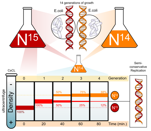
Enzymes at the replication fork
Several enzymes and proteins work at each fork. The names below are the E. coli ones, the standard on tests; eukaryotic equivalents follow.
| Protein | Job at the fork |
|---|---|
| Initiator protein (DnaA in E. coli) | Binds the origin and starts opening it |
| Helicase (DnaB in E. coli) | Unwinds the double helix by breaking the hydrogen bonds between base pairs; uses ATP |
| Single-strand binding proteins (SSB) | Coat the separated strands so they do not re-pair or fold back |
| Topoisomerase (gyrase in bacteria) | Relieves the twisting strain (positive supercoils) that builds up ahead of the fork by cutting, passing and rejoining DNA strands |
| Primase | Synthesizes a short RNA primer (about 5–10 nucleotides) from ribonucleotides, which gives DNA polymerase a 3′ end to extend |
| DNA polymerase III | Main elongation enzyme in bacteria: adds DNA nucleotides 5′ to 3′ onto the primer and proofreads (3′→5′ exonuclease) |
| Sliding clamp (β clamp) | Ring that holds DNA polymerase III on the template so it does not fall off (processivity) |
| DNA polymerase I | Removes RNA primers with its 5′→3′ exonuclease activity and replaces them with DNA |
| DNA ligase | Seals the nicks between Okazaki fragments by forming the last phosphodiester bond between a 3′-OH and the adjacent 5′-phosphate |
Ligase and DNA polymerase both form phosphodiester bonds; the difference is that polymerase adds a new nucleotide (powered by the dNTP), while ligase joins two existing strand ends and needs its own energy source (ATP in eukaryotes, NAD⁺ in E. coli). DNA polymerase II in E. coli is mainly a repair polymerase.
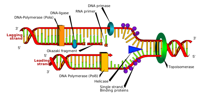
Leading and lagging strands
DNA polymerase can only add nucleotides to a 3′ end, so it builds only in the 5′-to-3′ direction while reading the template 3′ to 5′. The two strands at a fork are therefore synthesized differently. The leading strand is synthesized continuously toward the fork, needing only one primer per origin. The lagging strand runs the opposite way, so it is synthesized away from the fork in short pieces, each started by a new primer. These pieces are Okazaki fragments, about 1,000–2,000 nucleotides long in bacteria and 100–200 in eukaryotes. Once DNA polymerase I removes the primers and fills the gaps, ligase joins the fragments into one strand. An E. coli mutant with a temperature-sensitive ligase piles up short fragments at the restrictive temperature, which is the classic evidence for discontinuous synthesis.
Replication runs in both directions from an origin, so the two forks of a bubble mirror each other. A template strand that is copied continuously (leading) at the right-hand fork is copied in Okazaki fragments (lagging) at the left-hand fork, and vice versa.
Proofreading and fidelity
Proofreading is part of elongation. DNA polymerase checks each new base pair; when it adds a wrong nucleotide, the mismatched end does not fit the active site well, so the enzyme pauses, moves the new strand into a separate 3′→5′ exonuclease site, clips off the wrong nucleotide and resumes. Fidelity builds in layers: correct base selection alone gives roughly 1 error in 10⁴–10⁵ nucleotides; proofreading improves that about a hundredfold; mismatch repair after replication improves it about another 100- to 1,000-fold, for a final rate around 1 in 10⁹–10¹⁰ nucleotides per replication. A polymerase with no proofreading still replicates, but makes more mistakes.
Prokaryotic versus eukaryotic replication
| Feature | Bacteria (E. coli) | Eukaryotes |
|---|---|---|
| Origins | One (oriC) | Many per chromosome |
| Fork speed | About 1,000 nt/s | About 50 nt/s (roughly 20–100; slowed by nucleosomes) |
| Main polymerases | Pol III (elongation, both strands); Pol I (primer removal) | Pol α (with primase, starts each primer and adds a short DNA stretch); Pol ε (leading strand); Pol δ (lagging strand); Pol β (base excision repair); Pol γ (mitochondrial DNA) |
| Clamp, SSB | β clamp; SSB | PCNA; RPA |
| Primer removal | Pol I | RNase H and FEN1, then Pol δ fills |
| Okazaki fragments | 1,000–2,000 nt | 100–200 nt |
| Ends | Circular, none | Linear, telomeres; telomerase |
| Timing | Can start new rounds before division finishes | Once per cycle, only in S phase |
Telomeres and the end-replication problem
On a linear chromosome, the lagging strand cannot be finished at the very end: when the last RNA primer is removed there is no upstream 3′-OH for DNA polymerase to fill the gap. Each round of replication would therefore leave the new strand slightly shorter, with a single-stranded 3′ overhang on the parental strand. This is the end-replication problem.
Telomeres are long tandem repeats at chromosome ends (TTAGGG in humans, hundreds to thousands of copies) that contain no genes, so losing a little each division does no harm at first. They are also capped by proteins that stop the ends from being mistaken for broken DNA. Telomerase is an enzyme that carries its own RNA template and a reverse-transcriptase protein subunit (TERT): it extends the 3′ overhang by copying its RNA into new DNA repeats, after which primase and DNA polymerase fill in the complementary strand. Telomerase is active in germ cells, stem cells and most cancer cells; most somatic cells have little, so their telomeres shorten with each division until the cell stops dividing (the Hayflick limit, senescence). An organism without telomerase activity in its germline would see chromosomes shorten generation after generation.
Gene expression: transcription and translation
The central dogma describes how information flows from DNA to protein: DNA is transcribed to RNA, and RNA is translated into protein. DNA also copies itself (replication). The known exceptions add arrows rather than reversing the main one: reverse transcriptase makes DNA from an RNA template (retroviruses such as HIV; telomerase; the RT step of RT-PCR), and some RNA viruses copy RNA directly into RNA. Information never flows from protein back to nucleic acid. Most questions about gene expression ask you to name which molecule, enzyme or step does a job in one of the two stages.
Making an mRNA does not guarantee a protein. The amount of protein also depends on how fast the mRNA is degraded (its 5′ cap, poly-A tail and untranslated regions matter here; small regulatory RNAs called microRNAs can block translation or trigger degradation), on whether ribosomes initiate on it, and on how long the protein lasts. In eukaryotes the most important control point is still the start of transcription.
Genes, strands and RNA types
A protein-coding gene has a promoter (where RNA polymerase binds, upstream of the start), the transcribed region and a terminator. Inside the transcribed region of a eukaryotic gene are exons (kept in the mature mRNA; they include the coding sequence and the 5′ and 3′ untranslated regions, UTRs) and introns (removed by splicing). "Upstream" means toward the 5′ end of the coding strand; the first transcribed base is numbered +1, and promoter elements have negative numbers.
The two DNA strands have names that tests mix freely:
| Strand | Also called | Relation to the mRNA |
|---|---|---|
| Template strand | Antisense, non-coding, minus strand | Read by RNA polymerase; the mRNA is complementary to it |
| Coding strand | Sense, non-template, plus strand | Same sequence as the mRNA (5′→3′), with T in place of U |
The main kinds of RNA and their jobs:
| RNA | Job |
|---|---|
| mRNA (messenger) | Carries the coding sequence from DNA to the ribosome |
| tRNA (transfer) | Adapter: anticodon pairs with the codon, 3′ end carries the matching amino acid |
| rRNA (ribosomal) | Structural core of the ribosome and the catalyst of peptide bond formation; most of a cell's RNA by mass; its slow-changing sequence is used to build phylogenies |
| snRNA (small nuclear) | With proteins, forms the snRNPs of the spliceosome |
| miRNA (microRNA) | Short regulatory RNAs that pair with mRNAs and block their translation or cause their breakdown |
Transcription
Transcription makes an RNA copy of a gene. RNA polymerase reads the template strand 3′ to 5′ and builds RNA 5′ to 3′ from ribonucleoside triphosphates (ATP, GTP, CTP, UTP), releasing pyrophosphate with each addition. Unlike DNA polymerase, it does not need a primer, and it unwinds the DNA itself, opening a short transcription bubble that travels with it (no separate helicase is needed); the DNA rewinds behind it. The RNA sequence matches the coding (non-template) strand, with U in place of T. The three stages are:
- Initiation: RNA polymerase binds the promoter. In bacteria, a sigma factor recognizes promoter sequences about 10 and 35 bases upstream of the start. In eukaryotes, general transcription factors bind first (for example, at the TATA box) and recruit RNA polymerase II.
- Elongation: the enzyme moves along the template, adding RNA nucleotides complementary to it. RNA polymerase proofreads far less than DNA polymerase, so transcription is more error-prone (roughly one error per 10⁴–10⁵ nucleotides), but errors are not inherited because RNA is not the genetic material.
- Termination: in bacteria, either a GC-rich hairpin followed by a run of U's makes the polymerase fall off (rho-independent), or the Rho protein pulls the RNA away (rho-dependent). In eukaryotes, termination is linked to cleavage of the transcript after the polyadenylation signal.
Bacterial promoters and RNA polymerase. Bacteria have one RNA polymerase. Its core enzyme has five subunits (two α, β, β′ and ω) and can elongate, but it needs a sigma (σ) factor to find promoters; core plus σ is the holoenzyme. Sigma recognizes two short promoter elements, the −35 box (consensus TTGACA) and the −10 box or Pribnow box (consensus TATAAT), then is released once transcription is under way. Different sigma factors let bacteria switch on different gene sets (heat shock, starvation).
Eukaryotic promoters and RNA polymerases. Eukaryotes have three nuclear RNA polymerases: RNA polymerase I makes most rRNA (28S, 18S, 5.8S) in the nucleolus; RNA polymerase II makes mRNA (and most snRNAs and miRNAs); RNA polymerase III makes tRNA and 5S rRNA. RNA polymerase II cannot recognize a promoter on its own; general transcription factors must bind first. The core promoter often contains a TATA box (consensus TATAAA, also called the Goldberg–Hogness box) about 25–30 bases upstream of the start. The TATA-binding protein (TBP), part of TFIID, binds it first and bends the DNA; the other general transcription factors and RNA polymerase II then assemble into the preinitiation complex. A/T-rich DNA melts easily because A–T pairs have two hydrogen bonds, which helps the promoter open.
Regulation happens mostly at initiation. Activators bind DNA and help RNA polymerase bind the promoter, raising transcription. Repressors bind DNA (often an operator) and block RNA polymerase, lowering transcription. Bacterial operons are the standard examples.
Background beyond the rules' "activators and repressors": in eukaryotes, activators usually bind enhancers, DNA elements that can sit thousands of bases upstream or downstream, in either orientation; the DNA loops so the bound activator can contact the transcription machinery at the promoter (through a large Mediator complex). Silencers bind repressors. Elements that are part of the same DNA molecule as the gene they control (promoters, enhancers, operators) are cis-acting; diffusible proteins that can act on any copy of the target (transcription factors, repressors) are trans-acting. Transcription factors share a few DNA-binding shapes: helix-turn-helix (most bacterial repressors, including the lac repressor), zinc finger, leucine zipper and helix-loop-helix. Packing also matters: genes in tightly packed heterochromatin are hard to reach and usually silent (see DNA organization).

RNA processing in eukaryotes
RNA maturation happens in eukaryotes before the mRNA leaves the nucleus; the unprocessed transcript is pre-mRNA. Three changes are made, and tests ask for all three and their purposes:
- 5′ cap: a 7-methylguanosine (a modified guanine nucleotide) is attached to the first nucleotide by an unusual 5′–5′ triphosphate link, early in transcription. It protects the mRNA from 5′ exonucleases, helps export it from the nucleus, and is where the ribosome's small subunit attaches to start translation.
- 3′ poly-A tail: the transcript is cut about 10–35 nucleotides past a polyadenylation signal (AAUAAA) in the RNA, and poly(A) polymerase adds a tail of many adenines (often around 200) without a template. The tail protects the mRNA from degradation, aids export and helps translation.
- Splicing: the spliceosome (snRNPs, which are snRNA plus protein, along with other proteins) removes introns and joins exons. Introns almost always begin with GU at their 5′ end and end with AG at their 3′ end, with a branch-point A inside; the intron is released as a looped lariat. Alternative splicing, which includes or skips different exons, lets one gene make several proteins; it is a main reason humans make many more kinds of protein than they have genes. A mutation at a splice site can cause an exon to be skipped or an intron to be kept.
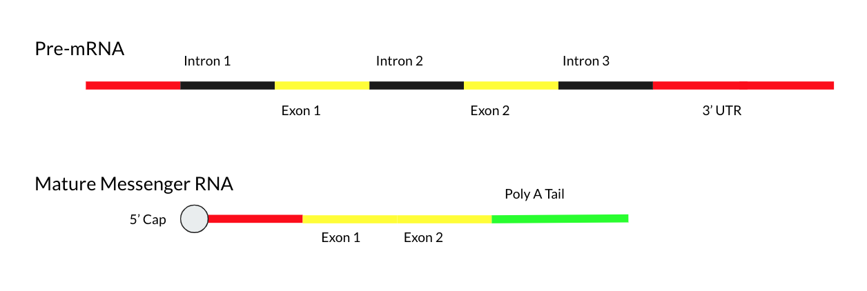
Bacteria do not process their mRNA this way, and they can translate an mRNA while it is still being transcribed. Because exons are kept and introns removed, a mature mRNA is much shorter than its gene: a 10,000-nucleotide transcription unit can encode a protein of only a few hundred amino acids.
| Feature | Prokaryotes | Eukaryotes |
|---|---|---|
| Location of transcription and translation | Both in the cytoplasm, coupled (ribosomes start on mRNA still being made) | Transcription in the nucleus, translation in the cytoplasm (free ribosomes or rough ER) |
| RNA polymerases | One (with sigma factors) | Three nuclear (I, II, III) |
| Promoter elements | −10 (Pribnow, TATAAT) and −35 boxes | TATA box (about −25 to −30), other elements, distant enhancers |
| Pre-mRNA processing | None | 5′ cap, poly-A tail, splicing |
| Introns | Rare | Common |
| Genes per mRNA | Often several (polycistronic, as in operons) | Usually one (monocistronic) |
| Ribosome | 70S (30S + 50S) | 80S (40S + 60S) |
| Ribosome finds start by | Shine–Dalgarno sequence pairing with 16S rRNA | 5′ cap, then scanning to the first AUG (Kozak sequence) |
| First amino acid | N-formylmethionine (fMet) | Methionine |
| Operons | Common | Rare |
Translation
Translation reads mRNA codon by codon and builds a polypeptide. Each codon (three bases) specifies one amino acid. AUG is the start codon, and it codes for methionine. UAA, UAG and UGA are stop codons. Each tRNA carries an anticodon that base-pairs with the codon, and its 3′ end carries the matching amino acid, attached by an aminoacyl-tRNA synthetase (tRNA charging, which uses ATP). The mRNA carries the message; the tRNAs are the adapters that turn codons into amino acids.
The genetic code
The code's properties are a favorite short-answer topic:
- Triplet: three bases per codon. Two-base codons would give only 4² = 16 combinations, too few for 20 amino acids; three give 4³ = 64.
- Non-overlapping and without punctuation: read in consecutive triplets from a fixed start, so an insertion or deletion shifts the reading frame.
- Degenerate (redundant): most amino acids have more than one codon, usually differing at the third base. Only methionine (AUG) and tryptophan (UGG) have one codon each.
- Unambiguous: each codon means only one amino acid.
- Nearly universal: the same code works in bacteria, plants and animals, which is why a firefly or jellyfish gene can be expressed in a pig or a bacterium, and is strong evidence of common ancestry. There are small exceptions, for example UGA codes for tryptophan in mitochondria.
Marshall Nirenberg and Heinrich Matthaei cracked the first codon in 1961: an artificial mRNA of only uracils (poly-U) made a protein of only phenylalanine, so UUU = Phe.
Read the table by the first base (left), the second base (column) and the third base (row within the block).
ReferenceThe standard genetic code (mRNA codons)· 16 rows
Find the first base in column 1 and the third base in column 2, then read across to the column for the second base. AUG is both methionine and the start signal.
No rows match.
| 1st | 3rd | 2nd = U | 2nd = C | 2nd = A | 2nd = G |
|---|---|---|---|---|---|
| U | U | UUU Phe | UCU Ser | UAU Tyr | UGU Cys |
| U | C | UUC Phe | UCC Ser | UAC Tyr | UGC Cys |
| U | A | UUA Leu | UCA Ser | UAA Stop | UGA Stop |
| U | G | UUG Leu | UCG Ser | UAG Stop | UGG Trp |
| C | U | CUU Leu | CCU Pro | CAU His | CGU Arg |
| C | C | CUC Leu | CCC Pro | CAC His | CGC Arg |
| C | A | CUA Leu | CCA Pro | CAA Gln | CGA Arg |
| C | G | CUG Leu | CCG Pro | CAG Gln | CGG Arg |
| A | U | AUU Ile | ACU Thr | AAU Asn | AGU Ser |
| A | C | AUC Ile | ACC Thr | AAC Asn | AGC Ser |
| A | A | AUA Ile | ACA Thr | AAA Lys | AGA Arg |
| A | G | AUG Met (start) | ACG Thr | AAG Lys | AGG Arg |
| G | U | GUU Val | GCU Ala | GAU Asp | GGU Gly |
| G | C | GUC Val | GCC Ala | GAC Asp | GGC Gly |
| G | A | GUA Val | GCA Ala | GAA Glu | GGA Gly |
| G | G | GUG Val | GCG Ala | GAG Glu | GGG Gly |
Tests write proteins with three-letter or one-letter abbreviations. The one-letter codes that are not simply the first letter are the ones to memorize:
| Amino acid | 3-letter | 1-letter | Amino acid | 3-letter | 1-letter |
|---|---|---|---|---|---|
| Alanine | Ala | A | Leucine | Leu | L |
| Arginine | Arg | R | Lysine | Lys | K |
| Asparagine | Asn | N | Methionine | Met | M |
| Aspartate (aspartic acid) | Asp | D | Phenylalanine | Phe | F |
| Cysteine | Cys | C | Proline | Pro | P |
| Glutamate (glutamic acid) | Glu | E | Serine | Ser | S |
| Glutamine | Gln | Q | Threonine | Thr | T |
| Glycine | Gly | G | Tryptophan | Trp | W |
| Histidine | His | H | Tyrosine | Tyr | Y |
| Isoleucine | Ile | I | Valine | Val | V |
tRNA and the wobble rule
A tRNA is a single RNA chain of about 70–90 nucleotides that folds, by internal base pairing (hydrogen bonds), into a cloverleaf in two dimensions and an L shape in three. One end carries the anticodon loop; the other is the 3′ end, which always ends in CCA, and the amino acid is attached to the terminal A. A cell usually has 20 aminoacyl-tRNA synthetases, one per amino acid; each attaches its amino acid to every matching tRNA, using ATP. Because the ribosome checks only codon–anticodon pairing, not the amino acid, a mischarged tRNA puts the wrong amino acid in, so synthetase accuracy (including proofreading) is essential. Overall, translation inserts a wrong amino acid roughly once per 10⁴ codons, which is less accurate than replication but acceptable because proteins are not inherited.
The anticodon pairs with the codon antiparallel: anticodon 3′-GAC-5′ reads codon 5′-CUG-3′ (Leu). Always line up 5′ of one with 3′ of the other.
Francis Crick's wobble hypothesis explains why a cell needs fewer than 61 tRNAs. The first two codon bases pair strictly, but the third codon base (paired with the first, 5′ base of the anticodon) can pair loosely: G in the anticodon can pair with C or U, and the modified base inosine (I) in the anticodon can pair with U, C or A. One tRNA can therefore read several codons for the same amino acid, which is another reason third-position changes are usually silent.
The ribosome
The ribosome has two subunits and three tRNA sites: the A site (aminoacyl), where a new charged tRNA enters; the P site (peptidyl), which holds the tRNA carrying the growing chain; and the E site (exit), from which the empty tRNA leaves. The peptide bond is made by the ribosomal RNA of the large subunit, so the ribosome is a ribozyme. Ribosomes are made of rRNA and protein (in bacteria roughly two-thirds rRNA by mass); their subunits are assembled in the nucleolus in eukaryotes.
Ribosome subunits and their rRNAs:
| Prokaryotic | Eukaryotic (cytoplasm) | |
|---|---|---|
| Whole ribosome | 70S | 80S |
| Small subunit | 30S (16S rRNA) | 40S (18S rRNA) |
| Large subunit | 50S (23S and 5S rRNA) | 60S (28S, 5.8S and 5S rRNA) |
S (Svedberg units) measure how fast a particle sediments, which depends on shape as well as mass, so subunit values do not add (30S + 50S = 70S). Mitochondria and chloroplasts have 70S-like ribosomes, one line of evidence that they descend from engulfed bacteria (endosymbiosis). Antibiotics such as streptomycin and tetracycline target the 70S ribosome, which is why they hit bacteria but not our cytoplasmic ribosomes.

Initiation, elongation and termination
-
Initiation: the small ribosomal subunit binds the mRNA first. In bacteria it binds at the Shine–Dalgarno sequence (a purine-rich stretch, consensus AGGAGG, about 8 bases upstream of the start codon), which base-pairs with the 3′ end of the 16S rRNA and so positions the AUG in the P site; initiation factors (IF1–IF3) help. In eukaryotes the small subunit, with initiation factors and the initiator tRNA, binds the 5′ cap, then scans 5′→3′ to the first AUG, usually in a favorable context called the Kozak sequence (consensus roughly GCCRCCAUGG, where R is a purine). The initiator tRNA is a special tRNA, different from the one that inserts internal methionines: in bacteria it carries N-formylmethionine (fMet-tRNA^fMet), in eukaryotes methionine (Met-tRNAi). It is the only tRNA that goes straight into the P site. The large subunit then joins, powered by GTP, forming the complete ribosome.
-
Elongation: a charged tRNA enters the A site, a peptide bond transfers the growing chain onto its amino acid, and the ribosome moves one codon along the mRNA (translocation). Each codon takes three steps:
- Delivery: an elongation factor (EF-Tu in bacteria) brings a charged tRNA to the A site, using GTP.
- Peptide bond: the peptidyl transferase activity of the large-subunit rRNA (23S in bacteria, 28S in eukaryotes) moves the growing chain from the P-site tRNA onto the amino acid on the A-site tRNA. A peptide bond is a condensation (dehydration) linkage, which is why tests name water as its byproduct; on the ribosome, the energy comes from the bond that held the chain to the P-site tRNA.
- Translocation: EF-G (with GTP) shifts the ribosome one codon toward the mRNA's 3′ end, moving the tRNAs from A to P and P to E.
The polypeptide grows from its N-terminus (amino end) to its C-terminus (carboxyl end). Each peptide bond costs about four high-energy phosphate bonds: two from ATP to charge the tRNA and one GTP each for delivery and translocation.
-
Termination: a stop codon enters the A site, no tRNA matches it, and release factors bind instead, causing the finished polypeptide to be released and the ribosome to come apart. Release factors are proteins shaped like a tRNA. In bacteria RF1 reads UAA and UAG, RF2 reads UAA and UGA, and RF3 is a GTP-binding protein that helps recycle them; in eukaryotes a single eRF1 reads all three stop codons. The release factor triggers hydrolysis of the bond between the chain and the P-site tRNA (water is added), freeing the polypeptide.
Many ribosomes can translate one mRNA at the same time, spaced along it; the cluster is a polyribosome (polysome). Proteins destined for secretion or membranes begin with an N-terminal signal sequence that directs the ribosome to the rough endoplasmic reticulum; others are made on free ribosomes. After translation, the chain folds (helped by chaperones) and may be modified, for example in the ER and Golgi.
The lac operon (State & National)
An operon is a cluster of bacterial genes under one promoter and one operator (the repressor's binding site), transcribed as one mRNA. The lac operon in E. coli carries genes for using lactose (lacZ, β-galactosidase; lacY, permease; lacA, transacetylase). It is inducible: normally off, switched on when its substrate appears.
The model was worked out by François Jacob and Jacques Monod (published 1961; Nobel Prize 1965). Its parts, in order along the DNA:
| Part | What it is | Job |
|---|---|---|
| lacI | Regulatory gene with its own promoter, upstream of the operon; expressed all the time (constitutively) at a low level | Encodes the Lac repressor, a protein that binds the operator |
| CAP site | DNA sequence just upstream of the promoter | Binds the cAMP–CAP activator |
| Promoter (P) | Where RNA polymerase binds | Start of transcription |
| Operator (O) | Short sequence overlapping or just downstream of the promoter, before lacZ | Where the repressor binds; a bound repressor blocks RNA polymerase |
| lacZ | Structural gene | β-galactosidase: splits lactose into glucose and galactose, and also converts some lactose to allolactose, the natural inducer |
| lacY | Structural gene | Permease: transports lactose into the cell |
| lacA | Structural gene | Transacetylase (thiogalactoside transacetylase); its role is minor |
- Without lactose: the LacI repressor binds the operator and blocks RNA polymerase.
- With lactose: allolactose, a lactose derivative, binds the repressor and changes its shape. The repressor releases the operator, and RNA polymerase can transcribe the genes.
- With glucose low: cyclic AMP rises and binds CAP (catabolite activator protein), which helps RNA polymerase bind the promoter. This is positive control, and it stacks with the repressor.
- Both lactose and glucose present: the repressor is off, but cAMP is low, so CAP does not help and transcription stays low. High transcription needs lactose present and glucose low.
The repressor is an allosteric protein: allolactose binds a site separate from its DNA-binding site and changes the protein's shape so it can no longer hold the operator. The lac promoter is weak on its own, so it relies on cAMP–CAP, which binds the CAP site and helps recruit RNA polymerase. Glucose keeps cAMP low (glucose transport inhibits adenylyl cyclase, the enzyme that makes cAMP), so the cell uses glucose first, its preferred sugar, before switching to lactose. This preference is catabolite repression.

The four combinations of glucose and lactose give these outcomes:
| Glucose | Lactose | Repressor | cAMP–CAP | Transcription |
|---|---|---|---|---|
| Present | Absent | Bound to operator | Low, not bound | Off |
| Absent | Absent | Bound to operator | High, bound | Off (repressor wins) |
| Present | Present | Released | Low, not bound | Low (weak) |
| Absent | Present | Released | High, bound | High (strong) |
Inducers in the lab (background). IPTG is a synthetic inducer: it binds the repressor like allolactose but is not broken down by β-galactosidase, so induction stays constant. X-gal is a colorless substrate that β-galactosidase cleaves into a blue product, so colonies making β-galactosidase turn blue.
Experimental questions often give mutant strains. A mutant repressor that cannot bind the operator, or an operator that cannot be bound, makes the operon constitutive (always on). A repressor that cannot bind allolactose keeps the operon off even with lactose. Because the repressor is a diffusible protein and the operator is a stretch of DNA, mutations behave differently when a second copy is added on a plasmid (a partial diploid):
| Mutation | Effect alone | In a partial diploid with a wild-type copy |
|---|---|---|
| lacI⁻ (repressor cannot bind operator) | Constitutive (on without lactose) | Recessive: the normal repressor from the other copy diffuses and binds both operators (trans-acting) |
| lacOᶜ (operator cannot bind repressor) | Constitutive | Cis-dominant: only the genes on the same DNA molecule as the bad operator stay on |
| lacIˢ ("super-repressor", cannot bind inducer) | Never induced, even with lactose | Dominant: the mutant repressor sits on both operators |
| lacZ⁻ | No functional β-galactosidase, so no lactose use (and no allolactose made from lactose) | Recessive |
| lacY⁻ | No permease, so lactose cannot enter | Recessive |
| CAP or adenylyl-cyclase mutant | Low expression even with lactose and no glucose | Recessive |
| Promoter mutation | Low or no transcription of lacZ, lacY, lacA | Cis-acting |
The trp operon (National only)
The trp operon makes the enzymes that synthesize the amino acid tryptophan (genes trpE, trpD, trpC, trpB and trpA). It is repressible: normally on, switched off when its product is plentiful. That makes sense for a biosynthetic pathway, since the cell should stop making tryptophan when it already has enough.

- Low tryptophan: the TrpR repressor is made in an inactive form that cannot bind the operator, so RNA polymerase transcribes the genes.
- High tryptophan: tryptophan acts as a corepressor. It binds the repressor and changes its shape so it can bind the operator, and transcription stops.
- Attenuation adds a second, finer control. The leader sequence before the first structural gene encodes a short peptide with two tryptophan codons in a row, and its mRNA can fold into alternative hairpins. When tryptophan is plentiful, the ribosome translating the leader moves quickly, and a terminator hairpin forms, so RNA polymerase stops before the structural genes. When tryptophan is scarce, the ribosome stalls at the Trp codons, an antiterminator hairpin forms instead, and transcription continues. Attenuation works only because bacteria translate mRNA while it is still being made.
The repressor is encoded by a separate gene, trpR, located elsewhere on the chromosome. The leader region (trpL) contains four segments that can pair in different ways: 1 with 2, 2 with 3 (the antiterminator) or 3 with 4 (the terminator, a GC-rich stem-loop followed by U's, a rho-independent terminator). Region 1 holds the two Trp codons (UGG UGG).
| Tryptophan level | Ribosome on the leader | Pairing | Result |
|---|---|---|---|
| High (charged Trp-tRNA plentiful) | Moves quickly past region 1 and covers region 2 | 3 pairs with 4 | Terminator forms; transcription stops early |
| Low (little charged Trp-tRNA) | Stalls on the Trp codons in region 1 | 2 pairs with 3 | Antiterminator; 3–4 cannot form; transcription continues |
| No translation of the leader at all | No ribosome | 1–2 and 3–4 | Terminator forms; transcription stops |

The two operons are mirror images:
| Feature | lac operon | trp operon |
|---|---|---|
| Pathway | Breaks down lactose (catabolic) | Builds tryptophan (anabolic) |
| Default state | Off (inducible) | On (repressible) |
| Repressor alone | Active, binds operator | Inactive, cannot bind operator |
| Small molecule | Allolactose (inducer) inactivates repressor | Tryptophan (corepressor) activates repressor |
| Extra control | CAP–cAMP activation when glucose is low | Attenuation in the leader region |
The general rule: catabolic (breakdown) operons are usually inducible, switched on by their substrate; anabolic (synthesis) operons are usually repressible, switched off by their end product. Control is negative when a repressor protein turns transcription off (both lac and trp repressors) and positive when an activator protein turns it on (CAP).
Population and evolutionary genetics
Population genetics moves from one family to a whole population. Instead of asking what one child will inherit, it asks how common each allele is in the gene pool (all the alleles in a population) and what makes those frequencies change. A change in allele frequencies over generations is microevolution. Individuals do not evolve; populations do.
Hardy-Weinberg equilibrium
For one gene with two alleles, let p be the frequency of the dominant allele and q the frequency of the recessive allele. If mating is random, the chance that a child gets two A alleles is p × p, two a alleles is q × q, and one of each is 2pq (either parent can supply the A). Those genotype frequencies stay constant from one generation to the next if the population meets five conditions. The principle was worked out independently in 1908 by the mathematician G. H. Hardy and the physician Wilhelm Weinberg, and it is a null model: a population that does not match it is evolving, or mating non-randomly, at that gene.
The five assumptions, and what breaks when each fails:
| Assumption | If it fails | Effect on the population |
|---|---|---|
| Very large population | Genetic drift | Random changes in allele frequency, strongest in small populations; alleles can be lost or fixed |
| No migration | Gene flow | Allele frequencies move toward those of the source population; populations become more similar |
| No mutation | Mutation | New alleles appear; slow change on its own |
| Random mating | Non-random mating (for example, inbreeding or choosing similar mates) | Genotype frequencies shift (inbreeding raises homozygotes) even if allele frequencies do not |
| No natural selection | Selection | Alleles that raise relative fitness become more common |

The most common test move is to start from the recessive phenotype, because it is the only class whose genotype you know for sure.
Getting allele frequencies from genotype counts. Count alleles, not people: each homozygote has two copies, each heterozygote one.
Testing whether a population is in equilibrium. When the question gives genotype counts, compute the allele frequencies from the counts first, then compare expected and observed genotype numbers.
X-linked genes. Males have one X, so the frequency of affected males equals the allele frequency q, and affected females are q². With q = 0.08 for red-green color blindness, about 8% of men but only 0.08² ≈ 0.6% of women are color-blind, and 2pq ≈ 15% of women are carriers.
Three alleles (ABO). With allele frequencies p (Iᴬ), q (Iᴮ) and r (i), p + q + r = 1 and the genotype frequencies come from (p + q + r)²: type A = p² + 2pr, type B = q² + 2qr, type AB = 2pq, type O = r². Start from type O: r = √(frequency of O).
Forces that change allele frequencies
Genetic drift is random change in allele frequency from one generation to the next, caused by chance in which individuals happen to reproduce. Its effect is larger in small populations, it does not adapt the population to anything, and it tends to reduce genetic variation by losing some alleles and fixing others. For a neutral allele, the chance it eventually becomes fixed equals its current frequency.
Two special cases of drift come up constantly:
- Founder effect: a few individuals start a new, isolated population, and its allele frequencies reflect whatever the founders happened to carry. Rare alleles can become common. The high frequency of Ellis–van Creveld syndrome (a form of dwarfism with extra fingers) among the Old Order Amish of Pennsylvania is the standard example.
- Bottleneck: a disaster or hunting sharply reduces a population's size, and the survivors carry only part of the original variation. Even if numbers recover, the diversity does not come back quickly. Northern elephant seals and cheetahs both show very low genetic diversity from past bottlenecks.
Migration (gene flow) moves alleles between populations. It makes populations more similar to one another and can bring new alleles into a population.
Mutation is the only force that creates brand-new alleles, but mutation rates per gene are low (very roughly 10⁻⁵ to 10⁻⁶ per gene per generation), so on its own it changes frequencies very slowly; it supplies the variation that the other forces act on.
Natural selection needs three things: variation in a trait, heritability of that variation, and differential survival and reproduction linked to the trait. The peppered moth is the classic case: when soot darkened tree bark during England's industrial revolution, dark moths were better camouflaged from birds and became common; after clean-air laws, light moths returned.
Relative fitness (w) measures how well a genotype survives and reproduces compared with the best genotype, which is set to w = 1. The selection coefficient is s = 1 − w. Selection changes allele frequency by weighting each genotype by its fitness before the next generation forms.
Heterozygote advantage (overdominance) keeps two alleles in a population when the heterozygote has the highest fitness. Sickle cell is the textbook case: in regions with malaria, SS homozygotes suffer sickle cell disease and AA homozygotes are more vulnerable to malaria, while AS carriers are protected from severe malaria without having the disease, so the S allele stays common. (In carriers, red cells infected by the malaria parasite tend to sickle and are removed by the spleen, and the parasite grows poorly in them.) If relative fitnesses are given as, say, 0.3 for FF, 1.0 for Ff and 0.2 for ff, the heterozygote is fittest and both alleles are maintained. This is one form of balancing selection.
Modes of selection on a continuous trait:
| Mode | Which phenotypes are favored | Effect on the distribution | Example |
|---|---|---|---|
| Directional | One extreme | Mean shifts toward that extreme | Dark peppered moths on sooty bark; antibiotic resistance |
| Stabilizing | The intermediate | Mean stays; variation shrinks | Human birth weight |
| Disruptive (diversifying) | Both extremes over the middle | Distribution splits toward two peaks | Seed-cracking finches with small or large beaks, but not medium |

Non-random mating changes genotype frequencies rather than allele frequencies. Inbreeding (mating between relatives) increases homozygosity at every gene, exposing harmful recessive alleles (inbreeding depression); this is why recessive disorders appear more often in children of consanguineous matings. Assortative mating (choosing mates with similar phenotypes) increases homozygosity for the genes behind that trait. Sexual selection favors traits that improve mating success, even at a survival cost (a peacock's tail).
Additive alleles and continuous variation (State & National)
Many traits, such as height, skin color and wheat kernel color, vary continuously instead of falling into neat classes. These are polygenic traits. The model the rules ask about is additive inheritance: several genes affect the trait, each "contributing" allele adds the same small amount, and the phenotype depends only on how many contributing alleles an individual carries. With many genes plus environmental effects, the classes blur into a smooth bell-shaped distribution, and most individuals fall near the average.
The classic experiment is Nilsson-Ehle's wheat crosses. A dark red × white cross gave an intermediate F1, and the F2 showed a range of shades with only a small fraction as extreme as either parent. Counting that fraction tells you how many genes are involved.
The distribution of classes follows the binomial coefficients (Pascal's triangle) for 2n alleles. With two genes (AaBb × AaBb, four alleles), the F₂ classes with 0, 1, 2, 3 and 4 contributing alleles occur in the ratio 1 : 4 : 6 : 4 : 1 (out of 16). The fraction with exactly k contributing alleles is .
Technology and techniques
PCR
The polymerase chain reaction copies a specific DNA segment millions to billions of times in a test tube (in vitro). Kary Mullis invented it in 1983 (Nobel Prize in Chemistry, 1993). It is replication stripped down to the essentials: heat replaces helicase, and two synthetic primers decide which segment is copied. One cycle has three steps:
- Denaturation (about 94–98 °C, typically 95 °C, the hottest step): heat breaks the hydrogen bonds and separates the double-stranded template into single strands.
- Annealing (typically around 50–65 °C, set by the primers): cooler temperature lets two short DNA primers bind their complementary sites on opposite strands, flanking the target.
- Extension (about 72 °C): a heat-stable DNA polymerase, such as Taq from a thermophilic bacterium, builds new DNA from each primer 5′ to 3′.
A run usually begins with a longer initial denaturation, repeats the three steps 25–35 times in a programmable thermocycler, and ends with a final extension so every product is finished.
| Component | Job |
|---|---|
| Template DNA | Contains the target sequence (can be tiny amounts: a hair root, a drop of blood) |
| Two primers (forward and reverse) | Short single-stranded DNA oligonucleotides (about 18–25 nt) that bind the 3′ ends of the target on opposite strands and point toward each other; they define the product's two ends |
| dNTPs (dATP, dTTP, dGTP, dCTP) | Building blocks and energy source for the new strands |
| Heat-stable DNA polymerase | Taq polymerase from Thermus aquaticus, a bacterium from Yellowstone hot springs; survives repeated 95 °C steps and works best near 72 °C |
| Buffer with Mg²⁺ | Keeps pH stable; magnesium ions are a required cofactor for DNA polymerase (too little: no product; too much: less specific) |
| Water | Brings the reaction to volume |
PCR does not need helicase (heat separates strands), primase (synthetic primers are supplied), ligase (no fragments to join), topoisomerase or RNA polymerase. Ordinary E. coli DNA polymerase would be destroyed in the first denaturation step, which is why the heat-stable enzyme made PCR practical. Taq has no 3′→5′ proofreading, so it makes more errors than polymerases such as Pfu (from the archaeon Pyrococcus furiosus), which proofreads; labs still use Taq because it is cheap, fast and robust when perfect accuracy is not needed.

Exact-length products. The first cycles make strands that start at a primer but run past the target's far end, because nothing stops the polymerase. Strands of exactly the target length (from one primer's 5′ end to the other's) first appear in cycle 3; after that they dominate, because they double every cycle while the long products only increase by two per cycle. Starting from one double-stranded template, the number of target-length double-stranded products after n cycles is (cycle 3: 8 − 6 = 2). The product length equals the distance from the 5′ end of the forward primer to the 5′ end of the reverse primer, primers included.
Primer design and annealing temperature. Each primer is written 5′→3′ and matches (is identical to) the 5′ end of one strand of the target, so it binds the complementary 3′ end of the other strand; the two primers must bind opposite strands and point toward each other. Good primers are about 18–25 nucleotides, about 40–60% G+C, do not pair with themselves or each other, and have similar melting temperatures. For short primers, a quick estimate is the Wallace rule.
Temperature is the main control in PCR, and failed reactions usually come down to it or to a missing component:
| Problem | What you see | Why |
|---|---|---|
| Annealing temperature too low | Extra bands, wrong-size products | Primers bind partly mismatched sites (non-specific binding) |
| Annealing temperature too high | Little or no product | Primers cannot stay bound to the template |
| Denaturation temperature too low | Low or no yield | Strands do not fully separate, so templates are not available |
| Non-heat-stable polymerase | No product after the first cycle | Enzyme denatured at 95 °C |
| Missing primer, dNTPs, Mg²⁺ or template | No product | A required component is absent |
| Primers that pair with each other | Very short "primer-dimer" band | Primers extended on each other instead of the template |
| Contamination | Band in the no-template (negative) control | Stray DNA, often from earlier PCR products; labs separate pre- and post-PCR work |
Reading PCR results. Products are usually checked by gel electrophoresis: DNA is negatively charged, moves toward the positive electrode, and shorter fragments move farther. Comparing band size with a DNA ladder of known sizes tells you whether the target is present and how big it is.
What PCR can answer. Typical experimental questions: Is a specific gene, allele or pathogen present in a sample (for example, viral DNA in a patient, or a species in an environmental sample)? Does a person carry a known insertion, deletion or repeat expansion (seen as a different product size; a longer CAG-repeat region in the Huntington gene gives a bigger product)? Is there enough of a target to sequence or study further? Variants extend the idea: RT-PCR first copies RNA into DNA with reverse transcriptase, so it can detect RNA viruses or gene expression, and quantitative (real-time) PCR measures how fast product accumulates to estimate how much template was present. PCR alone does not tell you the exact sequence of the product; that needs sequencing.
Sanger sequencing (State & National)
Sanger sequencing, or chain-termination (dideoxy) sequencing, determines the exact base sequence of a DNA segment. Frederick Sanger published it in 1977 and shared the 1980 Nobel Prize for it (his first Nobel was for sequencing the protein insulin). Automated Sanger sequencing did most of the Human Genome Project (1990–2003) and is still the accuracy standard for checking a single gene, reading about 700–1,000 bases per reaction.
It looks like PCR (template, primer, DNA polymerase, dNTPs, cycles of denaturing, annealing and extending), but the key difference is the terminator. The reaction includes a small amount of dideoxynucleotides (ddNTPs), which lack the 3′ hydroxyl group that DNA polymerase needs to attach the next nucleotide (they have H at both 2′ and 3′). When a ddNTP is incorporated, the chain stops. ddNTPs are added at a much lower concentration than the normal dNTPs, so most of the time the polymerase adds a normal nucleotide and only occasionally a terminator; across billions of molecules, some chains stop at every position. The ddNTP:dNTP ratio sets the read length: too much ddNTP and most chains stop early (strong short fragments, no long ones); too little and few chains stop near the primer. Leaving out one ddNTP leaves a gap at every position of that base. Each of the four ddNTPs carries a different fluorescent dye, so each fragment is labeled by the base at its 3′ end.
Setting up the reaction. Mix the template DNA (often a purified PCR product or plasmid), one primer, a DNA polymerase, all four dNTPs, a small amount of the four dye-labeled ddNTPs, and buffer with Mg²⁺; then run the thermal cycles.
Steps:
- Denaturation separates the template strands.
- Annealing of one primer, which binds the template just 3′ of the region to be read (complementary to the template's 3′ side), so the polymerase copies toward the template's 5′ end.
- Extension with random termination, producing a nested set of fragments that all start at the primer and end at every possible position.
- Capillary electrophoresis separates the fragments by size with single-base resolution; a laser excites each dye as fragments pass a detector, producing a chromatogram (colored peaks).
- Reading: shortest fragment first gives the newly made strand 5′→3′. Its complement, read in reverse, is the template.
The original 1977 method used four separate reactions, each with one ddNTP and a radioactive label, run in four lanes of a polyacrylamide gel and read from the bottom (shortest) of the gel upward. A reaction run with only one kind of ddNTP gives only the fragments that end in that base.
Reading a chromatogram: one clean peak per position means one sequence. Two overlapping peaks of different colors at one position in DNA from a diploid person means the person is heterozygous there (one allele per peak). An "N" in a sequence file means the base could not be called.


Here is how Sanger differs from standard PCR, the comparison the rules ask for:
| Feature | Standard PCR | Sanger sequencing |
|---|---|---|
| Primers | Two (forward and reverse) | One |
| Growth of product | Exponential (both strands copied) | Linear (one strand copied per cycle) |
| Nucleotides | dNTPs only | dNTPs plus a small amount of fluorescent ddNTPs |
| Chain-terminating nucleotide | None | Dideoxynucleotides (ddNTPs), lacking the 3′-OH |
| Output | Many copies of one target segment, all the same length | Fragments of every length, read as a sequence |
| Read-out | Gel band: present or absent, and size | Capillary electrophoresis chromatogram: base-by-base sequence |
| Typical question | Is the target present, and how big is it? | What is the exact base sequence of this target? |
Sanger sequencing answers questions PCR cannot: exactly which base changed in a mutant (substitution, small insertion or deletion), whether a person is heterozygous at a site (two overlapping peaks), whether a cloned gene has the expected sequence, or which sequence variant a pathogen carries. PCR usually comes first, to make enough copies of the target to sequence.
Common pitfalls
- Confusing "dominant" with "common". A dominant trait is expressed when one allele is present. It is not more common in the population; allele frequency, not dominance, sets how common a trait is.
- Forgetting that a dihybrid ratio assumes independent assortment. Linked genes do not give 9:3:3:1. Check whether the question says the genes are on the same chromosome.
- Answering 1/2 for a conditional question. Conditional questions (such as carrier-given-unaffected) change the denominator. 50% is a frequent correct answer on past tests, but not for every probability question.
- Mixing up the two meiotic divisions. Homologs separate in meiosis I; sister chromatids separate in meiosis II. Nondisjunction in meiosis I makes all four gametes abnormal; in meiosis II, only half.
- Counting chromatids as chromosomes. Count centromeres for chromosomes. A human cell in metaphase has 46 chromosomes but 92 chromatids.
- Writing a complementary strand in the wrong direction. Complementary strands are antiparallel; label 5′ and 3′ on every answer, and reverse a template given 5′→3′ before reading codons.
- Calling a mutation silent if the protein changed. A silent mutation keeps the same amino acid. A frameshift changes everything after the mutation; a nonsense mutation shortens the protein.
- Using Chargaff's rules on single-stranded nucleic acid. A = T and G = C only hold for double-stranded DNA; unequal values mean a single-stranded genome.
- Mixing up operon controls. Lac is inducible (allolactose inactivates the repressor), and glucose acts only through cAMP–CAP; trp is repressible (tryptophan activates the repressor).
- Believing a karyotype finds point mutations. Karyotypes show chromosome number and large structural changes only; sickle cell or cystic fibrosis alleles need DNA tests.
How tournaments test this
These counts describe the scio.ly archive of past Heredity tests: 3,318 questions, almost all Division B (3,246, or 98%). Most come from the 2025-26 season (1,913 questions, 58%), with the rest from the 2018-19 to 2020-21 seasons and 2024-25. The question bank holds a further 10,862 prompts (9,640 after removing exact duplicates), mostly Division C (6,748), so the archive is the better model for Division B.
Where the questions fall. Sorting the 9,640 distinct bank questions for this event by topic (about 8,600 fall inside the 2027 topic list) gives a rough map of where the points are:
| Area | Share of in-scope bank questions | Biggest single topics |
|---|---|---|
| Mendelian genetics, pedigrees, probability, linkage | about 30% | Mendel's laws and vocabulary, linkage and mapping, Punnett crosses, pedigrees |
| DNA structure, organization, mutations, repair, replication | about 27% | DNA structure and classic experiments, replication enzymes, mutation types |
| Gene expression (transcription, translation, operons) | about 22% | Translation and the genetic code, transcription and RNA processing |
| Mitosis, meiosis and karyotypes | about 10% | Stages, chromosome counts, nondisjunction |
| PCR and Sanger sequencing | about 5% | PCR steps, temperatures, components |
| Population and evolutionary genetics | about 2% (new for 2027, so expect far more) | Hardy-Weinberg, drift, selection |
| General genetics not tied to one area | about 4% | Central dogma and vocabulary items |
Out-of-scope questions in the bank (about 6%) were mostly CRISPR and genome editing, recombinant cloning and restriction enzymes, and genomic imprinting and other epigenetic inheritance; skip those. The basic link between chromatin packing (acetylation, methylation) and gene activity, covered in DNA organization, is still fair game.
Question types. The archive is nearly split between free response and multiple choice:
| Type | Archive | Share |
|---|---|---|
| Free response (FRQ) | 1,536 | 46% |
| Multiple choice (MCQ) | 1,473 | 44% |
| True/false | 136 | 4% |
| Select all that apply (multi) | 120 | 4% |
| Numeric | 53 | 2% |
Task profile. Only about a quarter of archive questions carry a task label, so read these as the most common patterns: "which of the following" items about 9%, "explain or why" about 8%, calculate or compute about 4%, compare or contrast about 2%, definitions about 1%, and data interpretation about 1%. The 2027 rules explicitly shift weight toward calculation, experimental results and evidence-based conclusions, so expect the calculation and data share to grow compared with past tests.
Subtopic tags. Archive questions carry overlapping tags: Genetics (1,476 questions, 44%), DNA (1,326, 40%), Heredity (686, 21%), Cell Division (571, 17%) and Proteins (541, 16%). Population Genetics (25) and Evolution (28) are nearly absent, because the area was not listed last season. Since the 2027 rules ask for coverage without overemphasizing any topic, population and evolutionary genetics should take a much larger share of 2027 tests than the archive suggests.
Recurring words and answers. The most common terms are DNA, gene, dominant, protein, amino acid, offspring, sequence, RNA, recessive and chromosomes; frequent phrases include "DNA replication", "DNA polymerase", "phenotypic ratio", "RNA polymerase", "Punnett square", "Sanger sequencing" and "lac operon". The most frequent correct answers are 50%, codominance, incomplete dominance, 25%, metaphase, anaphase, helicase, mRNA, prophase I, tRNA and uracil. Many of these answer a short question about one stage or enzyme, so know each stage's defining event and each enzyme's job.
Typical question shapes. Match an enzyme to its job at the fork; name the stage from a description ("homologs separate"); classify a mutation from a before-and-after sequence; translate a template strand with the codon table; predict lac operon expression for a glucose/lactose condition or a mutant; give a cross's ratio or a probability; read a pedigree for its mode of inheritance; compute map distance or gene order from test-cross counts; do Hardy-Weinberg from a recessive phenotype frequency; name a scientist's experiment and its conclusion; and explain what goes wrong in a PCR with a bad temperature.
Test yourself
The questions below come from past tournaments, re-solved for this guide and limited to topics on the 2027 list. Archive questions link to their tournament test; the rest come from the scio.ly question bank with keys checked for this guide.
- 1
A pedigree shows unaffected mothers producing affected sons; affected males do not pass the trait to their sons. Most likely inheritance?
Practice more Heredity questions →
- 1
Labradoodles' fur color is determined by a fur pigment gene (B, dominant = black; b, recessive = brown) and an epistatic gene (E, dominant = pigment is expressed; e, recessive = no pigment expressed; yellow). What is the ratio of black to brown to yellow offspring when a dihybrid cross for these two genes is performed?
From: LISO Invitational 2019-2020, Division C
Practice more Heredity questions →
- 1
Which event does NOT occur in Meiosis?
Practice more Heredity questions →
- 1
What is the primary role of RNA primase in DNA replication
Practice more Heredity questions →
- 1
If in comparison of mutant to normal mRNA the sequence length is the same but the number of amino acids in the mutant protein is shorter then it is a:
From: Haslett Invitational 2020, Division C
Practice more Heredity questions →
- 1
Puromycin is a drug that resembles the tRNA molecule when it has tyrosine attached. However, when it is added to a growing amino acid chain, it stops translation. If you have the following amino acid sequence, what sequence would you get if puromycin is added? Tyr is the shortened three letter notation for tyrosine.
Met - Phe - Phe - Lys - Arg - Tyr - Ser - Lys
Practice more Heredity questions →
- 1
In translation, where does the tRNA holding the polypeptide chain stay?
From: UCR Highlander Invitational 2025-26, Division B
Practice more Heredity questions →
Condensed notesheet
A print-ready summary of the ratios, formulas and tables most worth carrying on your one allowed sheet. Everything here is explained in the chapters above.
| Mode of inheritance | Fast test |
|---|---|
| Autosomal recessive | Unaffected parents → affected child; both sexes equally |
| Autosomal dominant | Every generation; affected child has an affected parent; father → son possible |
| X-linked recessive | Mostly males; carrier mother → affected son; never father → son |
| X-linked dominant | Affected father → all daughters, no sons |
| Y-linked | Father → all sons only |
| Stage | Defining event |
|---|---|
| S phase | DNA replication (2C → 4C) |
| Prophase / prometaphase | Chromosomes condense / envelope breaks down, kinetochores attach |
| Metaphase (mitosis, II) | Chromosomes single file on the plate |
| Metaphase I | Homologous pairs on the plate (independent assortment) |
| Anaphase I | Homologs separate |
| Anaphase (mitosis, II) | Sister chromatids separate (separase cuts cohesin) |
| Prophase I substages | Leptotene, zygotene (synapsis), pachytene (crossing over), diplotene (chiasmata), diakinesis |
| Enzyme | Job |
|---|---|
| Helicase | Unwinds DNA (breaks H-bonds) |
| SSB | Keeps strands apart |
| Topoisomerase / gyrase | Relieves supercoiling ahead of the fork |
| Primase | Makes RNA primer |
| DNA pol III / δ, ε | Main elongation, 5′→3′, proofreads 3′→5′ |
| DNA pol I | Removes RNA primers, fills gaps |
| Ligase | Seals nicks (phosphodiester bond) |
| Telomerase | Extends telomeres with its own RNA template |
| RNA polymerase | Transcription; no primer; reads template 3′→5′ |
| Aminoacyl-tRNA synthetase | Charges tRNA with its amino acid (ATP) |
| Peptidyl transferase (rRNA) | Forms peptide bonds |
| Mutation | Effect |
|---|---|
| Silent | Same amino acid |
| Missense | Different amino acid |
| Nonsense | Stop codon (UAA, UAG, UGA), truncated protein |
| Frameshift | Indel not a multiple of 3; downstream codons all change |
| Transition / transversion | Purine↔purine or pyrimidine↔pyrimidine / purine↔pyrimidine |
| Condition | Karyotype |
|---|---|
| Down / Edwards / Patau | +21 / +18 / +13 |
| Turner | 45,X |
| Klinefelter | 47,XXY |
| Triple X / Jacobs | 47,XXX / 47,XYY |
| Cri du chat | 46,XX or XY,del(5p) |
| lac (inducible) | Glucose high | Glucose low |
|---|---|---|
| Lactose absent | Off | Off |
| Lactose present | Low | High |
| Mitosis | Meiosis | |
|---|---|---|
| Result | 2 identical 2n | 4 different n |
| Pairing, crossing over | No | Prophase I |
| PCR step | Temperature | Event |
|---|---|---|
| Denaturation | ~94–98 °C | Strands separate |
| Annealing | ~50–65 °C | Primers bind |
| Extension | ~72 °C | Taq extends 5′→3′ |
| Operon | Type | Small molecule | Repressor alone | Extra control |
|---|---|---|---|---|
| lac | Inducible (catabolic) | Allolactose inactivates repressor | Active | cAMP–CAP when glucose low |
| trp | Repressible (anabolic) | Tryptophan activates repressor (corepressor) | Inactive | Attenuation |
| PCR | Sanger | |
|---|---|---|
| Primers | Two | One |
| Special nucleotides | None | Dye-labeled ddNTPs (no 3′-OH) |
| Product | Exponential copies of one target | Linear fragments of every length |
| Answers | Is it there? How big? | Exact sequence |
| 1st/3rd | 2nd U | 2nd C | 2nd A | 2nd G |
|---|---|---|---|---|
| U·U, U·C | Phe | Ser | Tyr | Cys |
| U·A | Leu | Ser | Stop | Stop |
| U·G | Leu | Ser | Stop | Trp |
| C·any | Leu | Pro | His (U, C) / Gln (A, G) | Arg |
| A·U, A·C, A·A | Ile | Thr | Asn (U, C) / Lys (A, G) | Ser (U, C) / Arg (A, G) |
| A·G | Met (start) | Thr | Lys | Arg |
| G·any | Val | Ala | Asp (U, C) / Glu (A, G) | Gly |
Read the compact codon table as first base · third base. Example: AAG = first A, third G, second A → Lys.
References and further reading
- Science Olympiad, 2027 Division B rules manual, Heredity. Check the current rules and any clarifications on soinc.org before each tournament.
- SciOly Wiki, Heredity page for past event discussion. Use it as a link only: scioly.org/wiki/Heredity.
- OpenStax, Biology 2e (free online textbook). The chapters on Mendelian genetics, DNA structure and replication, transcription and translation, gene regulation, the cell cycle, meiosis and the evolution of populations cover nearly all of the event: openstax.org/details/books/biology-2e.
- National Human Genome Research Institute, Genetics Glossary, for definitions of alleles, genotype, phenotype, pedigree, karyotype and related terms: genome.gov/genetics-glossary.
- MedlinePlus Genetics for plain-language pages on inheritance patterns, chromosome disorders and xeroderma pigmentosum: medlineplus.gov/genetics.
- Griffiths et al., An Introduction to Genetic Analysis and Alberts et al., Molecular Biology of the Cell (older editions free on the NCBI Bookshelf) for linkage mapping, chi-square, replication enzymes, DNA repair and the lac and trp operons: ncbi.nlm.nih.gov/books.
- Nature Education Scitable for readable primers on Mendel, Morgan, Meselson–Stahl and Hardy-Weinberg: nature.com/scitable.
- Figure sources are credited under each figure and link to their Wikimedia Commons pages.
Official references
Sample notesheet
Download a printable, rule-compliant sample notesheet. Customize with your notes.