Anatomy and Physiology - Digestive System
On this page(22 sections)
The digestive system is a long muscular tube with glands attached to it. Food enters at the mouth and leaves as feces at the anus. Along the way it is broken into smaller pieces, cut by enzymes into units small enough to absorb, taken up across a single layer of cells and pushed along by smooth muscle. Most digestive questions come down to four ideas: each region has a main job and a lining built for it, each enzyme cuts one kind of bond in one place, each nutrient is absorbed in a particular segment, and nerves and hormones match secretion and motility to the meal. If you can follow a bite of food along the tube and say what happens at each step, and then say what goes wrong in each listed disorder, you can handle most of the test.
What the rules cover
In 2027 Anatomy & Physiology covers three systems: respiratory, digestive and immune. Divisions B and C have the same event format:
| Item | 2027 rule (B and C) |
|---|---|
| Team | Up to 2 students |
| Time | About 50 minutes |
| Format | A written test, or a rotation of lab-practical stations (models, specimens, illustrations, data to analyze, problems to solve) |
| Calculators | Up to two Class II calculators per team |
| Notes | One 8.5 × 11 inch sheet per team. You may write on both sides, and it may be laminated or placed in a sheet protector. Affixed labels and extra sheets are not allowed |
| Scoring | Highest score wins; preselected questions break ties |
The digestive outline differs by division. Treat Regional as the base list; each higher level adds to it.
| Topic | Division B | Division C |
|---|---|---|
| Functions and gross anatomy of the canal and accessory organs | Regional | Regional |
| Abdominal wall layers; intraperitoneal vs retroperitoneal organs | Not listed | Regional |
| Four layers of the canal wall; linings of esophagus, stomach, small and large intestine compared | Regional | Regional |
| Motility and its control by hormones and the enteric nervous system | Not listed as its own item | Regional |
| Mechanical vs chemical digestion; chemical digestion of proteins, fats and carbohydrates | Regional | Regional |
| Absorption of nutrients, vitamins, minerals and water; role of fiber | Not listed as its own item | Regional |
| Effects of exercise and obesity | Regional | Regional |
| Liver and pancreas function, including Kupffer cells | State & National | State & National (at the cellular level, plus glucose homeostasis and HDL/LDL cholesterol balance) |
| Metabolic syndrome and six named toxins acting on liver and pancreas | Not listed | State & National |
| Intestinal microbes in digestion and defense | Not listed | State & National |
| Treatment and prevention of the listed diseases | State & National | National only (with diagnosis) |
Disorders are split by level too, and several sit at different levels in the two divisions:
| Disorder group | Division B | Division C |
|---|---|---|
| Stomach and duodenal ulcers, diarrhea, cancers of the digestive system (open list) | Regional | Ulcers appear as peptic ulcer disease (Regional); diarrhea is not named; cancers are limited (below) |
| Lactose intolerance, appendicitis | Regional | Regional |
| Hepatitis | Regional | State & National |
| GERD, peptic ulcer disease, ulcerative colitis, Crohn's disease, celiac disease, diverticular disease | State & National | Regional (diverticular disease limited to the named forms) |
| Pancreatitis, MASLD, cirrhosis; colorectal, gastric and hepatic cancer | Not named (B's cancers are open-ended) | State & National |
| Achalasia, Hirschsprung disease; cholecystitis and biliary disorders; esophageal, pancreatic and neuroendocrine cancer | Not named | National only |
The tract at a glance
Digestion has four stages: ingestion, digestion, absorption and elimination. Mechanical digestion (chewing, gastric churning, intestinal segmentation, emulsification by bile) makes particles smaller without breaking chemical bonds, which raises the surface area enzymes can reach. Chemical digestion is hydrolysis by enzymes: water is added across a bond, splitting polymers into monomers that can cross the epithelium. Both kinds happen in the mouth and the stomach. The esophagus only transports, and the small intestine does nearly all of the chemical digestion and absorption.
The alimentary canal (GI tract) runs mouth → pharynx → esophagus → stomach → small intestine (duodenum, jejunum, ileum) → large intestine (cecum, colon, rectum, anal canal). The accessory organs sit outside the tube and send secretions into it: teeth and tongue, salivary glands, liver, gallbladder and pancreas.

Abdominal wall and peritoneum (Division C)
Division C asks where the organs sit relative to each other, which means knowing the wall that encloses them and the membrane that wraps them.
Layers of the anterolateral abdominal wall, from outside in: skin → superficial fascia (a fatty outer layer, Camper's fascia, and a membranous inner layer, Scarpa's fascia) → muscles → transversalis fascia → extraperitoneal fat → parietal peritoneum. Laterally there are three flat muscles stacked with fibers in different directions, like plywood: external oblique (fibers run down and forward, "hands in pockets"), internal oblique (roughly at right angles to it) and transversus abdominis (horizontal). In front, the vertical rectus abdominis sits in the rectus sheath, formed by the aponeuroses of the three flat muscles, which meet in the midline at the linea alba. Below the arcuate line all three aponeuroses pass in front of the rectus, so its back surface touches only transversalis fascia.
The peritoneum is a serous membrane. The parietal layer lines the abdominal wall, and the visceral layer covers organs (it is the serosa of the gut wall). Between them is the peritoneal cavity, a potential space with a film of fluid. Double folds of peritoneum carry vessels and nerves to the gut: the mesentery of the small intestine, the transverse and sigmoid mesocolons, the greater omentum (an apron hanging from the stomach's greater curvature) and the lesser omentum (stomach and duodenum to liver; its free edge carries the portal vein, hepatic artery and common bile duct).
- Intraperitoneal organs are almost completely covered by visceral peritoneum and hang on a mesentery, so they can move: stomach, the first 2 to 3 cm of the duodenum (the duodenal cap), jejunum, ileum, cecum and appendix, transverse and sigmoid colon, liver, gallbladder and spleen.
- Retroperitoneal organs lie behind the peritoneum, covered on the front only. Primary retroperitoneal organs developed there (kidneys, adrenal glands, ureters, aorta, inferior vena cava, most of the rectum). Secondary retroperitoneal organs started with a mesentery and later fused to the back wall: duodenum (parts 2 to 4), pancreas (except the tail), and ascending and descending colon.
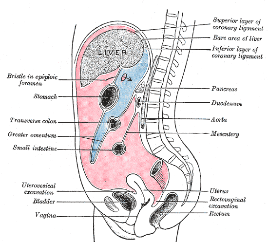
Spatial questions also use the four quadrants. The right upper quadrant holds most of the liver, the gallbladder, much of the duodenum and the head of the pancreas. The left upper quadrant holds the stomach, the spleen and the body and tail of the pancreas. The right lower quadrant holds the cecum and appendix, and the left lower quadrant holds the descending and sigmoid colon. Pain location in a vignette usually points to one of these groups.
The wall and its linings
From the esophagus to the anus the wall has the same four layers, from the lumen outward:
| Layer | What it contains | Why it matters |
|---|---|---|
| Mucosa | Epithelium, lamina propria (loose connective tissue with capillaries, lymphatics and immune cells), muscularis mucosae | Secretion and absorption; most ulcers and cancers start here |
| Submucosa | Dense connective tissue, larger vessels, submucosal (Meissner) plexus, glands in some regions | Nerve control of secretion and local blood flow |
| Muscularis externa | Inner circular and outer longitudinal smooth muscle, myenteric (Auerbach) plexus between them | Peristalsis and segmentation |
| Serosa or adventitia | Visceral peritoneum (serosa) on intraperitoneal organs; loose connective tissue (adventitia) on the esophagus and retroperitoneal parts | Anchors the organ |

The lining changes with the job of each region, and comparing them is a favorite question:
| Region | Epithelium | Special features |
|---|---|---|
| Esophagus | Non-keratinized stratified squamous (resists abrasion) | Mucous glands in the submucosa; muscle is skeletal in the upper third, mixed in the middle, smooth below; adventitia instead of serosa for most of its length |
| Stomach | Simple columnar, every surface cell secretes mucus | Gastric pits and glands; rugae; an extra inner oblique muscle layer for churning |
| Small intestine | Simple columnar absorptive cells (enterocytes) with microvilli, plus goblet cells | Circular folds, villi and crypts; Brunner's glands in the duodenal submucosa make alkaline mucus; Peyer's patches in the ileum |
| Large intestine | Simple columnar with many goblet cells | No villi; straight deep crypts; taeniae coli; the anal canal changes to stratified squamous |
The sharp change from stratified squamous to simple columnar at the gastroesophageal junction explains why reflux hurts the esophagus: its lining is built for friction, not acid.
Mouth, salivary glands and esophagus
Mechanical digestion starts in the mouth. Adults usually have 32 teeth: incisors cut, canines tear, premolars and molars grind. The tongue mixes the food with saliva and shapes it into a bolus.
Saliva is about 99% water; an adult makes roughly 1 to 1.5 L a day, mostly from three paired glands: the parotid (largest, watery serous secretion, duct opening beside the upper second molar), the submandibular and the sublingual. Small lingual glands at the back of the tongue add lingual lipase, which works in the acidic stomach. Saliva also carries mucus, IgA, lysozyme and bicarbonate.
Salivary amylase cleaves internal α-1,4 bonds of starch, producing maltose and short chains. It needs near-neutral pH, so it stops once stomach acid penetrates the bolus. Saliva has no protease, so the mouth does not start protein digestion.
Swallowing has an oral phase (voluntary), a pharyngeal phase (involuntary: the soft palate seals the nasal cavity, the larynx rises, the epiglottis covers the airway, the upper esophageal sphincter opens) and an esophageal phase (a peristaltic wave). The esophagus is about 25 cm long and secretes only mucus. The upper esophageal sphincter keeps air out, and the lower esophageal sphincter (LES) keeps stomach contents from refluxing; it relaxes as each swallow arrives.
The stomach
The stomach has a cardia, fundus, body, and antrum ending at the pylorus and pyloric sphincter. When empty, its mucosa folds into rugae. Gastric pits lead into glands that contain several cell types:
| Cell | Secretes | Function |
|---|---|---|
| Surface mucous cells | Thick alkaline mucus with bicarbonate | Main barrier against acid and pepsin |
| Mucous neck cells | Thin mucus | Protects the gland neck |
| Parietal cells | HCl and intrinsic factor | Acid lowers pH to about 2, denatures proteins and kills microbes; intrinsic factor is needed for vitamin B₁₂ absorption |
| Chief cells | Pepsinogen and gastric lipase | Acid converts pepsinogen to pepsin |
| G cells (antrum) | Gastrin | Stimulates acid secretion and motility |
| D cells | Somatostatin | Inhibits gastrin and acid |
| ECL cells | Histamine | Stimulates parietal cells through H₂ receptors |
Parietal cells pump H⁺ into the lumen with the H⁺/K⁺-ATPase (the proton pump) and release bicarbonate into the blood (the "alkaline tide" after a meal). Acid secretion has three phases. The cephalic phase starts at the sight, smell or thought of food through the vagus nerve. The gastric phase responds to stretch and to peptides in the stomach. The intestinal phase starts as chyme enters the duodenum and mostly inhibits the stomach, so the duodenum is not flooded with acid.
The stomach avoids digesting itself through overlapping defenses: a mucus-bicarbonate gel that keeps the surface near neutral, tight junctions that block back-leak of H⁺, rapid epithelial renewal, and prostaglandins (especially PGE₂) that boost mucus, bicarbonate and mucosal blood flow. When these fail, an ulcer forms (see Disorders).
Gastric motility has three parts. Receptive relaxation lets the fundus and body stretch to hold a meal without a pressure rise. Contractions then sweep toward the pylorus, and most chyme is pushed back (retropulsion) against a nearly closed sphincter, grinding food to a slurry. Small squirts pass into the duodenum, and emptying slows when fat, acid or concentrated chyme reaches the duodenum.
Small intestine
The small intestine is about 3 to 5 m long in a living adult (longer in a cadaver, where muscle tone is lost). The duodenum (about 25 cm) receives chyme, bile and pancreatic juice; bile and pancreatic juice enter together at the hepatopancreatic ampulla (of Vater), guarded by the sphincter of Oddi. The jejunum is the main absorptive segment; it has tall villi, prominent circular folds and the richest blood supply. The ileum absorbs bile salts and B₁₂ and ends at the ileocecal valve.
Surface area is multiplied at three scales: circular folds (plicae circulares), villi and microvilli (the brush border). Each villus has a capillary network that takes sugars and amino acids to the portal vein and a central lacteal that takes up fat. The crypts between villi hold stem cells, enteroendocrine cells, goblet cells and Paneth cells, which secrete defensins and lysozyme. M cells over Peyer's patches sample antigens for the immune system.
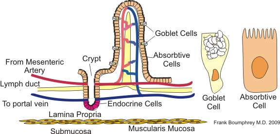
The brush border carries enzymes that finish digestion: maltase, sucrase-isomaltase (sucrose, and the branched α-limit dextrins amylase leaves behind), lactase, and peptidases. Enterokinase (enteropeptidase), made by the duodenal lining, converts trypsinogen to trypsin, which then activates the other pancreatic proteases.
Large intestine
The large intestine has no villi. It comprises the cecum (with the appendix), the ascending, transverse, descending and sigmoid colon, the rectum and the anal canal. Its outer longitudinal muscle is gathered into three bands, the taeniae coli, which pucker the wall into pouches called haustra. Its job is to reclaim water and electrolytes, house bacteria, and store and eliminate feces.
Slow haustral contractions mix the contents, and a few times a day a mass movement sweeps them toward the rectum, often after a meal (the gastrocolic reflex). Stretch of the rectum triggers the defecation reflex. The internal anal sphincter is smooth muscle and involuntary; the external anal sphincter is skeletal muscle and voluntary. The appendix is a blind pouch rich in lymphoid tissue and can become obstructed.
Motility and its control
Two patterns move the contents. Peristalsis is a wave in which the muscle contracts behind a bolus and relaxes in front of it, pushing it forward. Segmentation is a set of stationary ring contractions that chop and mix chyme against the mucosa with little net movement.

Interstitial cells of Cajal act as pacemakers, producing slow waves (the basal electrical rhythm) that set the maximum contraction frequency. The stomach runs at about 3 per minute; the duodenum is the fastest (about 11 to 12 per minute), with the rate falling toward the ileum (about 8 to 9). Textbooks differ by a cycle or two for the intestine, but the stomach is consistently the slowest. Between meals, the migrating motor complex sweeps residue and bacteria from the stomach through the ileum roughly every 90 to 120 minutes; it is triggered by the hormone motilin and stopped by eating.
The enteric nervous system (ENS) is a network of hundreds of millions of neurons in the gut wall that can run reflexes with no input from the brain. The myenteric plexus mainly controls motility and the submucosal plexus mainly controls secretion and local blood flow. In the peristaltic reflex, stretch causes contraction above the bolus (acetylcholine, substance P) and relaxation below it (nitric oxide, VIP). The autonomic system adjusts the ENS: parasympathetic (mostly vagal) input increases motility and secretion, and sympathetic input reduces them and constricts gut blood vessels, which is why stress and hard exercise slow digestion.
Hormones from enteroendocrine cells coordinate the organs:
| Hormone | Made by | Stimulus | Main effects |
|---|---|---|---|
| Gastrin | G cells (antrum) | Peptides, stretch, vagal input | ↑ acid; ↑ gastric motility; ↑ ECL histamine |
| Secretin | S cells (duodenum) | Acid in the duodenum | ↑ bicarbonate from pancreatic and bile ducts; ↓ acid |
| CCK | I cells (duodenum, jejunum) | Fatty acids, peptides | ↑ pancreatic enzymes; gallbladder contracts; sphincter of Oddi relaxes; ↓ gastric emptying; satiety |
| GIP | K cells (duodenum, jejunum) | Glucose, fatty acids | ↑ insulin; ↓ gastric acid |
| GLP-1 | L cells (ileum, colon) | Nutrients reaching the distal gut | ↑ insulin; ↓ glucagon; slows gastric emptying; satiety |
| Motilin | M cells (duodenum) | Fasting | Starts the migrating motor complex |
| Somatostatin | D cells | Acid | ↓ gastrin, acid and most GI secretion |
Chemical digestion of proteins, fats and carbohydrates
Carbohydrates. Dietary carbohydrate is mostly starch plus sucrose, lactose and maltose. Salivary and pancreatic amylase cut starch into maltose, maltotriose and α-limit dextrins. Brush-border enzymes finish the job: maltase gives glucose, sucrase gives glucose + fructose, lactase gives glucose + galactose. Glucose and galactose enter enterocytes with sodium through SGLT1 (secondary active transport); fructose enters by facilitated diffusion through GLUT5; all three leave through GLUT2 into portal blood. Because each disaccharide has its own enzyme, a missing enzyme blocks only its substrate, and the undigested sugar draws water into the gut and is fermented by colon bacteria.
Proteins. Pepsin starts protein digestion in the stomach. In the duodenum, trypsin, chymotrypsin, elastase (endopeptidases that cut inside chains) and carboxypeptidase (an exopeptidase that trims from the end) make short peptides, and brush-border peptidases finish them. Amino acids enter by sodium-linked carriers; di- and tripeptides enter by PEPT1 and are split inside the cell. Absorbed amino acids travel to the liver in the portal vein.
Fats. Bile salts emulsify triglyceride droplets. Pancreatic lipase, anchored by colipase, cuts triglycerides into 2-monoglyceride and free fatty acids, which pack into bile-salt micelles that ferry them to the brush border. Inside the enterocyte they are rebuilt into triglycerides and packaged with apolipoproteins as chylomicrons, which enter the lacteals, travel up the thoracic duct and join the blood at the left subclavian vein, bypassing the liver at first. Short- and medium-chain fatty acids go straight into portal blood.
| Nutrient | Enzymes | Where | Absorbed as |
|---|---|---|---|
| Starch | Salivary and pancreatic amylase; brush-border maltase and isomaltase | Mouth, duodenum, jejunum | Glucose (SGLT1) |
| Protein | Pepsin; trypsin, chymotrypsin, elastase, carboxypeptidase; brush-border peptidases | Stomach, duodenum, jejunum | Amino acids, di- and tripeptides |
| Triglyceride | Lingual and gastric lipase (minor); pancreatic lipase + colipase | Stomach (minor), duodenum | Fatty acids and monoglycerides in micelles → chylomicrons |
Absorption of water, minerals and vitamins
About 9 L of fluid reaches the small intestine each day: 1 to 2 L from drinking and the rest from saliva, gastric juice, bile, pancreatic juice and intestinal secretions. The small intestine reabsorbs most of it, the colon takes up most of what is left (it can absorb several liters a day if pushed), and only about 0.1 to 0.2 L leaves in feces. Water is never pumped. It follows solutes osmotically, so absorbing water means absorbing sodium: through Na⁺-glucose cotransport (SGLT1), Na⁺/H⁺ exchange, and, in the distal colon, aldosterone-regulated ENaC channels. Oral rehydration solution works because glucose drives sodium uptake through SGLT1 even when a toxin is making the gut secrete.
| Substance | Main site | How |
|---|---|---|
| Iron | Duodenum | Fe³⁺ reduced to Fe²⁺, taken up by DMT1; exported by ferroportin; hepcidin from the liver blocks export when iron is high |
| Calcium | Duodenum (active), rest of small intestine (passive) | Active uptake needs vitamin D (calcitriol) |
| Water-soluble vitamins (B, C) | Jejunum mostly | Carrier-mediated |
| Vitamin B₁₂ | Terminal ileum | Bound to intrinsic factor; receptor-mediated endocytosis |
| Fat-soluble vitamins (A, D, E, K) | Jejunum and ileum | Carried in micelles, so they are lost when fat absorption fails |
| Bile salts | Terminal ileum | Active transport back to the liver |
| Sodium, chloride, water | Whole small intestine; colon finishes | Na⁺ transport, water follows |
Fiber is plant carbohydrate that human enzymes cannot digest. Soluble fiber (oats, beans, fruit pectin) forms a gel that slows gastric emptying and glucose absorption, binds bile acids so the liver must use cholesterol to replace them (lowering LDL), and is fermented by colon bacteria into short-chain fatty acids. Insoluble fiber (wheat bran, vegetable skins) holds water and adds bulk, speeding transit and softening stool, which lowers colonic pressure and is linked to less diverticular disease.
Liver and gallbladder
The liver, the largest internal organ (about 1.5 kg), sits under the right diaphragm. About three-quarters of its blood comes from the portal vein (nutrient-rich, oxygen-poor blood from the gut and spleen) and the rest from the hepatic artery. Its anatomical unit is the hexagonal lobule: plates of hepatocytes radiating from a central vein, with a portal triad (hepatic artery branch, portal vein branch, bile duct) at the corners. Blood runs through the sinusoids toward the central vein; bile runs the other way in bile canaliculi toward the bile ducts.

Liver cells (State & National)
| Cell | Location | Job |
|---|---|---|
| Hepatocytes | Plates between sinusoids | Metabolism, bile secretion, plasma proteins (albumin, clotting factors), detoxification |
| Sinusoidal endothelial cells | Line sinusoids; fenestrated, no basement membrane | Let plasma reach hepatocytes through the space of Disse |
| Kupffer cells | Inside sinusoids | Resident macrophages: remove bacteria and endotoxin arriving from the gut, old red cells and debris; release cytokines that drive liver inflammation |
| Stellate (Ito) cells | Space of Disse | Store vitamin A; when activated by injury they become collagen-making myofibroblasts, causing fibrosis and cirrhosis |
| Cholangiocytes | Bile ducts | Modify bile, adding water and bicarbonate (secretin-driven) |
The lobule also has zones. Zone 1, near the portal triad, gets the most oxygen and does most gluconeogenesis and fatty-acid oxidation. Zone 3, around the central vein, gets the least oxygen and has the most cytochrome P450 drug-metabolizing enzymes, so it is injured first by low blood flow and by acetaminophen overdose.
Energy metabolism and glucose homeostasis
The liver buffers blood glucose. After a meal, insulin (from pancreatic beta cells) tells hepatocytes to store glucose as glycogen (glycogenesis) and convert the excess into fatty acids, exported as VLDL. Between meals, glucagon (alpha cells) drives glycogenolysis, which covers the first day or so of fasting, and then gluconeogenesis from lactate, amino acids (especially alanine) and glycerol. The liver can release free glucose because it has glucose-6-phosphatase; skeletal muscle lacks it, so muscle glycogen fuels only the muscle itself. In prolonged fasting the liver also makes ketone bodies from fatty acids for the brain and heart. The liver also makes urea from ammonia, which is why liver failure raises blood ammonia.
Cholesterol balance
The liver makes cholesterol (rate-limited by HMG-CoA reductase, the target of statins), takes it up from LDL and chylomicron remnants, and gets rid of it by converting it to bile acids or secreting it into bile. Lipoproteins carry it in blood:
| Lipoprotein | Made by | Carries | Clinical meaning |
|---|---|---|---|
| Chylomicron | Enterocytes | Dietary triglyceride to tissues | Remnants go to the liver |
| VLDL | Liver | Triglyceride made by the liver | Becomes IDL, then LDL, as triglyceride is removed |
| LDL | From VLDL in blood | Cholesterol to tissues; cleared by liver LDL receptors | High LDL deposits in artery walls ("bad") |
| HDL | Liver and intestine | Collects cholesterol from tissues back to the liver (reverse cholesterol transport) | Higher HDL is protective ("good") |
Fewer hepatic LDL receptors means higher blood LDL. Anything that forces the liver to make more bile acid (soluble fiber, bile-acid sequestrant drugs) pulls cholesterol out of the blood.
Bile, bilirubin and the gallbladder
Hepatocytes make about 0.5 to 1 L of bile a day, containing bile salts (amphipathic cholesterol derivatives), phospholipid, cholesterol, bicarbonate and bilirubin. Bile salts emulsify fat and form micelles; about 95% are reabsorbed in the terminal ileum and return in portal blood, the enterohepatic circulation.

Bilirubin comes from heme. Macrophages convert heme to unconjugated (indirect) bilirubin, which travels bound to albumin. Hepatocytes conjugate it with glucuronic acid using the enzyme UGT1A1, making water-soluble conjugated (direct) bilirubin for bile. Colon bacteria turn it into urobilinogen and stercobilin (the brown of feces). Jaundice has three sources: too much hemolysis (unconjugated bilirubin rises), liver-cell disease or a conjugation defect, or blocked bile flow (conjugated bilirubin rises, with pale stools and dark urine).
The gallbladder stores bile and concentrates it by absorbing water. CCK contracts it and relaxes the sphincter of Oddi. Gallstones form when bile is supersaturated with cholesterol or short of bile salts; obesity, rapid weight loss and pregnancy raise the risk.
Pancreas
The exocrine pancreas (acinar cells in grape-like clusters) makes digestive juice; the endocrine islets of Langerhans make hormones. Ducts join into the main pancreatic duct, which meets the common bile duct at the ampulla.
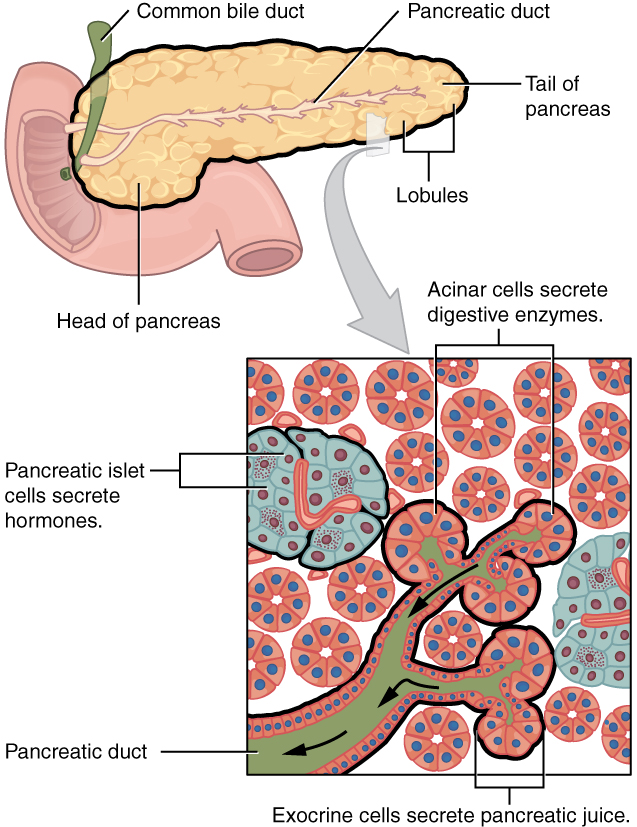
At the cellular level, acinar cells store enzymes in apical zymogen granules and release them under CCK and vagal stimulation. Duct cells secrete bicarbonate under secretin: CFTR channels let chloride out, and a Cl⁻/HCO₃⁻ exchanger swaps it for bicarbonate, which is why cystic fibrosis thickens pancreatic secretions. The juice contains amylase, lipase with colipase, nucleases, and the inactive zymogens trypsinogen, chymotrypsinogen, proelastase and procarboxypeptidase. Making proteases inactive, plus a trypsin inhibitor in the juice, protects the gland; pancreatitis is what happens when trypsin is activated inside it.
| Islet cell | Hormone | Main effect |
|---|---|---|
| Beta (most numerous) | Insulin | Lowers glucose: uptake into muscle and fat, storage as glycogen and fat |
| Alpha | Glucagon | Raises glucose: glycogenolysis and gluconeogenesis in the liver |
| Delta | Somatostatin | Inhibits insulin and glucagon locally |
| PP (F) | Pancreatic polypeptide | Modulates pancreatic and gallbladder secretion |
A beta cell senses glucose by metabolizing it: rising ATP closes ATP-sensitive K⁺ channels, the membrane depolarizes, Ca²⁺ enters and insulin is released. Gut hormones (GIP and GLP-1) add to this response, which is why oral glucose triggers more insulin than the same glucose given intravenously (the incretin effect).
Intestinal microbes (Division C, State & National)
The colon holds the densest microbial community in the body, mostly anaerobic bacteria. The stomach and upper small intestine have far fewer because acid, bile and the migrating motor complex keep numbers down. The microbes help digestion by fermenting fiber and resistant starch into short-chain fatty acids: butyrate is the main fuel of colon cells, and acetate and propionate travel to the liver. They also make vitamin K and some B vitamins and convert primary bile acids into secondary bile acids.
They help defense in three ways. Colonization resistance: resident bacteria occupy niches and nutrients, so pathogens such as Clostridioides difficile cannot take hold. Barrier support: butyrate strengthens tight junctions and mucus production. Immune training: microbial signals sampled through M cells and dendritic cells drive IgA production and regulatory T cells. Broad-spectrum antibiotics disturb this community (dysbiosis), which is why they can cause diarrhea, C. difficile colitis or lower vitamin K. An altered microbiome is also linked to inflammatory bowel disease, obesity and MASLD.
Effects of exercise and obesity
During hard exercise, sympathetic activity diverts blood from the gut to working muscle, and splanchnic blood flow can fall by more than half. Gastric emptying and absorption slow, and endurance athletes commonly get nausea, cramps or "runner's diarrhea"; very long efforts can injure the gut lining from low blood flow. Eating a large or fatty meal just before exercise worsens this.
Regular moderate exercise helps the system: it shortens colonic transit time (less constipation), and active people have lower rates of colon cancer, diverticular disease, gallstones and MASLD. Exercise also improves insulin sensitivity, so the liver and pancreas handle glucose more easily.
Obesity affects almost every organ in the tract:
| Organ | Effect of obesity |
|---|---|
| Esophagus | Higher abdominal pressure pushes acid upward: GERD, hiatal hernia, Barrett's esophagus and esophageal adenocarcinoma are more common |
| Gallbladder | Cholesterol-supersaturated bile → gallstones and cholecystitis |
| Liver | Fat builds up in hepatocytes → MASLD, which can progress to cirrhosis |
| Pancreas | Fat infiltration; more severe pancreatitis; insulin resistance strains beta cells |
| Colon | Higher risk of colorectal cancer and diverticulitis |
Obesity is also tied to high triglycerides, high LDL and low HDL, the lipid pattern of metabolic syndrome.
Metabolic syndrome and toxins (Division C, State & National)
Metabolic syndrome is a cluster of risk factors that share a root in insulin resistance and excess visceral fat. Under the widely used NCEP ATP III definition, a person has it with any three of: large waist circumference, high triglycerides (150 mg/dL or more), low HDL, blood pressure of 130/85 mmHg or more, and fasting glucose of 100 mg/dL or more. In the liver, insulin resistance keeps fat flowing from adipose tissue to hepatocytes and keeps the liver making new fat, causing steatosis (MASLD). In the pancreas, beta cells must secrete more insulin to compensate until they fail (type 2 diabetes), and very high triglycerides can trigger pancreatitis.
The 2027 Division C list names six toxins:
| Toxin | How it injures the liver or pancreas |
|---|---|
| Acetaminophen | An overdose saturates normal processing, so more is converted by P450 enzymes to NAPQI; once glutathione runs out, NAPQI kills zone 3 hepatocytes. The antidote N-acetylcysteine restores glutathione |
| Anabolic steroids | Impair bile flow (cholestatic jaundice) and are linked to liver adenomas, blood-filled cysts (peliosis hepatis) and liver cancer |
| Alcohol | Metabolism makes acetaldehyde and shifts the liver toward fat synthesis: fatty liver → alcoholic hepatitis → cirrhosis. A leading cause of acute and chronic pancreatitis |
| Heavy metals (lead, mercury, cadmium, arsenic) | Bind protein sulfur groups and cause oxidative stress; the liver concentrates many of them; arsenic exposure is linked to liver fibrosis and cancer |
| Bisphenols (such as BPA) | Endocrine disruptors; animal and population studies link exposure to fatty liver and insulin resistance |
| Polychlorinated biphenyls (PCBs) | Persistent, fat-stored industrial chemicals that induce liver enzymes; exposure is associated with fatty liver disease and diabetes |
Formulas and worked calculations
Disorders
Rows follow the order of the 2027 lists: the shared Regional list first, then the State and National additions. The level column gives B / C. In Division B, treatment and prevention are tested at State and National; in Division C, prevention, diagnosis and treatment are National only.
| Disorder | Level (B / C) | Cause / mechanism | Signs and symptoms | Diagnosis | Treatment / prevention |
|---|---|---|---|---|---|
| Stomach (gastric) and duodenal ulcers; peptic ulcer disease | Regional (as ulcers) and S&N (as PUD) / Regional | H. pylori (urease makes ammonia; inflammation weakens the barrier) or NSAIDs (block COX → less prostaglandin) | Burning epigastric pain: gastric ulcer pain worse with food, duodenal often eased by food; bleeding (black stool) | Urea breath test or stool antigen for H. pylori; endoscopy with biopsy | Antibiotics plus a proton-pump inhibitor; stop NSAIDs; surgery for perforation |
| Cancers of the digestive system | Regional (any) / see below | Accumulated mutations; risk factors vary by organ | Weight loss, bleeding, anemia, obstruction | Endoscopy or imaging with biopsy | Surgery, chemotherapy, radiation; screening |
| Diarrhea | Regional / not separately listed | Osmotic (unabsorbed solute, e.g. lactose), secretory (toxins raise cAMP and open CFTR chloride channels, as in cholera), inflammatory (damaged mucosa) or fast transit | Frequent loose stool, dehydration, low potassium | History; stool tests; secretory diarrhea continues when fasting | Oral rehydration solution; treat the cause; sanitation and handwashing prevent infectious causes |
| Lactose intolerance | Regional / Regional | Low lactase; lactose reaches the colon and is fermented | Bloating, gas, osmotic diarrhea after dairy | Hydrogen breath test | Less lactose, lactase tablets, lactose-free products |
| Hepatitis | Regional / S&N | Inflammation of the liver: viruses (A and E fecal-oral; B, C and D by blood and body fluids), alcohol, drugs, autoimmunity | Jaundice, fatigue, dark urine, right upper quadrant pain | Raised ALT and AST; viral antigens and antibodies | Vaccines for A and B; antivirals for B; curative antivirals for C; avoid alcohol |
| Appendicitis | Regional / Regional | Lumen blocked (fecalith in adults; lymphoid hyperplasia in children) → bacterial overgrowth, swelling, ischemia | Pain near the navel moving to McBurney's point (right lower quadrant), fever, vomiting | Exam, raised white count, ultrasound or CT | Appendectomy; antibiotics; perforation causes peritonitis |
| Diverticular disease | S&N / Regional | Pouches of mucosa push through weak points in the colon wall where vessels enter; linked to low fiber, high pressure, obesity | Ranges from none to left lower quadrant pain and fever | CT for diverticulitis; colonoscopy after recovery | Fiber; antibiotics or rest for uncomplicated disease; drainage or surgery for complications |
| GERD | S&N / Regional | LES weak or relaxing too often; hiatal hernia and obesity contribute | Heartburn, regurgitation, cough; complications include esophagitis, stricture and Barrett's esophagus | Symptoms and response to PPIs; endoscopy; pH monitoring | Lifestyle changes; antacids, H₂ blockers, PPIs; fundoplication |
| Ulcerative colitis | S&N / Regional | Autoimmune inflammation of the mucosa, starting at the rectum and spreading continuously | Bloody diarrhea, urgency, cramping | Colonoscopy and biopsy (crypt abscesses, no granulomas) | 5-ASA drugs, steroids, immunosuppressants, biologics; colectomy is curative |
| Crohn's disease | S&N / Regional | Autoimmune transmural inflammation anywhere from mouth to anus, most often the terminal ileum, with skip lesions | Pain, diarrhea, weight loss, fistulas, strictures, B₁₂ deficiency | Colonoscopy and biopsy (granulomas, cobblestoning); imaging | Steroids, immunosuppressants, biologics; surgery for complications; stop smoking |
| Celiac disease | S&N / Regional | Gluten peptides trigger a T-cell attack in people with HLA-DQ2 or DQ8 → villous atrophy | Diarrhea, bloating, weight loss, iron-deficiency anemia, itchy rash (dermatitis herpetiformis) | Anti-tissue transglutaminase IgA; duodenal biopsy | Lifelong gluten-free diet |
| Pancreatitis | not named / S&N | Trypsin activated inside the gland; gallstones and alcohol are the top causes, then very high triglycerides | Severe epigastric pain radiating to the back, vomiting | Lipase at least 3 times normal; CT | Fluids, pain control, early feeding; remove gallstones; stop alcohol |
| MASLD | not named / S&N | Fat in over 5% of hepatocytes plus a cardiometabolic risk factor (formerly NAFLD); can progress to inflammation (MASH) and fibrosis | Usually silent; fatigue; enlarged liver | Ultrasound, liver enzymes, elastography | Weight loss of 7 to 10%, exercise; resmetirom for MASH with fibrosis |
| Cirrhosis | not named / S&N | Chronic injury → stellate-cell fibrosis and regenerating nodules | Jaundice, ascites, varices, bleeding, confusion (high ammonia) | Imaging, elastography, low albumin, prolonged clotting time | Treat the cause; manage complications (diuretics, lactulose); transplant |
| Colorectal, gastric and hepatic cancer | covered by "cancers" / S&N | Colorectal: polyp-to-cancer sequence; gastric: H. pylori, salted foods; hepatic: cirrhosis, hepatitis B or C | Blood in stool, anemia; early satiety; liver mass | Colonoscopy, endoscopy, imaging, biopsy | Surgery, chemotherapy; colonoscopy screening; H. pylori treatment; hepatitis B vaccine |
The diverticular forms named in the Division C list form a spectrum:
| Form | What it means |
|---|---|
| Diverticulosis | Pouches present, no inflammation; often found by chance |
| Symptomatic uncomplicated diverticular disease | Ongoing pain or bowel changes with diverticula but no clear inflammation |
| Uncomplicated diverticulitis | Inflamed diverticula without abscess, perforation, fistula or obstruction |
| Complicated diverticulitis | Diverticulitis with an abscess, perforation, fistula, obstruction or peritonitis |
| Segmental colitis associated with diverticulosis | Chronic mucosal inflammation limited to the diverticula-bearing segment, usually the sigmoid; the rectum is spared |
| Smoldering diverticulitis | Persistent low-grade inflammation and symptoms for weeks to months after an attack that never fully settles |
| Disorder | Cause / mechanism | Key finding |
|---|---|---|
| Achalasia | Loss of inhibitory neurons in the esophageal myenteric plexus: LES cannot relax and peristalsis is lost | Difficulty swallowing solids and liquids; "bird's beak" on barium swallow; treated by dilation or myotomy |
| Hirschsprung disease | Neural crest cells fail to reach the distal colon, so it has no ganglion cells and stays contracted | Newborn does not pass meconium; dilated colon above the narrow segment; rectal biopsy shows no ganglia; surgical removal |
| Cholecystitis | A stone blocks the cystic duct → gallbladder inflammation | Right upper quadrant pain after fatty meals, fever, Murphy's sign; ultrasound; cholecystectomy |
| Choledocholithiasis | Stone in the common bile duct | Obstructive jaundice (conjugated bilirubin, raised ALP); removed by ERCP |
| Ascending cholangitis | Infection above a blocked bile duct | Fever, jaundice and right upper quadrant pain (Charcot's triad); antibiotics and urgent drainage |
| Mirizzi syndrome | Stone stuck in the cystic duct or gallbladder neck compresses the common hepatic duct | Obstructive jaundice without a stone in the duct itself; surgery |
| Primary biliary cholangitis | Autoimmune destruction of small bile ducts inside the liver, mostly middle-aged women | Itching, fatigue, raised ALP, anti-mitochondrial antibodies; ursodeoxycholic acid |
| Primary sclerosing cholangitis | Inflammation and scarring of bile ducts inside and outside the liver; strongly linked to ulcerative colitis | "Beaded" ducts on MRCP; raises cholangiocarcinoma risk; transplant |
| Gilbert syndrome | Reduced UGT1A1 activity | Mild unconjugated jaundice during fasting, illness or stress; harmless |
| Crigler-Najjar syndrome | Absent (type I) or severely reduced (type II) UGT1A1 | Severe unconjugated jaundice in infancy with risk of brain damage (kernicterus); phototherapy, transplant |
| Esophageal cancer | Squamous cell (smoking, alcohol) or adenocarcinoma (GERD, Barrett's, obesity) | Progressive difficulty swallowing, weight loss |
| Pancreatic cancer | Usually ductal adenocarcinoma; smoking, diabetes, chronic pancreatitis | Painless jaundice when in the head; often found late |
| Neuroendocrine tumors | Tumors of enteroendocrine (hormone-making) cells | May secrete hormones; liver spread can cause carcinoid syndrome (flushing, diarrhea) |
Common pitfalls
How tournaments test this
The Scio.ly archive holds 1,754 digestive questions from two past seasons: 967 from 2017-18 and 787 from 2022-23. Division C accounts for 1,174, Division B for 530, and 50 are tagged for both. The practice bank adds 3,066 questions.
- Format. Free response is the largest type at 875 questions (49.9%), then multiple choice at 693 (39.5%), multiple-select at 105 (6.0%) and true/false at 80 (4.6%).
- Task profile. The most common tasks are "which of the following" (9.6%), naming a structure in a figure (5.9%), explain or why (4.9%) and patient scenarios (4.0%). Calculation is rare (0.5%). Figure identification is common, so practice unlabeled diagrams of the tract, the gut wall, the villus and the liver lobule.
- Topics. Intestines are tagged on 787 archived questions, stomach on 488, liver on 280, mouth on 263 and pancreas on 186.
- Recurring terms. "Small intestine" appears in 364 prompts, "large intestine" in 151, "chemical digestion" in 80, "lactose intolerance" in 60, "mechanical digestion" in 41, "celiac disease" in 39, "parietal cells" in 34, "chief cells" in 31, "lamina propria" in 30 and "intrinsic factor" in 27.
- Answers that recur. As the correct option, "small intestine" appears 35 times, "stomach" 34, "mouth" 31, "liver" 30 and "duodenum" 28; secretin (16), CCK (10) and gastrin (9) are frequent hormone answers, and submucosa (17) and mucosa (15) frequent layer answers.
- What the archive does not show. Past tests predate several 2027 additions: abdominal wall layers, intraperitoneal versus retroperitoneal organs, gut microbes, metabolic syndrome, the six toxins, MASLD and the National-only biliary list. Expect new questions on these, likely as labeled diagrams and patient cases.
Test yourself
These are real questions from public tournament tests in the Scio.ly archive. Click an answer to check it; each question links to its full test.
- 1
The Lamina Propria is a layer of the
Practice more Anatomy and Physiology questions →
- 1
Through which of the following pancreatic ducts does both bile and pancreatic secretions empty into the duodenum?
From: Columbia University (CUSO) Invitational, 2022-23, Division C
Practice more Anatomy and Physiology questions →
- 1
Which of the following statements is true concerning the role of NSAIDs in causing gastric ulcers? I. They inhibit the enzyme cyclooxygenase II. They decrease the synthesis of inflammatory prostaglandins III. They artificially increase secretion of gastrin by G cells in the antrum IV. They contain proteases that directly damage the mucosa and mucosal cells
Practice more Anatomy and Physiology questions →
References and further reading
- OpenStax, Anatomy and Physiology 2e, chapter 23 (The Digestive System): https://openstax.org/books/anatomy-and-physiology-2e/pages/23-introduction
- OpenStax, Anatomy and Physiology 2e, chapter 24 (Metabolism and Nutrition), for glucose homeostasis and lipoproteins: https://openstax.org/books/anatomy-and-physiology-2e/pages/24-introduction
- NIDDK, digestive diseases (GERD, peptic ulcers, celiac disease, diverticular disease, IBD, cirrhosis, MASLD, gallstones, pancreatitis): https://www.niddk.nih.gov/health-information/digestive-diseases
- NIDDK, Definition and facts for NAFLD and NASH (now MASLD and MASH): https://www.niddk.nih.gov/health-information/liver-disease/nafld-nash/definition-facts
- National Heart, Lung, and Blood Institute, Metabolic syndrome: https://www.nhlbi.nih.gov/health/metabolic-syndrome
- NIH LiverTox database, for acetaminophen, anabolic steroids and other liver toxins: https://www.ncbi.nlm.nih.gov/books/NBK547852/
- SciOly Wiki, Anatomy and Physiology (Digestive): https://scioly.org/wiki/index.php/Anatomy_and_Physiology/Digestive
- Practice more: the archived Anatomy & Physiology tests in the Scio.ly test archive.