Anatomy and Physiology - Immune System
On this page(16 sections)
The immune system is the body's defense network: barriers, white blood cells, signaling proteins and organs that find and remove pathogens, damaged cells and tumor cells. In 2027 it is one of the three systems Anatomy & Physiology tests, alongside respiratory and digestive. Almost every Immune question comes down to three ideas. Innate defenses respond fast and the same way every time. Adaptive defenses respond more slowly, target one specific antigen and remember it. And the same tools that protect you cause disease when they are aimed at the wrong target, too weak, or too strong, which is what hypersensitivity, autoimmunity, immunodeficiency and transplant rejection are. Keep those ideas in view and the cell names, antibody classes and disorders sort themselves out.
What the rules cover
Under the 2027 rules, teams of up to two take a written test or rotate through lab-practical stations (models, specimens, illustrations, data to analyze) in about 50 minutes. Each team may bring writing utensils, two Class II calculators and one 8.5" × 11" sheet of notes. The sheet may be written on both sides, laminated or kept in a sheet protector, but extra sheets and stuck-on labels are not allowed. The 2027 event covers the respiratory, digestive and immune systems only. The highest score wins, and preselected questions break ties.
The immune outline (Division B section 3.c, Division C section 3.c of the 2027 rules manual) is summarized below. Levels are cumulative: State tests everything Regional tests, and Nationals tests everything.
| Topic | Division B | Division C |
|---|---|---|
| Organs: bone marrow, thymus, spleen, lymph nodes, skin | Regional | Regional |
| Innate immunity: barriers, complement, Toll-like receptors, inflammation, innate cells | Regional | Regional (adds antimicrobial proteins and fever) |
| Adaptive immunity | Regional | Regional (B and T cells, antigen recognition, maturation, selection, tolerance, presentation, activation, memory) |
| The immune response and how innate and adaptive arms work together | Regional (with allergic reactions) | Regional |
| Active vs passive immunity, immunization | Not listed separately | Regional |
| The five antibody classes: structure and roles | Not listed separately | Regional |
| Cytokines, interferons, JAK-STAT and cGAS-STING; constant-region variation; IgG and IgA subclasses | Not listed | State & National |
| Transplant types and preventing rejection | State & National (allograft, autograft) | State & National (adds rejection types and graft-versus-host disease) |
| Cancer immunotherapies | State & National (antibodies, CAR-T) | National only, inside tumor immunology |
| Vaccine routes and adjuvants | Not listed | National only |
| Immunological imprinting and antibody-dependent enhancement | Not listed | National only |
| Treatment and prevention of every listed disorder | State & National | National only (with diagnosis) |
The disorder lists differ by division and level:
| Level | Division B | Division C |
|---|---|---|
| Regional | HIV/AIDS, CVID, SCID; multiple sclerosis, rheumatoid arthritis, Hashimoto thyroiditis, Graves' disease; anaphylaxis, urticaria, myasthenia gravis, Arthus reaction, serum sickness, contact dermatitis | The same, plus Sjögren's syndrome, and asthma and celiac disease as hypersensitivity examples |
| State & National | Systemic lupus erythematosus, dermatomyositis, scleroderma, psoriasis, psoriatic arthritis | The same, plus type 1 diabetes, erythroblastosis fetalis, DiGeorge syndrome, Bruton agammaglobulinemia, Job syndrome, Bloom syndrome, Wiskott-Aldrich syndrome, MHC class I and II deficiency |
| National only | None | Acute lymphoblastic, acute myeloid, chronic lymphocytic and chronic myeloid leukemia; severe congenital neutropenia; cyclic neutropenia; staph enterotoxin food poisoning and toxic shock syndrome (superantigens) |
Immune organs and tissues
Immune cells are made in one place, trained in another and put to work in a third, so the organs fall into two groups. Primary lymphoid organs are where lymphocytes develop: the red bone marrow makes every blood cell and is where B cells mature, and the thymus, in the upper mediastinum behind the sternum, is where T cells mature. The thymus is largest relative to body size in childhood and is gradually replaced by fat after puberty. Its cortex is packed with immature T cells; its medulla holds the epithelial cells that display self-proteins during negative selection.
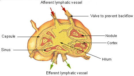
Secondary lymphoid organs are where mature lymphocytes meet antigen. Lymph nodes filter lymph: afferent vessels bring fluid and dendritic cells from tissues, B cells wait in follicles of the outer cortex, T cells sit in the deeper paracortex, and the efferent vessel carries lymph out at the hilum. The spleen does the same job for blood: its white pulp is lymphoid tissue around central arterioles, and its red pulp removes old red cells and platelets. Losing the spleen leaves a person vulnerable to encapsulated bacteria such as Streptococcus pneumoniae, because splenic macrophages and IgM-producing B cells clear those bacteria from blood. Mucosa-associated lymphoid tissue (MALT), including the tonsils and the Peyer's patches of the small intestine, guards the airways and gut.
The skin is listed as an immune organ because it is both barrier and sentinel. Keratinized epidermis blocks entry, sweat and sebum keep the surface acidic, and Langerhans cells (epidermal dendritic cells) carry antigen from the skin to the regional lymph nodes.
Innate immunity
Barriers and antimicrobial proteins
The first line of defense is physical and chemical: intact skin, mucus and the cilia that sweep it upward in the airways, stomach acid, tears and the normal flora that crowd out pathogens. Many surfaces add antimicrobial proteins: lysozyme in tears and saliva breaks down bacterial cell walls, defensins and cathelicidin punch holes in microbial membranes, and lactoferrin binds iron that bacteria need. Interferons and complement are sometimes grouped here too, since they are proteins that act against any microbe.
Pattern recognition and Toll-like receptors
The second line works inside the body. Its cells do not recognize one particular antigen; they recognize pathogen-associated molecular patterns (PAMPs), molecules that microbes share and human cells lack, using pattern-recognition receptors. The best-known family is the Toll-like receptors (TLRs). Those on the cell surface detect microbial surfaces; those inside endosomes detect nucleic acids from microbes that have been engulfed.
| Receptor | Location | Detects |
|---|---|---|
| TLR4 | Cell surface | Lipopolysaccharide (LPS) of gram-negative bacteria |
| TLR2 (with TLR1 or TLR6) | Cell surface | Lipoproteins and peptidoglycan components of bacteria |
| TLR5 | Cell surface | Bacterial flagellin |
| TLR3 | Endosome | Double-stranded RNA (many viruses) |
| TLR7 and TLR8 | Endosome | Single-stranded viral RNA |
| TLR9 | Endosome | Unmethylated CpG DNA, common in bacteria and DNA viruses |
Binding a TLR sets off a chain inside the cell: most TLRs signal through the adaptor MyD88, which activates the transcription factor NF-κB → genes for inflammatory cytokines (IL-1, IL-6, TNF-α) switch on → inflammation and fever. TLR3 (and TLR4 in part) use a second adaptor, TRIF, that turns on type I interferons. TLR activation also makes dendritic cells mature and travel to lymph nodes, which is how innate recognition starts the adaptive response.
Innate cells
| Cell | Main job | Key facts |
|---|---|---|
| Neutrophil | First responder to bacterial infection | The most common white cell (50–70% of WBCs); multi-lobed nucleus; the main cell in pus |
| Macrophage | Engulfs debris and microbes; presents antigen | Tissue form of the monocyte; releases IL-1 and TNF-α |
| Dendritic cell | Carries antigen from tissue to lymph nodes | The key antigen-presenting cell for naive T cells |
| Natural killer (NK) cell | Kills virus-infected and tumor cells | Detects "missing self" (little or no MHC class I); releases perforin and granzymes |
| Mast cell | Triggers inflammation and allergy | Stores preformed histamine and heparin; lives in connective tissue |
| Eosinophil | Attacks large parasites; part of allergic late phase | Two-lobed nucleus, red-orange granules |
| Basophil | Circulating relative of the mast cell | Releases histamine; dark blue granules |
Phagocytosis follows the same steps every time: the phagocyte binds the microbe, wraps pseudopods around it, pinches it into a phagosome, and fuses that vesicle with a lysosome full of enzymes and reactive oxygen species.

The complement system
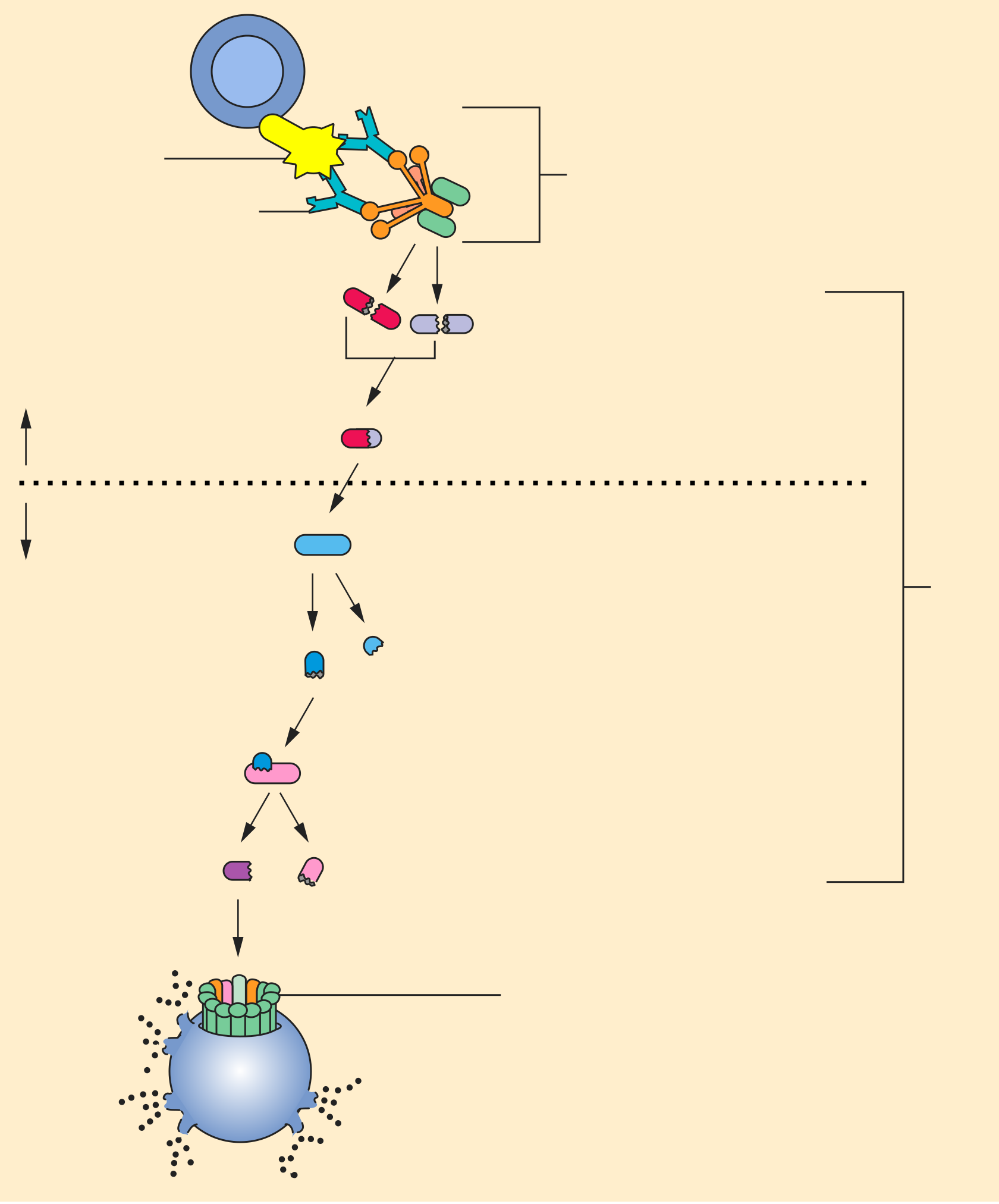
Complement is a set of about 30 plasma proteins that circulate inactive. The classical pathway starts when C1 binds antibody already attached to antigen; the lectin pathway starts when mannose-binding lectin attaches to microbial sugars; the alternative pathway starts from slow spontaneous breakdown of C3 on surfaces that lack the body's protective proteins. All three make a C3 convertase that cuts C3 into C3a and C3b. C3b coats the microbe (opsonization) so phagocytes grab it; C3a and C5a recruit phagocytes and trigger mast cells (inflammation); and C5b–C9 assemble the membrane attack complex, which punches holes in the microbe.
Inflammation and fever
Inflammation brings defenders to an injury and walls it off. Its classic signs are redness, heat, swelling and pain, with loss of function sometimes added. Mast cells release histamine → arterioles dilate → more blood flow → redness and heat. Histamine also makes capillaries leaky → plasma and protein leave the vessel → swelling. Prostaglandins and bradykinin sensitize nerve endings → pain. Phagocytes then arrive in a fixed order: leukocytosis (marrow releases more white cells), margination (cells roll along the vessel wall), diapedesis (they squeeze between endothelial cells) and chemotaxis (they follow gradients of C5a and chemokines to the site). Neutrophils arrive first, within hours; macrophages follow.
Fever is a regulated rise in the hypothalamic set point. Macrophages release pyrogens (IL-1, IL-6, TNF-α) → the hypothalamus makes prostaglandin E2 → the set point rises → shivering and vasoconstriction raise body temperature until it matches. Aspirin and ibuprofen lower fever by blocking prostaglandin synthesis.
Adaptive immunity
Adaptive immunity is built from lymphocytes, each carrying receptors for one specific antigen. Only a few cells share any receptor, so a response starts small. When antigen binds a matching lymphocyte, that cell divides into a clone (clonal selection). Most clone members become effector cells; some become long-lived memory cells.
| Feature | B cell | T cell |
|---|---|---|
| Matures in | Bone marrow | Thymus |
| Receptor | B-cell receptor (membrane antibody) | T-cell receptor |
| Recognizes | Intact antigen, directly | Peptide fragments only when displayed on MHC |
| Main effector | Plasma cell secreting antibody | Helper T cell (cytokines) or cytotoxic T cell (killing) |
| Surface markers | CD19, CD20 | CD3, plus CD4 or CD8 |
Maturation, selection and tolerance
Receptor diversity comes from gene rearrangement. In the pro-B cell stage the heavy-chain D segment joins a J segment, then a V segment joins DJ; the light chain rearranges next, and the immature B cell displays IgM. Immature B cells that bind self-antigen strongly are deleted or edit their receptor. T cells rearrange their receptor genes in the thymus and then face two tests:
- Positive selection (thymic cortex): a T cell survives only if its receptor binds self MHC weakly. This step also decides CD4 (binds MHC II) or CD8 (binds MHC I).
- Negative selection (medulla): a T cell that binds self-peptide on MHC too strongly dies. The transcription factor AIRE lets medullary cells display proteins from other organs, such as insulin, so self-reactive cells against them are removed.
Deletion in the thymus and marrow is central tolerance. Peripheral tolerance handles escapees: regulatory T cells (Tregs) suppress them, and a T cell that sees antigen without costimulation becomes unresponsive (anergy). Autoimmune disease is a failure of tolerance.
Antigen presentation and activation
T cells see antigen only as fragments displayed on major histocompatibility complex (MHC) proteins, called HLA in humans.
| MHC class I | MHC class II | |
|---|---|---|
| Found on | Almost every nucleated cell | Antigen-presenting cells: dendritic cells, macrophages, B cells |
| Displays | Peptides from proteins made inside the cell (including viral) | Peptides from material taken in from outside |
| Loading route | Proteasome → TAP transporter → endoplasmic reticulum | Endosome, after the invariant chain is removed |
| Read by | CD8 cytotoxic T cells | CD4 helper T cells |

A naive T cell needs two signals: signal 1 is its receptor binding peptide–MHC, and signal 2 is costimulation, mainly CD28 on the T cell binding B7 (CD80/86) on an activated antigen-presenting cell. Cytokines then act as a third signal that steers what the cell becomes. Activated cytotoxic (CD8) T cells kill infected or tumor cells with perforin and granzymes; they remove the virus factory, not free virus. Helper (CD4) T cells release cytokines, and a B cell that has captured a protein antigen needs help from a CD4 cell that recognizes the same antigen on its MHC II before it switches class and makes large amounts of antibody.

The integrated immune response
A typical infection runs as one chain. Barriers are breached → macrophages and dendritic cells detect PAMPs through TLRs → cytokines start inflammation, and neutrophils and complement attack → dendritic cells carry antigen to a lymph node → naive T cells are activated → helper T cells drive B cells to make antibody and activate macrophages, while CD8 cells kill infected cells → antibody returns the favor to innate immunity by opsonizing microbes and triggering the classical complement pathway.
Helper T cells specialize according to the cytokines present when they are activated:
| Subset | Driven by | Signature cytokines | Main job |
|---|---|---|---|
| Th1 | IL-12, IFN-γ | IFN-γ | Activate macrophages against intracellular microbes |
| Th2 | IL-4 | IL-4, IL-5, IL-13 | Parasites; IgE class switching; allergy |
| Th17 | IL-6, TGF-β, IL-23 | IL-17, IL-22 | Recruit neutrophils against extracellular bacteria and fungi |
| Treg | TGF-β, IL-2 | IL-10, TGF-β | Suppress responses; maintain tolerance |
The primary response to a new antigen has a lag of about a week while naive cells find antigen and clone; IgM appears first, then class switching produces IgG, IgA or IgE. The secondary response comes from memory cells, so it starts sooner, rises higher, lasts longer and is mostly high-affinity IgG. Humoral immunity (antibody) handles extracellular bacteria, toxins and free virus; cell-mediated immunity (T cells) handles infected cells, tumor cells and transplanted tissue.
Allergic reactions are the immune response aimed at a harmless antigen. On first exposure, Th2 cells push B cells to make IgE, which binds Fc receptors on mast cells (sensitization). On re-exposure, the allergen cross-links neighboring IgE molecules → the mast cell degranulates within minutes → histamine causes vasodilation, leaky vessels, bronchoconstriction and itching. Hours later, recruited eosinophils and Th2 cells, with the leukotrienes and cytokines they release, produce a late phase, which is why asthma attacks can recur after the first wave.
Antibodies
An antibody (immunoglobulin, Ig) is a glycoprotein made by plasma cells: two identical heavy chains and two identical light chains (κ or λ) joined by disulfide bonds into a Y. The tips of the arms (Fab) carry the variable regions that bind antigen; the stem (Fc) is built from heavy-chain constant regions and decides what happens next: which Fc receptors bind it, whether it activates complement, and whether it crosses the placenta.

| Class | Heavy chain | Form | Where | What it does |
|---|---|---|---|---|
| IgG | γ | Monomer | ~75% of serum Ig; blood and tissue fluid | Opsonizes, activates complement, neutralizes; only class that crosses the placenta (via FcRn); dominant in secondary responses |
| IgA | α | Monomer in serum; dimer with J chain in secretions | Tears, saliva, mucus, breast milk | Blocks microbes from attaching to mucosa; most abundant antibody made each day |
| IgM | μ | Pentamer with J chain | Blood; also a monomer on naive B cells | First antibody in a primary response; the most efficient class at activating complement |
| IgD | δ | Monomer | Surface of naive B cells | B-cell receptor alongside IgM |
| IgE | ε | Monomer | Bound to mast cells and basophils; lowest serum level | Allergy and defense against parasites |
Constant-region variation and subclasses
Antibodies vary at three levels. Isotypes are the classes and subclasses, defined by the heavy-chain constant region and shared by every healthy person. Allotypes are small inherited differences in constant regions between individuals (for example the Gm markers of IgG). Idiotypes are the unique shapes of one clone's variable region, so they are not a constant-region feature at all. Each heavy chain has one variable domain plus three constant domains (IgG, IgA, IgD) or four (IgM, IgE, which lack a hinge); the flexible hinge in IgG, IgA and IgD lets the arms spread.
| Subclass | Share of its class | Distinctive features |
|---|---|---|
| IgG1 | About 60–70% of IgG | Main response to protein antigens; strong complement and Fc-receptor binding |
| IgG2 | About 20–25% | Main response to bacterial polysaccharide capsules; weak complement activation; crosses the placenta least well |
| IgG3 | About 5–8% | Longest hinge; the strongest complement activator among IgG subclasses; shortest half-life (about 1 week vs 3 weeks) |
| IgG4 | About 4% | Does not activate complement; rises with long repeated exposure (allergen immunotherapy "blocking" antibody) |
| IgA1 | About 90% of serum IgA | Long hinge cut by bacterial IgA1 proteases (Neisseria, Haemophilus, pneumococcus) |
| IgA2 | Larger share in colon secretions | Short hinge resists those proteases |
Secretory IgA crosses epithelium by transcytosis: the polymeric Ig receptor carries the dimer through the cell and part of it stays attached as the secretory component, which protects the antibody from gut enzymes.
Active and passive immunity, and immunization
Immunity is active when your own body made it (with memory) and passive when you received ready-made antibody (immediate, temporary, no memory). Each can be natural or artificial.
| Type | Example | Memory? |
|---|---|---|
| Naturally acquired active | Recovering from measles | Yes |
| Artificially acquired active | Vaccination | Yes |
| Naturally acquired passive | Maternal IgG crossing the placenta; IgA in breast milk | No |
| Artificially acquired passive | Antivenom, rabies or tetanus immune globulin, monoclonal antibodies | No |
Immunization uses this logic. A vaccine presents antigen safely (live attenuated, inactivated, subunit or toxoid, conjugate, viral vector or mRNA) so the body builds memory B and T cells, and the next exposure triggers a secondary response before disease develops. When enough of a population is immune, transmission chains break and people who cannot be vaccinated are protected (herd immunity). Live vaccines are avoided in people with SCID and other severe deficiencies because the vaccine strain can cause disease.
Vaccine routes and adjuvants
| Route | Examples | What it favors |
|---|---|---|
| Intramuscular | Most inactivated, subunit and mRNA vaccines (influenza shot, hepatitis B, tetanus, COVID-19) | Well-vascularized muscle; strong systemic IgG; fewer local reactions to adjuvants |
| Subcutaneous | MMR and varicella (traditionally) | Systemic response; slower absorption |
| Intradermal | BCG | Small dose into skin rich in dendritic cells |
| Oral | Rotavirus, oral polio, oral typhoid, cholera | Mucosal secretory IgA in the gut, where the pathogen enters |
| Intranasal | Live attenuated influenza (nasal spray) | Mucosal IgA in the upper airway |
The principle: mucosal routes give mucosal IgA at the portal of entry; injected vaccines give strong blood IgG. An adjuvant is an ingredient that boosts the response to the antigen, usually by acting as a danger signal that activates innate immunity. Aluminum salts (alum), used for about a century, hold antigen at the injection site and activate inflammasomes. Oil-in-water squalene emulsions (MF59, AS03) recruit antigen-presenting cells and are used in some influenza vaccines. AS01 combines a TLR4 agonist (MPL) with the saponin QS-21 and is used in the recombinant shingles vaccine. CpG 1018, a TLR9 agonist, is in a two-dose hepatitis B vaccine, and Matrix-M (a saponin) is in a protein COVID-19 vaccine. Adjuvants let a smaller antigen dose give a stronger, longer response.
Suboptimal antibodies
Immunological imprinting (once called "original antigenic sin") means the first strain of a virus you meet, especially influenza, shapes later responses. When a related strain appears, memory B cells against the shared epitopes respond faster than naive cells against the new ones, so the response is biased toward the first strain and may fit the new strain poorly. Imprinting can also help: people imprinted on one hemagglutinin group are better protected against new bird-flu strains from the same group.
Antibody-dependent enhancement (ADE) happens when antibodies bind a virus without neutralizing it. The antibody's Fc region then docks on Fc receptors of monocytes and macrophages, carrying the virus into cells it infects. The textbook case is dengue: antibody from a first infection with one serotype binds but does not neutralize a second serotype → more infected cells → higher risk of severe dengue. That is why the first dengue vaccine is given only to people with a prior infection.
Immune signaling molecules and pathways
Cytokines are small proteins that carry messages between immune cells. Interleukins act mainly between leukocytes, chemokines direct cell movement, TNF-α drives inflammation, and interferons coordinate antiviral defense.
| Molecule | Main source | Key effect | Connection to disease or therapy |
|---|---|---|---|
| IL-1, IL-6, TNF-α | Macrophages | Fever, acute-phase proteins, inflammation | TNF inhibitors treat RA and psoriasis; anti-IL-6 receptor treats CAR-T cytokine release syndrome |
| IL-2 | Activated T cells | T-cell proliferation | Calcineurin inhibitors block its production; high-dose IL-2 is a cancer therapy |
| IL-4 | Th2 cells | Class switch to IgE | Allergy and asthma |
| IL-12, IFN-γ | Dendritic cells; Th1 and NK cells | Th1 differentiation; macrophage activation; more MHC | Defense against intracellular microbes |
| IL-17, IL-23 | Th17 cells; dendritic cells | Neutrophil recruitment | Psoriasis drugs target IL-17 and IL-23; Job syndrome lacks Th17 cells |
| IL-10, TGF-β | Tregs | Suppress inflammation | Tumors secrete them to evade immunity |
| Type I IFN (α, β) | Infected cells, plasmacytoid dendritic cells | Antiviral state in neighboring cells | High in lupus; anifrolumab blocks its receptor |
Interferons come in three types. Type I (IFN-α and IFN-β) is made by almost any virus-infected cell and tells neighbors to make antiviral proteins that block viral replication and spread. Type II (IFN-γ) comes from T and NK cells and activates macrophages. Type III (IFN-λ) protects epithelial surfaces.
JAK-STAT is how many cytokine receptors, including the interferon receptors, send their message to the nucleus. Cytokine binds its receptor → two receptor chains come together → the attached Janus kinases (JAKs) phosphorylate each other and the receptor's tails → STAT proteins dock on the phosphorylated tails and are phosphorylated → STATs pair up, enter the nucleus and switch on target genes. Medicine uses both ends of this chain: JAK inhibitors (tofacitinib, baricitinib) treat rheumatoid and psoriatic arthritis, and defects in the chain cause disease. A missing JAK3 or common gamma chain (shared by several interleukin receptors) causes SCID, and a faulty STAT3 causes Job syndrome.
cGAS-STING detects DNA in the wrong place. DNA belongs in the nucleus and mitochondria, so DNA in the cytosol signals a virus, a bacterium or a damaged cell. The enzyme cGAS binds cytosolic double-stranded DNA and makes the messenger cGAMP → cGAMP binds STING on the endoplasmic reticulum → STING recruits the kinase TBK1 → the transcription factor IRF3 is activated → type I interferons are made. The pathway helps detect DNA viruses, retroviruses such as HIV and tumor cells, but if self-DNA is not cleared it drives the interferon excess seen in lupus.
Hypersensitivity
Hypersensitivity is an immune response that harms the host. Four types are named by mechanism (Division B writes them as types 1–4).
| Type | Mechanism | Timing | Listed examples |
|---|---|---|---|
| I (immediate) | Allergen cross-links IgE on mast cells → histamine, leukotrienes | Minutes | Anaphylaxis, urticaria, asthma (C) |
| II (cytotoxic, antibody to cell surface) | IgG or IgM binds antigens on cells or receptors → complement, phagocytosis, or receptor blocking/stimulation | Hours to days | Myasthenia gravis, erythroblastosis fetalis (C), Graves' disease |
| III (immune complex) | Soluble antigen–antibody complexes deposit in vessels, joints and kidneys → complement and neutrophils | Hours (Arthus) to 1–2 weeks (serum sickness) | Arthus reaction, serum sickness, lupus (State & National) |
| IV (delayed, T-cell mediated) | Sensitized T cells and macrophages | 24–72 hours | Contact dermatitis, celiac disease (C), tuberculin skin test |
The Arthus reaction is a local type III reaction: an injected antigen meets high levels of existing antibody in the skin, forming complexes in the vessel walls of that spot. Serum sickness is the systemic version: about 1–2 weeks after a foreign protein (such as antivenom or some drugs) the body's new antibodies form complexes throughout the circulation, causing fever, rash, joint pain and low complement levels. Skin tests show sensitization: a wheal within minutes means type I IgE, and induration after 48–72 hours means type IV T cells. Neither proves a current infection.
Transplantation and rejection
| Graft | Donor | Rejection risk |
|---|---|---|
| Autograft | Yourself (skin graft, your own stem cells) | None |
| Isograft | Identical twin | None |
| Allograft | Another human | Yes: different HLA |
| Xenograft | Another species (pig heart valves) | Highest |
Rejection is an immune response to foreign HLA (MHC) and ABO blood-group antigens. Division C classifies it by timing:
| Rejection | Timing | Mechanism | Prevention or treatment |
|---|---|---|---|
| Hyperacute | Minutes to hours | Preformed recipient antibodies (anti-ABO or anti-HLA) bind graft vessels → complement → clotting; a type II reaction | Prevented by ABO matching and crossmatch testing; cannot be reversed |
| Acute | Days to months | Recipient T cells (and new antibodies) attack the graft | Increase immunosuppression, corticosteroid pulses |
| Chronic | Months to years | Ongoing low-grade immune injury → thickened vessel walls and fibrosis | Hard to treat; the main cause of late graft loss |
Rejection is prevented by matching (ABO, HLA typing, crossmatch) and by lifelong immunosuppression: calcineurin inhibitors (tacrolimus, cyclosporine) block IL-2 production; antiproliferatives (mycophenolate, azathioprine) stop lymphocyte division; mTOR inhibitors (sirolimus) block the IL-2 growth signal; corticosteroids broadly suppress inflammation; and induction antibodies (anti-thymocyte globulin, basiliximab) deplete or block T cells at the time of transplant. The cost is a higher risk of infection and some cancers.
Graft-versus-host disease (GVHD) reverses the direction: immune cells in the graft attack the recipient. It is a risk mainly after bone-marrow (hematopoietic stem cell) transplants, because the graft contains donor T cells and the recipient's immune system has usually been destroyed and cannot fight back. The skin (rash), liver (jaundice) and gut (diarrhea) are the main targets. HLA matching, T-cell depletion and preventive immunosuppression reduce it.
Tumor immunology and cancer immunotherapy
Immune surveillance is the constant search for cancer cells by NK cells, CD8 T cells and IFN-γ. Tumors that survive have evaded it by lowering MHC class I, losing the antigens T cells see, expressing PD-L1 that switches off T cells through PD-1, secreting suppressive TGF-β and IL-10, and recruiting Tregs.
| Therapy | How it works | Examples |
|---|---|---|
| Therapeutic cancer vaccines (C) | Train the immune system against an existing tumor | Sipuleucel-T (patient's antigen-presenting cells loaded with a prostate-cancer antigen); BCG instilled into the bladder |
| Cytokine therapies (C) | Boost lymphocyte growth or activation | High-dose IL-2 for metastatic melanoma and kidney cancer; interferon-α |
| Antibody therapies | Mark tumor cells for destruction, or release the brakes on T cells | Rituximab (anti-CD20); trastuzumab (anti-HER2); checkpoint inhibitors pembrolizumab and nivolumab (anti-PD-1) and ipilimumab (anti-CTLA-4) |
| CAR T-cell therapy | Patient's T cells given an engineered receptor against a tumor surface protein | Anti-CD19 CAR-T for relapsed B-cell ALL and lymphomas; anti-BCMA for myeloma |
A chimeric antigen receptor (CAR) joins an antibody-style binding piece outside the cell to the T-cell receptor's CD3ζ signaling chain and a costimulatory domain (CD28 or 4-1BB) inside, so the T cell recognizes the tumor without needing MHC and supplies both activation signals itself. T cells are collected, given the CAR gene with a viral vector, expanded and infused. The main side effects are cytokine release syndrome (fever and low blood pressure, treated with the anti-IL-6 receptor antibody tocilizumab), neurotoxicity, and loss of normal B cells because they also carry CD19.
Disorders
Rows follow the order of the 2027 lists. Treatment and prevention columns are tested at State & National in Division B and at Nationals in Division C.
Regional (both divisions)
| Disorder | Cause / mechanism | Signs and symptoms | How a test shows it | Treatment | Prevention |
|---|---|---|---|---|---|
| HIV/AIDS | Retrovirus binds CD4 plus CCR5 or CXCR4 and destroys helper T cells | Flu-like acute illness, long latency, then opportunistic infections, weight loss, lymphadenopathy | Antigen/antibody test, viral load; AIDS when CD4 falls below 200 cells/µL or an AIDS-defining illness appears | Antiretroviral therapy | Condoms, PrEP, clean needles, treating pregnant mothers |
| Common variable immunodeficiency (CVID) | B cells fail to become plasma cells | Recurrent sinus and lung infections, often first diagnosed in adults; more autoimmunity and lymphoma | Low IgG with low IgA and/or IgM; poor vaccine response | Immunoglobulin replacement (IVIG/SCIG) | Infection prevention; avoid live vaccines |
| Severe combined immunodeficiency (SCID) | Inherited block in T-cell development (often X-linked common gamma chain; also JAK3, ADA) | Severe infections, thrush, diarrhea and failure to thrive in infancy | Very low T cells; low TREC on newborn screening; no thymic shadow | Stem-cell transplant; enzyme replacement or gene therapy for ADA | Newborn screening; no live vaccines |
| Multiple sclerosis | T cells and antibodies attack CNS myelin | Optic neuritis, numbness, weakness, relapsing course | MRI white-matter lesions; oligoclonal bands in spinal fluid | Interferon-β, anti-CD20 (ocrelizumab), steroids for relapses | No proven prevention |
| Rheumatoid arthritis | Autoimmune synovitis erodes cartilage and bone | Symmetric small-joint pain and swelling, morning stiffness over an hour | Rheumatoid factor, anti-CCP antibodies; X-ray erosions | Methotrexate, TNF inhibitors, JAK inhibitors | Not smoking lowers risk |
| Hashimoto thyroiditis | T cells and anti-TPO/anti-thyroglobulin antibodies destroy the thyroid | Hypothyroidism: fatigue, cold intolerance, weight gain, goiter | High TSH, low free T4, anti-TPO antibodies | Levothyroxine | No proven prevention |
| Graves' disease | Antibodies stimulate the TSH receptor | Hyperthyroidism, diffuse goiter, bulging eyes | Low TSH, high T4, TSH-receptor antibodies | Methimazole, radioactive iodine, surgery; beta-blockers for symptoms | No proven prevention |
| Sjögren's syndrome (C) | Lymphocytes infiltrate tear and salivary glands | Dry eyes, dry mouth | Anti-Ro/SSA and anti-La/SSB; Schirmer test; lip biopsy | Artificial tears, pilocarpine, hydroxychloroquine | No proven prevention |
| Asthma (C, as type I) | IgE/Th2 airway inflammation, bronchoconstriction | Wheeze, cough, chest tightness | Reversible obstruction on spirometry | Inhaled steroids, short-acting β2 agonists | Avoid triggers |
| Anaphylaxis | Systemic type I reaction (foods, drugs, venom) | Hives, airway swelling, wheeze, low blood pressure | Clinical diagnosis; raised tryptase; specific IgE testing | Intramuscular epinephrine first, then antihistamines and steroids | Avoid the trigger; carry auto-injector; venom immunotherapy |
| Urticaria | Mast-cell histamine release in the dermis | Itchy raised wheals that fade within 24 hours | Clinical; trigger history | Second-generation antihistamines; omalizumab (anti-IgE) for chronic cases | Avoid triggers |
| Myasthenia gravis | Type II: antibodies block and destroy acetylcholine receptors | Weakness that worsens with use; drooping eyelids, double vision | Anti-AChR antibodies; decreasing response on repeated nerve stimulation | Pyridostigmine, immunosuppression, thymectomy | No proven prevention |
| Arthus reaction | Local type III immune complexes | Painful swelling at an injection site after 4–12 hours | History of repeated injections (for example, tetanus boosters) | Usually self-limited; NSAIDs | Space out booster doses |
| Serum sickness | Systemic type III after a foreign protein | Fever, rash, joint pain 1–2 weeks after exposure | Low C3/C4; protein in urine | Stop the trigger; antihistamines, steroids | Avoid animal-derived antisera when possible |
| Contact dermatitis | Type IV reaction to a hapten (nickel, urushiol) | Itchy red vesicular rash 1–3 days after contact | Patch test | Topical steroids | Identify and avoid the allergen |
| Celiac disease (C, as type IV) | T cells react to gluten peptides modified by tissue transglutaminase (HLA-DQ2/DQ8) | Diarrhea, bloating, weight loss, anemia | Anti-tTG IgA; villous atrophy on biopsy | Lifelong gluten-free diet | None known |
A hapten is a small molecule, such as nickel or urushiol, that is not immunogenic alone but becomes an antigen after binding a self-protein, which is how contact dermatitis starts.
State & National
| Disorder | Cause / mechanism | Signs and symptoms | How a test shows it | Treatment | Prevention |
|---|---|---|---|---|---|
| Systemic lupus erythematosus | Autoantibodies to nuclear antigens; immune complexes; type I interferon excess | Malar rash, photosensitivity, arthritis, kidney disease; mostly women | ANA (sensitive), anti-dsDNA and anti-Smith (specific), low complement | Hydroxychloroquine, steroids, belimumab, anifrolumab | Sun protection reduces flares |
| Dermatomyositis | Autoimmune injury to small vessels of muscle and skin | Proximal muscle weakness, purple eyelid rash, Gottron papules; linked to cancer | High creatine kinase; muscle biopsy; myositis antibodies | Steroids, methotrexate, IVIG | Cancer screening after diagnosis |
| Scleroderma | Autoimmunity and vessel injury → excess collagen | Tight skin, Raynaud phenomenon, lung fibrosis, kidney crisis | Anti-centromere (limited) or anti-Scl-70 (diffuse) | Organ-specific: ACE inhibitors for kidney crisis, mycophenolate for lung disease | No proven prevention |
| Psoriasis | IL-23/IL-17 T-cell signaling drives keratinocyte overgrowth | Sharp red plaques with silvery scale on elbows, knees, scalp | Clinical; biopsy if unclear | Topical steroids, vitamin D analogs, phototherapy, anti-IL-17/IL-23 or anti-TNF biologics | Avoid triggers (skin injury, smoking) |
| Psoriatic arthritis | Same pathway affecting joints and tendon insertions | Fingertip-joint arthritis, sausage-shaped digits, nail pitting | Rheumatoid factor negative; "pencil-in-cup" X-ray | NSAIDs, methotrexate, biologics, JAK inhibitors | Early treatment of psoriasis |
| Type 1 diabetes (C) | Autoreactive T cells destroy pancreatic beta cells | Thirst, frequent urination, weight loss, ketoacidosis | High glucose; GAD65 and insulin autoantibodies; low C-peptide | Insulin | Teplizumab (anti-CD3) can delay onset in high-risk people |
| Erythroblastosis fetalis (C) | Type II: Rh-negative mother's anti-D IgG crosses the placenta and destroys Rh-positive fetal red cells | Fetal anemia, jaundice, swelling (hydrops) | Positive maternal antibody screen; positive direct Coombs test in the baby | Intrauterine transfusion, phototherapy, exchange transfusion | Anti-D immune globulin at about 28 weeks and after delivery |
| DiGeorge syndrome (C) | 22q11.2 deletion → third and fourth pharyngeal pouches fail → thymus and parathyroids missing | Infections, low-calcium seizures, heart defects, facial features | Low T cells, low calcium, FISH or microarray for the deletion | Calcium and vitamin D, heart surgery, thymus transplant if complete | Genetic counseling |
| Bruton agammaglobulinemia (C) | X-linked BTK mutation stops B cells at the pre-B stage | Bacterial infections in boys after about 6 months, when maternal IgG fades; tiny tonsils | Almost no B cells (CD19); all Ig classes very low | Lifelong IVIG | No live vaccines; genetic counseling |
| Job syndrome (C) | Dominant-negative STAT3 mutation → few Th17 cells | Cold staph abscesses, eczema, lung cysts, retained baby teeth | Very high IgE, eosinophilia | Long-term antibiotics, skin care | Infection prevention |
| Bloom syndrome (C) | Recessive BLM helicase mutation → genomic instability | Short stature, sun-sensitive facial rash, infections, early cancers | Increased sister-chromatid exchange; low Ig | Cancer surveillance; treat infections | Avoid unnecessary radiation; genetic counseling |
| Wiskott-Aldrich syndrome (C) | X-linked WAS mutation disrupts the actin cytoskeleton of blood cells | Eczema, bleeding, recurrent infections | Low platelet count with small platelets; low IgM, high IgA and IgE | Stem-cell transplant, gene therapy | Genetic counseling |
| MHC class I deficiency (C) | Usually TAP1 or TAP2 mutation → little MHC I on cells → few CD8 T cells | Chronic bacterial lung infections, skin lesions | Low surface HLA class I on flow cytometry | Treat infections, airway care | Genetic counseling |
| MHC class II deficiency (C) | Mutated regulators of MHC II genes (such as CIITA) → no MHC II → few CD4 T cells | Severe infections and diarrhea in infancy | No HLA-DR on B cells and monocytes; low antibody | Stem-cell transplant | Genetic counseling |
National only (Division C)
| Disorder | Cause / mechanism | Signs and symptoms | How a test shows it | Treatment | Prevention |
|---|---|---|---|---|---|
| Acute lymphoblastic leukemia | Immature lymphoid blasts fill the marrow | Most common childhood cancer: anemia, bleeding, infection, bone pain | Blasts on smear and marrow; TdT positive | Multi-agent chemotherapy; anti-CD19 CAR-T if relapsed | None known |
| Acute myeloid leukemia | Immature myeloid blasts; mostly older adults | Fatigue, infections, bleeding | Myeloblasts with Auer rods; at least 20% blasts | Chemotherapy, stem-cell transplant; ATRA and arsenic for the APL subtype | Avoid benzene, smoking, unnecessary radiation |
| Chronic lymphocytic leukemia | Slowly growing mature-looking B cells; older adults | Often found on routine blood work; lymph nodes, infections | Lymphocytosis, smudge cells, CD5+ CD20+ B cells | Watchful waiting; BTK inhibitors (ibrutinib), venetoclax | None known |
| Chronic myeloid leukemia | Philadelphia chromosome t(9;22) makes BCR-ABL kinase | Very high WBC with all granulocyte stages, enlarged spleen | BCR-ABL by PCR or cytogenetics | Imatinib and other tyrosine-kinase inhibitors | None known |
| Severe congenital neutropenia | Mutations (often ELANE, also HAX1) stop neutrophil maturation in the marrow | Severe bacterial infections from infancy | ANC persistently below 500 cells/µL | G-CSF (filgrastim); stem-cell transplant | Monitor for progression to AML |
| Cyclic neutropenia | ELANE mutation; neutrophil output oscillates | Fever, mouth ulcers and infections about every 21 days | Serial counts two to three times a week for six weeks | G-CSF | Anticipate low phases |
| Staph enterotoxin food poisoning | Preformed, heat-stable enterotoxin (a superantigen) in food | Sudden vomiting and cramps 1–6 hours after eating; gone within a day | Clinical; toxin in leftover food | Fluids and support | Refrigerate food; hand hygiene (reheating does not destroy the toxin) |
| Toxic shock syndrome | TSST-1 (or streptococcal toxins) cross-links MHC II and T-cell receptors → cytokine storm | High fever, sunburn-like rash, low blood pressure, later peeling of palms and soles | Clinical criteria; source such as a tampon or packed wound | Remove the source, fluids, antibiotics including clindamycin | Change tampons often; wound care |
A superantigen skips antigen processing: it clamps the outside of MHC class II to the variable β region of the T-cell receptor, so it activates every T cell with a matching Vβ, up to about 20% of all T cells instead of the 1 in 10,000 or fewer that a normal antigen activates. The flood of IL-2, TNF-α and IFN-γ causes the shock.
Common pitfalls
How tournaments test this
Our archive holds 1,620 Immune questions from the 2017-18 season (899) and the 2022-23 season (721). The practice bank holds 3,880 immune-related questions, including 2,133 tagged Immunity and 946 tagged White Blood Cells.
- Format. Multiple choice is 47% of archived questions (766), free response 41% (660), true/false 6% (101) and select-all 6% (90).
- Division. Division C has 1,127 questions, Division B 453, and 40 appear in both.
- Subtopic tags. Immunity (864), White Blood Cells (625), Antibodies (355), Inflammation (252) and Allergies (96).
- Task profile. About 13% are "which of the following" items, 4% ask you to explain, 3% are case scenarios and 3% ask you to identify a structure in a figure. Calculations are under 1%.
- Answers that keep coming back. Neutrophils, IgE, helper T cells, macrophages, IgG, B cells, IgA, thymus, bone marrow, natural killer cells, IgM, spleen and dendritic cells.
The archived tests were written to older outlines, so they cover the Regional core well (cells, organs, antibody classes, inflammation, hypersensitivity) but contain almost nothing on signaling pathways, IgG subclasses, transplant rejection, vaccine adjuvants, ADE or the Division C State and National disorder lists. Expect 2027 writers to add case vignettes for those new disorders and mechanism questions on JAK-STAT and cGAS-STING; the tables above are built to answer them.
Test yourself
These are real questions from public tournament tests in our archive. Click an answer to check it; each links to the full test.
- 1
Of the following selections, the one that includes only nonspecific defenses is:
Practice more Anatomy and Physiology questions →
- 1
Which cell type is effective against intracellular pathogens such as viruses?
Practice more Anatomy and Physiology questions →
- 1
The specific type of acquired immunity that a fetus obtains from maternal antibodies that cross the placenta is called:
Practice more Anatomy and Physiology questions →
References and further reading
- OpenStax, Anatomy and Physiology 2e, chapter 21, the lymphatic and immune system: https://openstax.org/books/anatomy-and-physiology-2e/pages/21-introduction
- OpenStax, Microbiology, chapters on innate and adaptive immunity, vaccines and hypersensitivities: https://openstax.org/details/books/microbiology
- NCBI Bookshelf, Janeway's Immunobiology (5th edition, free online): https://www.ncbi.nlm.nih.gov/books/NBK10757/
- National Institute of Allergy and Infectious Diseases, primary immune deficiency diseases: https://www.niaid.nih.gov/diseases-conditions/primary-immune-deficiency-diseases-pidds
- CDC, vaccine administration routes and adjuvants: https://www.cdc.gov/vaccines/basics/
- CDC, HIV basics: https://www.cdc.gov/hiv/
- National Cancer Institute, CAR T-cell therapy and immunotherapy: https://www.cancer.gov/about-cancer/treatment/types/immunotherapy
- MedlinePlus, autoimmune and blood-cancer topics: https://medlineplus.gov/
- SciOly Wiki, Anatomy and Physiology: https://scioly.org/wiki/index.php/Anatomy_and_Physiology
- Practice more: the archived Anatomy & Physiology tests in the Scio.ly test archive.