Designer Genes
2027 season
On this page(18 sections)
Designer Genes is a written, quantitative genetics event for Division C. Teams work through problems in classical (Mendelian) genetics, population and evolutionary genetics, and molecular biology. The 2027 rules say outright that questions will lean on process skills: quantitative reasoning, calculations, reading experimental results and drawing conclusions from evidence. Expect fewer pure-vocabulary items and more "here is a data set, what does it show and why." Points are awarded for reasoning as well as answers, so always show the setup. Teams that lose points usually know the material but skip a step, mix up two similar terms or misread which strand a question is about.
This guide covers the whole event. The sections below cover format and strategy first, then the six topic areas of the 2027 rules in order, and finish with pitfalls and how tournaments write the test.
Event format
| Rule | Detail |
|---|---|
| Team | Up to 2 students |
| Time | About 50 minutes |
| Format | A written test, which may be run as stations |
| Calculators | Up to two Class II calculators per team |
| Notes | One 8.5 × 11 in sheet, both sides, any form and any source. It may be laminated or put in a sheet protector. Attached labels and extra sheets (even inside one protector) are not allowed |
| Other | Writing utensils |
| Scoring | High score wins. Points for accuracy, quality of supporting reasoning and proper scientific method. Preselected questions break ties |
| Division | Division C |
The notes rule is broad: formulas, a codon table, epistasis ratios, a χ² critical-value row, the steps of each technique and a worked template for each calculation type all fit. Keep it to what you will actually use, since searching a crowded sheet costs more than it saves.
What the rules cover
The 2027 rules split the event into six topic areas, paraphrased below. Items marked State or National are not part of Invitational or Regional tests. The rules call the topic list exhaustive and say no single topic should be overemphasized, so spread your preparation.
| Area | Every level (Invitational and Regional) | State and National adds |
|---|---|---|
| Mendelian genetics | Mendel's laws and the patterns that break them (linkage, incomplete dominance and codominance, complementation); mono-, di- and trihybrid Punnett squares; probability rules applied to data; reading and building pedigrees; epistasis | Mapping genes from recombination data: two genes at State, three genes at National |
| Mitosis and meiosis | Stages and key structures of both divisions and how they compare; effects of nondisjunction; reading karyotypes for abnormalities | Somatic recombination: V(D)J recombination and immunoglobulin class switching |
| Population and evolutionary genetics | Hardy–Weinberg assumptions, calculations and violations; drift, founder effects, bottlenecks, migration and relative fitness; additive alleles and the number of genes behind a trait; gene duplication with homologs, orthologs and paralogs; reading phylogenies and the steps of building one from sequences | Heritability: broad-sense, narrow-sense and realized |
| Molecular biology of DNA | Nucleotide structure, strand direction and base pairing; replication from pre-replication complex to termination in bacteria, archaea and eukaryotes; polymerase fidelity and proofreading; plasmids, chromatin and chromosome organization; damage from UV, ionizing radiation and intercalators and its repair (NER, NHEJ, HDR) in bacteria and eukaryotes; mutation classes and protein effects | The end-replication problem and telomere synthesis |
| Eukaryotic gene expression and regulation | The central dogma; transcription stages and RNA polymerase; cis and trans regulatory elements including epigenetics and riboswitches, and yeast two-hybrid screening; RNA maturation (capping, polyadenylation, splicing); translation stages and its regulation | Mobile genetic elements: retroviruses, retrotransposons and DNA transposons, and the enzymes that move them |
| Technology and techniques | PCR and its applications (Sanger sequencing, error-prone PCR, RT-qPCR); Illumina vs Nanopore sequencing; molecular cloning (plasmids, restriction enzymes, PCR, Gibson and Golden Gate assembly, stable lines and transient transfection); knockout, knockdown and complementation logic for necessity and sufficiency | How knockdowns and knockouts are made (RNAi, homologous recombination including Cre-Lox, CRISPR); ChIP-seq, Hi-C and RNA-seq (single-cell vs bulk) |
Strategy
Splitting the work
Two students means two parallel streams of calculations. A workable split is one partner on inheritance (Mendelian ratios, pedigrees, probability, mapping, Hardy–Weinberg, heritability) and the other on molecules and techniques (replication, repair, expression, cloning, sequencing). Both read the whole test first and flag questions that need the other person, then check each other's arithmetic. Agree on notation up front (RF as a percentage or in cM, how you write genotypes) so a single answer never mixes conventions.
Time and the answer sheet
With 50 minutes, answer the short items first, then the multi-step calculations and data questions. For every calculation, write the setup on one line before the number: the cross, the table, or the formula with its variables defined. For every experiment question, give a claim, the evidence from the data and a mechanism, and say what control or follow-up experiment would test it. Because the rules reward supporting reasoning and proper scientific method, a short answer that names the control and the mechanism beats a long one that restates the question.
Mendelian genetics and pedigrees
Mendel's first law, segregation, says that each gamete carries only one allele of each gene, so the two alleles in a diploid parent separate during gamete formation. The second law, independent assortment, says that genes on different chromosomes sort independently. Both laws describe what happens in meiosis I, which is why the cell-division and Mendelian material are tested together. Departures from the classic ratios happen when genes are linked (on the same chromosome and close enough that they usually travel together), when alleles are incompletely or co-dominant, or when two genes interact, so that the combination of genotypes at two loci makes a phenotype neither locus makes alone (gene interaction and epistasis; the complementation test, a different idea, is defined under "Complementation and epistasis").
The baseline dihybrid ratio is 9:3:3:1 from a cross of two heterozygotes at two unlinked loci. A monohybrid cross of two heterozygotes gives 3:1, and a testcross of a heterozygote with a homozygous recessive gives 1:1. Punnett squares for three genes (trihybrid crosses) have 64 boxes, so the per-gene product method is faster: treat each gene as its own monohybrid cross, then multiply. For AaBbCc × AaBbCc, the chance of the A_B_C_ phenotype is ¾ × ¾ × ¾ = 27/64, and the chance of aabbcc is (¼)³ = 1/64. The full phenotypic ratio with complete dominance and independent assortment is 27:9:9:9:3:3:3:1.
Terms and Mendel's experiments
Many points on every test are vocabulary in disguise: "which term describes…", "what is the genotype of…". Learn the words so that a calculation question never fails at the first step.
| Term | Meaning |
|---|---|
| Gene / locus | A heritable unit that affects a trait / its fixed position on a chromosome |
| Allele | One version of a gene; a diploid carries two alleles per autosomal locus |
| Genotype / phenotype | The alleles present (Aa) / the observable trait (purple flowers). There are usually more possible genotypes than phenotypes |
| Homozygous / heterozygous | Two identical alleles (AA or aa) / two different alleles (Aa). A heterozygote for a recessive disease allele is a carrier |
| Hemizygous | Only one copy of the locus, as for X-linked genes in XY males |
| Dominant / recessive | The dominant allele's phenotype shows in the heterozygote; the recessive phenotype shows only in the homozygote |
| True-breeding (pure line) | Homozygous; selfing gives offspring identical to the parent |
| P, F1, F2 | Parental generation; first filial generation (their offspring); second filial generation (F1 × F1 or F1 selfed) |
| Monohybrid / dihybrid / trihybrid | Heterozygous at one / two / three loci, or a cross between such individuals |
| Testcross | Cross to a homozygous recessive to reveal the unknown genotype of a dominant-phenotype individual: all dominant offspring → AA; about 1:1 → Aa |
| Backcross | Cross of an F1 to either parent (a testcross is a backcross to the recessive parent) |
| Reciprocal cross | The same cross with the sexes of the parents swapped; different results flag sex linkage, maternal effect or organelle inheritance |
| Wild type | The common, "standard" phenotype or allele in natural populations, often written + |
Mendel's work. Gregor Mendel crossed garden peas (Pisum sativum) in the 1850s–60s and published in 1866. Peas suited him because they have clear either/or traits, are easy to grow in large numbers with a short generation time, normally self-pollinate (so true-breeding lines exist), and can be cross-pollinated by hand. He followed seven traits: seed shape (round/wrinkled), seed color (yellow/green), flower color (purple/white), pod shape (inflated/constricted), pod color (green/yellow), flower position (axial/terminal) and stem length (tall/dwarf). Crossing true-breeding parents gave a uniform F1 showing only one trait (the dominant one); the hidden trait reappeared in the F2 in about one quarter of plants. That reappearance is why he needed the F2: it showed the recessive "factor" was carried unchanged through the F1, not blended away. His work was rediscovered in 1900 (by de Vries, Correns and von Tschermak), and Sutton and Boveri's chromosome theory of inheritance (1902–03) explained it: genes sit on chromosomes, and chromosomes behave in meiosis exactly as Mendel's factors do. Thomas Hunt Morgan's white-eyed Drosophila (1910) placed a specific gene on a specific chromosome (the X).
Counting shortcuts. For an individual heterozygous at n independent loci, the number of gamete types is 2ⁿ. For a cross between two such individuals, the number of genotype classes is 3ⁿ, and with complete dominance the number of phenotype classes is 2ⁿ; the Punnett square has (2ⁿ)² = 4ⁿ boxes (each parent contributes 2ⁿ gamete types, and the square crosses every one of the mother's with every one of the father's). A dihybrid self therefore gives 4 gamete types, 16 boxes, 9 genotypes and 4 phenotypes. A human, with 23 chromosome pairs, can make 2²³ ≈ 8.4 million chromosome combinations in gametes before crossing over is counted, so one couple could produce about 7 × 10¹³ different zygotes.
Probability
Most calculations use two rules. The product rule applies to independent events joined by "and": multiply. The sum rule applies to mutually exclusive outcomes joined by "or": add. Write out the connecting word before you choose the rule.
Conditional probability is the step most students miss. When the question tells you something already known about a person, remove the outcomes that information rules out, then re-divide.
Testing data against a ratio
Because the 2027 rules emphasize evaluating experimental results, expect a cross with real counts and the question "do these data fit the expected ratio?" The standard tool is the chi-square goodness-of-fit test.
| Degrees of freedom | p = 0.10 | p = 0.05 | p = 0.01 |
|---|---|---|---|
| 1 | 2.71 | 3.84 | 6.63 |
| 2 | 4.61 | 5.99 | 9.21 |
| 3 | 6.25 | 7.81 | 11.34 |
| 4 | 7.78 | 9.49 | 13.28 |
| 5 | 9.24 | 11.07 | 15.09 |
Read the table this way: if your χ² is smaller than the value under p = 0.05, the p-value is greater than 0.05 and you fail to reject the null hypothesis (the deviation could be chance; the data fit the ratio). If it is larger, reject the null hypothesis: something other than chance (linkage, lethality, epistasis, selection, a wrong model) explains the deviation. Always state the null hypothesis ("the genes assort independently and give 9:3:3:1"), use counts rather than percentages, and remember that "fail to reject" does not prove the hypothesis.
Extensions of Mendelian inheritance
Mendel's traits were chosen to be simple. Most real genes are not, and tests love the cases where a ratio "looks wrong". Each pattern below has a signature ratio or clue.
| Pattern | What happens | Signature or classic example |
|---|---|---|
| Complete dominance | Heterozygote looks like the dominant homozygote | 3:1 in an F2 |
| Incomplete dominance | Heterozygote is intermediate | Snapdragon or four-o'clock flowers: red × white → all pink F1; F2 1 red : 2 pink : 1 white (phenotype ratio = genotype ratio) |
| Codominance | Both alleles are fully and separately expressed | AB blood type (both A and B antigens); MN blood group; roan cattle (red and white hairs side by side) |
| Multiple alleles | More than two alleles exist in the population (each person still has two) | ABO: Iᴬ and Iᴮ codominant, both dominant to i. Rabbit coat: C (full) > cʰᶜʰ (chinchilla) > cʰ (Himalayan) > c (albino), a dominance series |
| Lethal alleles | One genotype dies before it can be counted | Yellow mice (Aʸ) and Manx cats: the allele is dominant for the visible trait but recessive lethal, so heterozygote × heterozygote gives 2:1 among survivors (the 1/4 homozygous class is missing) |
| Late-onset lethal | Kills after reproductive age, so selection barely removes it | Huntington disease (autosomal dominant, onset usually 30–50 years) |
| Incomplete penetrance | Not everyone with the genotype shows the phenotype; penetrance = % of carriers of the genotype who show it | Polydactyly; BRCA1 mutations raise breast-cancer risk but not to 100% |
| Variable expressivity | Individuals with the same genotype show the phenotype to different degrees | Neurofibromatosis type 1; Marfan syndrome |
| Pleiotropy | One gene affects several unrelated traits | Cystic fibrosis (lungs, pancreas, sweat, fertility); Marfan; PKU |
| Sex-limited | Autosomal gene expressed in only one sex | Milk yield in cattle, beard growth, plumage in some birds |
| Sex-influenced | Autosomal gene whose dominance depends on sex | Pattern baldness: dominant in males, recessive in females |
| Environmental effects | Phenotype depends on conditions | Himalayan rabbits and Siamese cats: a temperature-sensitive enzyme makes dark pigment only at the cool extremities. Hydrangea color depends on soil pH |
| Phenocopy | An environmental change mimics a genetic phenotype | Thalidomide limb defects resembling inherited ones |
| Genetic anticipation | A disease gets more severe or appears earlier in each generation | Trinucleotide-repeat expansions (Huntington, fragile X, myotonic dystrophy) |
Dominance at the molecular level. Most loss-of-function alleles are recessive because one working copy makes enough protein (the gene is haplosufficient). A loss-of-function allele is dominant when half the protein is not enough (haploinsufficiency), or when the faulty protein poisons the good one in a multi-subunit complex (dominant negative). Gain-of-function alleles (a protein that is overactive or acquires a new job) are usually dominant. H. J. Muller classified alleles by what they do to the gene's activity:
| Muller's class | Meaning | Usual dominance |
|---|---|---|
| Amorph | Null allele, no activity | Recessive (unless haploinsufficient) |
| Hypomorph | Reduced activity ("leaky") | Recessive |
| Hypermorph | Increased activity of the normal function | Often dominant |
| Antimorph | Opposes the normal allele (dominant negative) | Dominant |
| Neomorph | A new function the normal gene does not have | Dominant |
A mutant that behaves like a weaker version of wild type (larvae that move slowly rather than not at all) is a hypomorph; if the phenotype gets worse when the mutant is placed over a deletion of the gene, the allele still had some activity.
Sex determination and sex linkage
Sex is decided by different systems in different organisms, and the system decides who is hemizygous.
| System | Female | Male | Examples and notes |
|---|---|---|---|
| XX/XY | XX | XY | Mammals. The Y-linked SRY gene triggers testis development, so XXY is male and X (45,X) is female |
| XX/XY (ratio-based) | XX | XY | Drosophila: sex follows the X : autosome ratio, so XXY flies are female and XO flies are sterile males |
| ZZ/ZW | ZW | ZZ | Birds, butterflies and moths, some reptiles. The female is the heterogametic sex; dose of the Z-linked DMRT1 gene drives male development in birds |
| XX/XO | XX | XO | Grasshoppers, crickets; C. elegans (XX hermaphrodites, XO males) |
| Haplodiploidy | Diploid (fertilized) | Haploid (unfertilized) | Bees, wasps, ants. Males have one allele per gene, so recessive harmful alleles are exposed and purged; sisters share ¾ of their genes |
| Environmental | — | — | Temperature during incubation sets sex in many turtles and all crocodilians |
X-linked crosses. Write genotypes with the chromosome: XᴬXᵃ, XᵃY. A male receives his X from his mother and his Y from his father, so a father never passes an X-linked allele to a son, and gives it to every daughter. For an X-linked recessive allele in a carrier mother × normal father: daughters ½ normal, ½ carrier, none affected; sons ½ affected. Morgan's white-eyed fly showed this: red-eyed F1 females crossed to red-eyed F1 males gave an F2 in which all the white-eyed flies were male. Questions ask for different denominators, so read carefully: "the chance that a child is an affected son" (¼) is not "the chance that a son is affected" (½).
Dosage compensation and X inactivation. Mammalian females silence one X in each somatic cell early in development, so both sexes have one active X. The inactive X is coated by the long noncoding RNA XIST (transcribed from the X that will be silenced), becomes facultative heterochromatin with H3K27 methylation and DNA methylation, and is visible as a dense Barr body at the nuclear edge. The number of Barr bodies is the number of X chromosomes minus one (XXY has one; 45,X has none). The choice is random in each early cell and then inherited by its descendants, so carrier females are mosaics: calico and tortoiseshell cats show orange and black patches because the coat-color gene is X-linked, which is why calico cats are almost always female (or XXY males), and why a cloned calico (CC, 2001) did not have the same coat pattern as its donor. A female carrier of an X-linked disease can be mildly affected if, by chance, most of her cells inactivated the normal X (skewed inactivation). Other animals compensate differently. In Drosophila the single male X is transcribed about twice as strongly: the MSL complex, with the roX long noncoding RNAs, binds the male X, and its subunit MOF acetylates H4K16, loosening the chromatin. In C. elegans XX hermaphrodites halve the expression of both X chromosomes with a condensin-like dosage compensation complex, matching XO males. All three solutions equalize X-linked gene products between the sexes; birds (ZW) compensate only partially.
Pedigrees
A pedigree problem asks you to decide the mode of inheritance from which individuals are affected and how the trait passes between sexes and generations. Work through the pattern systematically rather than guessing from one generation.
| Mode | What the pedigree shows |
|---|---|
| Autosomal dominant | Affected individuals in every generation; two affected parents can have an unaffected child; an affected parent passes the trait to about half of children; both sexes equally affected; an affected father with an affected son rules out X-linked dominant |
| Autosomal recessive | Often skips generations; affected children of unaffected carrier parents; both sexes equally affected |
| X-linked recessive | Many more affected males; an affected son usually has a carrier mother; an affected father passes the allele to all daughters and no sons |
| X-linked dominant | An affected father passes the trait to all daughters and none of his sons; an affected mother passes it to about half of all children |
| Y-linked | Every son of an affected male is affected; no daughters are affected |
| Mitochondrial | Passed from mother to all children; an affected father passes it to none of his children |
A pedigree construction question hands you a case study and asks you to draw it. Use squares for males, circles for females, filled symbols for affected individuals and a horizontal line for a mating; half-filled symbols mark carriers if the key says so.
Pedigree symbols and numbering. Generations are Roman numerals (I at the top), individuals in each generation are numbered left to right (II-3). A square is male, a circle female, a diamond sex unknown; filled = affected; a dot in the center or half-filling = carrier (when the key says so); a slash through a symbol = deceased; an arrow marks the proband (the person through whom the family came to attention). A horizontal line joins mates; a double line is a consanguineous mating (relatives), which raises the chance that both parents carry the same rare recessive allele. Offspring hang from a vertical line in birth order; identical (monozygotic) twins share one branch point joined by a bar, fraternal twins share a branch point without the bar.
Human genetic disorders
Tests use named disorders as the "case" for pedigree, probability, mutation and karyotype questions, so you need the mode of inheritance and the kind of mutation for the common ones. Use the table to classify a disorder, then apply the rules for that mode.
ReferenceHuman disorders by mode of inheritance· 22 rows
AD = autosomal dominant; AR = autosomal recessive; XR / XD = X-linked recessive / dominant; mt = mitochondrial.
No rows match.
| Disorder | Mode | Gene or cause | What to remember |
|---|---|---|---|
| Huntington disease | AD | HTT; CAG repeat expansion (≥ 40 repeats fully penetrant) | Late onset, so it escapes selection; anticipation, especially through fathers |
| Achondroplasia | AD | FGFR3 gain-of-function missense (one recurrent G→A change) | Most cases are new mutations; homozygotes do not survive |
| Marfan syndrome | AD | FBN1 (fibrillin-1) | Pleiotropy (tall, long limbs, lens dislocation, aortic aneurysm) and variable expressivity |
| Neurofibromatosis type 1 | AD | NF1 (a tumor suppressor) | Variable expressivity; café-au-lait spots |
| Familial hypercholesterolemia | AD | LDLR | Heterozygotes intermediate, homozygotes severe (incomplete dominance at the phenotype level) |
| Polydactyly (some forms) | AD | Several genes | Classic example of incomplete penetrance |
| Cystic fibrosis | AR | CFTR chloride channel; ΔF508 (an in-frame 3-bp deletion removing Phe508) is the most common allele | Most common severe AR disorder in people of European descent; pleiotropic |
| Sickle-cell disease | AR | HBB missense: GAG → GTG, Glu6Val in β-globin | Heterozygotes resist malaria (heterozygote advantage); codominant at the molecular level |
| Phenylketonuria (PKU) | AR | PAH (phenylalanine hydroxylase) | Treated by a low-phenylalanine diet: genotype plus environment sets phenotype |
| Tay–Sachs disease | AR | HEXA (hexosaminidase A) | Lipid builds up in neurons; higher frequency in Ashkenazi Jewish and some other founder populations |
| Albinism (oculocutaneous type 1) | AR | TYR (tyrosinase) | Epistatic to other pigment genes |
| Alkaptonuria | AR | HGD | Garrod's first "inborn error of metabolism" |
| Gaucher disease | AR | GBA | Lysosomal storage disease |
| Hemophilia A / B | XR | Clotting factor VIII / IX | Mostly males; Queen Victoria's descendants |
| Duchenne muscular dystrophy | XR | DMD (dystrophin), mostly large deletions | Frameshift deletions → Duchenne; in-frame deletions → milder Becker |
| Red–green color blindness | XR | Opsin genes on the X | About 8% of men of European descent |
| Fragile X syndrome | X-linked | FMR1; CGG repeat expansion (> 200) methylates and silences the gene | Most common inherited intellectual disability; anticipation through mothers |
| Hypophosphatemic (vitamin D–resistant) rickets | XD | PHEX | Affected fathers pass it to all daughters, no sons |
| Rett syndrome | XD | MECP2 | Mostly affected girls; usually lethal in hemizygous males |
| LHON (Leber hereditary optic neuropathy), MELAS | mt | mtDNA genes | Maternal transmission; MELAS severity tracks heteroplasmy, while LHON is usually homoplasmic with incomplete penetrance (more often affects males) |
| Prader–Willi / Angelman syndromes | Imprinting | Loss of the paternal / maternal copy of a 15q11–13 region | Same deletion, different syndrome depending on the parent of origin |
| Spina bifida, cleft lip, heart disease, diabetes | Multifactorial | Many genes plus environment | Do not fit any single-gene ratio |
Inheritance outside the nucleus and parent-of-origin effects
Three patterns break the rule that reciprocal crosses give the same result. Each has a different signature, and tests ask you to tell them apart.
- Mitochondrial (cytoplasmic) inheritance. Mitochondria and chloroplasts carry their own small circular genomes. The egg supplies almost all the cytoplasm and the sperm's mitochondria are destroyed after fertilization, so mtDNA comes from the mother: an affected mother passes the trait to all her children, an affected father to none. Cells hold hundreds of mtDNA copies; a mix of mutant and normal copies is heteroplasmy, and because organelles sort randomly at each division, siblings can be affected to different degrees. Leaf variegation in four-o'clock plants (Correns) is the plant example. Mitochondrial replacement therapy moves the nucleus of an affected mother's egg (or zygote) into a donor egg with healthy mitochondria, giving a child with nuclear DNA from both parents and mtDNA from the donor.
- Maternal effect. The offspring's phenotype is set by the mother's genotype, not its own, because she loads mRNAs and proteins into the egg. In snails, shell coiling direction follows the mother's genotype, so a dd snail whose mother was Dd coils right (dextral) even though its own genotype says left; the effect of its genotype shows only in its own offspring. Drosophila bicoid is the developmental example.
- Genomic imprinting. Some genes are expressed from only one parent's copy because the other copy was silenced by DNA methylation in that parent's germ line. The imprint is erased and reset in each generation according to the sex of the parent. Deleting 15q11–13 on the paternal chromosome causes Prader–Willi syndrome (the paternally expressed genes such as SNRPN are lost); the same deletion on the maternal chromosome causes Angelman syndrome (the maternally expressed UBE3A is lost). Uniparental disomy, inheriting both copies of a chromosome from one parent, causes the same syndromes. IGF2 (paternally expressed) and H19 (maternally expressed) are the classic mouse pair.
Complementation and epistasis
A complementation test asks whether two recessive mutants with the same phenotype carry mutations in the same gene. Cross the two homozygous mutants. If the offspring are normal, the mutations are in different genes, because each parent supplies a working copy of the gene the other lacks. If the offspring are still mutant, the mutations are in the same gene (they fail to complement).
Epistasis happens when one gene masks or modifies another. Start from the 9:3:3:1 baseline, merge the classes the interaction makes identical, and name the resulting ratio.
| Ratio | Interaction type | What it means | Classic example |
|---|---|---|---|
| 9:7 | Complementary genes | A homozygous recessive at either locus blocks the pathway | Sweet-pea flower color (two enzymes in one pathway) |
| 9:3:4 | Recessive epistasis | Homozygous recessive at one locus masks the other locus | Labrador coat color: ee dogs are yellow whatever the B/b genotype (E/e decides whether pigment is deposited in the hair); otherwise B_ black, bb brown. Also mouse agouti/albino |
| 12:3:1 | Dominant epistasis | A dominant allele at one locus masks the other locus | Summer-squash color: dominant W gives white regardless; yellow : green below it |
| 15:1 | Duplicate dominant genes | Any dominant allele at either locus gives the same phenotype | Shepherd's-purse seed-capsule shape |
| 13:3 | Dominant suppression | A dominant allele at one locus suppresses the other's dominant phenotype | White leghorn × white wyandotte chickens (dominant inhibitor I suppresses color) |
| 9:6:1 | Duplicate genes with cumulative effect | One dominant allele at either locus gives an intermediate phenotype; dominant alleles at both give the full one | Summer-squash fruit shape: disc (A_B_) : sphere (A_bb, aaB_) : long (aabb) |
| 9:3:3:1 with new phenotypes | Two genes interact to make four distinct phenotypes | No classes merge, but the four phenotypes are not simply "A trait plus B trait" | Chicken combs: walnut, rose, pea, single |
Gene interaction vocabulary. The gene that does the masking is epistatic; the gene being masked is hypostatic. Epistasis is between different genes; dominance is between alleles of one gene. The albino genotype (cc) in mammals is epistatic to all other coat-color genes.
Complementation groups. When many recessive mutants with the same phenotype are crossed in all pairs, mutants that fail to complement each other fall in the same complementation group, and each group is one gene. Complementation only works with recessive alleles: a dominant mutant shows its phenotype in any cross, so it cannot be tested this way.
Linkage and mapping (State and National)
Genes on the same chromosome are linked. During prophase I, homologs exchange segments by crossing over, and crossovers between two linked genes create recombinant gametes. The recombination frequency (RF) is the proportion of recombinant progeny. Genes farther apart are more likely to be separated, so RF rises with distance up to a cap of 50%, the value for unlinked genes. Two-gene mapping is a State task and three-gene mapping a National task.
In three-point mapping the most common classes are parental and the rarest are double crossovers. The middle gene is the one whose allele differs between the double-crossover and parental classes. Each adjacent distance is (single crossovers in that interval + double crossovers) / total, because a double crossover counts in both intervals.
Coupling and repulsion. A double heterozygote can carry its alleles two ways. In coupling (cis), the two dominant alleles sit on one homolog: AB/ab. In repulsion (trans), each homolog has one: Ab/aB. The phase decides which progeny classes are parental (most common) and which are recombinant (least common), so always identify the two largest classes in a testcross first; they tell you the parent's arrangement. With complete linkage (no crossing over), an AB/ab parent makes only AB and ab gametes.
RF from an F2 instead of a testcross. When an F1 (AB/ab, coupling) is intercrossed rather than testcrossed, recombinants cannot be counted directly, because most F2 phenotype classes mix several genotypes. Use the double-recessive class, which can only come from two ab gametes: each parent makes ab gametes at frequency (1 − r)/2, so freq(aabb) = [(1 − r)/2]² and r = 1 − 2√freq(aabb). If the F1 was in repulsion (Ab/aB), ab is a recombinant gamete made at r/2, so freq(aabb) = (r/2)² and r = 2√freq(aabb). Example: an F2 from AB/ab with 16% aabb gives √0.16 = 0.4 = (1 − r)/2, so r = 0.2, 20 cM. Unlinked genes give aabb = 1/16 (r = 0.5) either way.
Why RF never exceeds 50%. A single crossover involves only two of the four chromatids in a bivalent, so even when a crossover always occurs between two genes, half the products are recombinant. Genes far apart on one chromosome undergo so many crossovers that recombinants and parentals become equally common, and the genes look unlinked (RF ≈ 50%). That is why genes far apart on the same chromosome must be mapped through intermediate genes, adding up short intervals. It also explains why map distances over about 20–30 cM are underestimates: double crossovers between the outer genes restore the parental combination and go uncounted.
Map distance is not physical distance. Crossing over is not uniform: it is suppressed near centromeres and in heterochromatin, concentrated in recombination hotspots, and differs between the sexes (in humans the female map is longer; in Drosophila males there is no meiotic crossing over at all). So cM convert to base pairs only on average (in humans roughly 1 cM ≈ 1 Mb).
History. Morgan's student Alfred Sturtevant made the first genetic map in 1913 from Drosophila X-linked genes, reasoning that crossover frequency reflects distance. Fly genes fell into four linkage groups, matching the four chromosome pairs, which supported the chromosome theory.
Is it linked? Test with χ². For a dihybrid testcross, the null hypothesis of independent assortment predicts four equal classes (1:1:1:1). A χ² well above the critical value (7.81 at 3 df, p = 0.05) with an excess of two classes says the genes are linked; the two excess classes are the parental ones. Human geneticists, who cannot set up crosses, use the LOD score instead: the log₁₀ of the odds that the family data arose with linkage at a given RF versus with no linkage (RF = 0.5). A LOD of 3 or more (1,000 : 1 odds) is accepted as evidence of linkage; −2 or less excludes it.
Mitosis, meiosis and nondisjunction
Mitosis produces two genetically identical diploid cells. Meiosis produces four haploid gametes that differ because of crossing over in prophase I and random orientation of homolog pairs at metaphase I.
| Feature | Mitosis | Meiosis |
|---|---|---|
| Divisions | One | Two (I and II) |
| Daughter cells | Two, diploid | Four, haploid |
| Homologs pair | No | Yes, in prophase I (synapsis) |
| Crossing over | No | Yes, in prophase I |
| What separates at anaphase | Sister chromatids | Homologs at anaphase I; sister chromatids at anaphase II |
| Genetic result | Identical to the parent cell | Different from each other |
The cell cycle and its checkpoints
A dividing cell alternates between interphase (about 90% of the cycle in a typical cultured cell) and the M phase (mitosis plus cytokinesis). Interphase has three parts: G1 (growth, normal function; G stands for "gap"), S (DNA synthesis: every chromosome is replicated into two sister chromatids) and G2 (growth and preparation for division). Cells that stop dividing leave G1 for G0, a resting state; mature neurons and muscle cells sit in G0 permanently, while liver cells can re-enter the cycle when needed.
The cycle is driven by cyclin-dependent kinases (CDKs), which are active only when bound to a cyclin. Cyclin levels rise and fall each cycle: G1 cyclins (cyclin D, then E) push the cell into S phase, cyclin A acts in S, and cyclin B with CDK1 forms MPF (maturation- or M-phase-promoting factor), which triggers mitosis. Cyclins are removed by ubiquitination and destruction in the proteasome; the anaphase-promoting complex (APC/C) tags cyclin B and securin for destruction, which releases separase to cut cohesin and starts anaphase. A cyclin still present after its phase is over is the kind of protein that gets ubiquitinated.
| Checkpoint | What it checks | Key proteins | If it fails |
|---|---|---|---|
| G1/S (restriction point) | Cell size, nutrients, growth factors, DNA damage | Rb holds E2F transcription factors until cyclin D–CDK4/6 phosphorylates it; p53 induces p21, a CDK inhibitor | Cell enters G0, pauses for repair or undergoes apoptosis |
| G2/M | Has all DNA been replicated, and is it undamaged? | ATM/ATR kinases, Chk1/Chk2, cyclin B–CDK1 | Mitosis delayed until repair |
| Spindle assembly (metaphase/anaphase) | Is every kinetochore attached to the spindle under tension? | Mad2, BubR1 inhibit APC/C | Anaphase delayed; failure causes aneuploidy |
p53, the "guardian of the genome", is a transcription factor that accumulates after DNA damage. It turns on p21 (arresting the cycle), DNA-repair genes and, if damage is severe, apoptosis genes. Loss of p53 is found in about half of human cancers.
Cancer genetics: oncogenes and tumor suppressors
Cancer is uncontrolled division caused by an accumulation of somatic mutations in genes that control the cell cycle, apoptosis and DNA repair.
| Feature | Proto-oncogene → oncogene | Tumor suppressor gene |
|---|---|---|
| Normal job | Promotes division (the accelerator): growth factors, receptors, signaling proteins, transcription factors | Restrains division or repairs damage (the brakes): cell-cycle inhibitors, repair proteins, apoptosis triggers |
| Cancer-causing mutation | Gain of function (point mutation, amplification, translocation, viral promoter insertion) | Loss of function |
| Genetics | Dominant at the cell level: one mutant copy is enough | Recessive at the cell level: both copies must be lost (Knudson's two-hit hypothesis) |
| Examples | RAS (stuck in the GTP-bound "on" state), MYC, HER2/ERBB2 (amplified), BCR-ABL (Philadelphia chromosome, t(9;22), in chronic myeloid leukemia) | TP53, RB1 (retinoblastoma), BRCA1/2 (homologous recombination repair), APC (colon), NF1 |
Inherited cancer syndromes such as hereditary retinoblastoma and BRCA-linked breast and ovarian cancer pass on one mutant tumor-suppressor allele: every cell already has the first hit, so the second somatic hit is very likely somewhere, and the predisposition behaves as autosomal dominant in pedigrees even though the gene acts recessively in cells. Cancer cells also typically reactivate telomerase, evade apoptosis and often lose DNA repair, which is why DNA-damaging radiation and chemotherapy hit many tumors harder than normal tissue: tumor cells replicate more often and cannot pause to repair. Tumors with intact repair resist them, and the same damage in healthy dividing tissue (marrow, gut lining, hair follicles) causes the side effects. Hanahan and Weinberg summarized what a tumor has to acquire as the hallmarks of cancer: sustained growth signaling, evading growth suppressors, resisting cell death, replicative immortality (telomerase), inducing angiogenesis (new blood vessels), activating invasion and metastasis, reprogramming energy metabolism (aerobic glycolysis, the Warburg effect) and evading the immune system, enabled by genome instability and tumor-promoting inflammation. Small RNAs matter too: some microRNA genes sit at deletion or translocation breakpoints in cancers (miR-15a and miR-16-1 are deleted at 13q14 in many chronic lymphocytic leukemias, removing a brake on the anti-apoptotic protein BCL2), so a miRNA can act as a tumor suppressor or, when amplified, as an oncogene ("oncomiR").
Stages and key structures
| Stage | What happens | Key structures |
|---|---|---|
| Prophase | Chromatin condenses into chromosomes of two sister chromatids; centrosomes move apart | Centrosomes, condensin, cohesin |
| Prometaphase | Nuclear envelope breaks down; microtubules attach to chromosomes | Kinetochores on centromeres |
| Metaphase | Chromosomes align; the spindle assembly checkpoint confirms every kinetochore is attached | Spindle, metaphase plate |
| Anaphase | Separase cleaves cohesin; sister chromatids move to opposite poles | Shortening kinetochore microtubules |
| Telophase and cytokinesis | Nuclear envelopes re-form; cytoplasm divides (cleavage furrow in animals, cell plate in plants) | Actin–myosin contractile ring |
Meiosis II resembles mitosis in a haploid cell. Meiosis I is the division that differs:
| Feature | Mitosis (and meiosis II) | Meiosis I |
|---|---|---|
| Pairing | None; each chromosome acts alone | Homologs pair as bivalents, held together by chiasmata |
| Sister kinetochores | Face opposite poles | Face the same pole (co-oriented) |
| What separates at anaphase | Sister chromatids | Whole homologs (the physical basis of the law of segregation) |
| DNA replication before it | Yes, in S phase (none before meiosis II) | Yes, in the premeiotic S phase |

Prophase I has five substages:
- Leptotene: chromosomes begin to condense.
- Zygotene: homologs pair and synapsis begins.
- Pachytene: synapsis is complete along the synaptonemal complex; crossing over happens.
- Diplotene: the synaptonemal complex disassembles while chiasmata hold homologs together.
- Diakinesis: condensation is complete and the bivalents are ready for the spindle.
Why cohesin is cut twice. Cohesion is released in two steps. At anaphase I, separase cleaves the meiotic cohesin (with its Rec8 subunit) along the chromosome arms, which frees the chiasmata so homologs can part, while shugoshin protects the cohesin at the centromeres, keeping sister chromatids together. At anaphase II the centromeric cohesin is cleaved and the sisters separate. Loss of this protection, or slow decay of cohesin in long-arrested oocytes, lets sisters separate early and causes aneuploidy.
Counting chromosomes, chromatids and DNA
Questions often give 2n and ask for the count at a stage. Count chromosomes by centromeres (or kinetochore pairs), whether or not each has one or two chromatids. Write the DNA content as c, where 1c is the DNA in one haploid set of unreplicated chromosomes.
| Stage (organism 2n = 46) | Chromosomes | Chromatids | DNA content | Ploidy |
|---|---|---|---|---|
| G1 | 46 | 46 | 2c | 2n |
| G2, prophase to metaphase of mitosis | 46 | 92 | 4c | 2n |
| Anaphase of mitosis (whole cell) | 92 | 92 | 4c | 2n per future daughter (the cell transiently holds two diploid sets) |
| Each daughter after mitosis | 46 | 46 | 2c | 2n |
| Prophase I / metaphase I | 46 (23 bivalents = 23 tetrads) | 92 | 4c | 2n |
| Each cell after meiosis I (through metaphase II) | 23 | 46 | 2c | n |
| Anaphase II (each cell) | 46 | 46 | 2c | n per future gamete (the cell transiently holds two haploid sets) |
| Each gamete | 23 | 23 | 1c | n |
So a 2n = 14 cell showing 7 chromosomes, each of two chromatids, is in meiosis II (prophase II or metaphase II). The number of kinetochores attached to the spindle at metaphase of mitosis is twice the chromosome number (one per sister chromatid): 92 in a diploid human cell, 276 in a hexaploid (6n = 138) cell. In metaphase I, each bivalent has 4 chromatids; there are n bivalents.
Sources of variation in meiosis. Independent orientation of bivalents at metaphase I gives 2ⁿ combinations of maternal and paternal chromosomes (2²³ ≈ 8.4 million in humans); crossing over in prophase I makes recombinant chromatids; random fertilization multiplies again. Mitosis makes no new combinations, so asexual reproduction and cloning (for example taking cuttings of a desirable plant) preserve a genotype exactly.
Gametogenesis. In males, spermatogonia divide by mitosis; a primary spermatocyte (2n) completes meiosis I to give two secondary spermatocytes (n), and meiosis II gives four spermatids that mature into sperm, continuously from puberty. In females, oogonia multiply before birth; primary oocytes start meiosis I in the fetus and arrest in prophase I (diplotene) until puberty. Each month one completes meiosis I, unequally, giving a large secondary oocyte and a small first polar body; the secondary oocyte arrests at metaphase II and finishes meiosis II only if fertilized, producing a second polar body. One meiosis therefore yields one egg (and two or three polar bodies). The decades-long arrest is the main reason nondisjunction rises with maternal age: cohesion between chromosomes weakens as cohesin deteriorates over time.
Chromosome anatomy. Each chromosome has a centromere, two arms (short arm p, long arm q) and two telomeres. By centromere position chromosomes are metacentric (middle), submetacentric (off-center), acrocentric (near one end, with a tiny p arm; human 13, 14, 15, 21, 22 and the Y) or telocentric (at the end; absent in humans, common in mice). Point centromeres (budding yeast) are defined by a short specific DNA sequence; regional centromeres (most eukaryotes) span long arrays of repetitive satellite DNA and are defined epigenetically by the histone H3 variant CENP-A. Holocentric chromosomes (C. elegans) have kinetochores along their whole length. The centromere is where sister chromatids stay joined longest (by cohesin) and where the kinetochore assembles; the spindle is built from microtubules organized by centrosomes, so colchicine, vincristine and nocodazole, which prevent microtubule assembly, stop chromosome movement, while taxol does the opposite: it stabilizes microtubules so they cannot shorten. Both kinds of drug arrest cells at metaphase, because the spindle-assembly checkpoint is never satisfied.
Cytokinesis differs by kingdom: animal cells pinch in with an actin–myosin contractile ring (cleavage furrow); plant cells build a cell plate from Golgi vesicles (the phragmoplast) that becomes the new cell wall. Bacteria divide by binary fission, copying the circular chromosome from one origin and splitting with the FtsZ ring, with no spindle.
Nondisjunction and karyotypes
Nondisjunction is a failure of chromosomes to separate. In meiosis I the homologs fail to separate, so two gametes are n + 1 and two are n − 1. In meiosis II one pair of sister chromatids fails, so one gamete is n + 1, one is n − 1 and two are normal. Counting gametes tells you which division failed.

A karyotype is written as the total count, the sex chromosomes, then any change. Trisomy 21 is 47,XX,+21 or 47,XY,+21; Klinefelter syndrome is 47,XXY; Turner syndrome is 45,X. Karyotypes also reveal structural changes such as deletions, inversions and translocations, written with the chromosome arm and band. A band address such as 9q34 reads chromosome 9, long arm (q; p is the short arm), region 3, band 4; regions and bands are numbered outward from the centromere, so q34 lies near the end of the long arm, and sub-bands add a decimal (9q34.1). So del(5p) is a deletion in the short arm of chromosome 5, and t(9;22)(q34;q11) is a translocation between chromosomes 9 and 22 with breakpoints at 9q34 and 22q11. A common example is a Robertsonian translocation, which fuses the long arms of two acrocentric chromosomes.
The same logic explains uniparental disomy (both copies of a chromosome from one parent; see "Inheritance outside the nucleus and parent-of-origin effects"). It usually arises when a trisomic zygote loses one copy ("trisomy rescue"). If the extra copy came from meiosis I nondisjunction, the two remaining copies from that parent are the parent's two different homologs (heterodisomy); if it came from meiosis II, they are copies of the same homolog (isodisomy), so any recessive allele on it becomes homozygous. Crossing over makes real cases partly iso and partly hetero.
Making and reading a karyotype. Cells that divide (usually white blood cells stimulated to divide; amniocytes or chorionic villus cells for prenatal tests) are cultured, arrested in metaphase with colchicine (chromosomes are most condensed then), swollen in a hypotonic salt solution so the chromosomes spread, fixed, dropped on a slide and stained, most often with Giemsa (G-banding, dark bands are AT-rich, gene-poor regions). The chromosomes are photographed and arranged in pairs by size, centromere position and band pattern, numbered 1 (largest) to 22, plus the sex chromosomes. The X is a medium-large submetacentric chromosome (about the size of chromosome 7) carrying around 800 protein-coding genes; the Y is small and acrocentric, with few genes. FISH (fluorescence in situ hybridization) adds color: a labeled probe for one region shows a missing or extra copy that banding cannot resolve, and centromere-repeat probes count chromosomes in interphase nuclei.
| Condition | Karyotype | Origin and features |
|---|---|---|
| Down syndrome | 47,XX,+21 or 47,XY,+21 | Trisomy 21, about 95% from nondisjunction, mostly maternal meiosis I; risk rises steeply with maternal age. About 4% are Robertsonian translocation cases (46 chromosomes, recurrence risk much higher) and a few are mosaics. Features: intellectual disability, flat facial profile, upslanting eyes with epicanthal folds, low muscle tone (hypotonia), a single palmar crease, short stature, heart defects (about half), and higher risk of leukemia and early Alzheimer disease |
| Edwards syndrome | 47,+18 | Severe, most die in the first year |
| Patau syndrome | 47,+13 | Severe, most die in infancy |
| Klinefelter syndrome | 47,XXY | Male (SRY present): tall, small testes, infertility, sometimes breast development; one Barr body |
| Turner syndrome | 45,X | Female: short stature, ovarian failure, webbed neck; no Barr body. The only viable human monosomy. Usually the paternal X or Y is the one missing (meiotic nondisjunction or loss of a sex chromosome after fertilization) |
| Triple X | 47,XXX | Female, usually mild; two Barr bodies |
| XYY (Jacobs) syndrome | 47,XYY | Male, tall, usually unremarkable; the extra Y comes from paternal meiosis II nondisjunction (YY sperm) or, rarely, a post-zygotic error |
| Cri-du-chat | 46,XX,del(5p) | Deletion of part of 5p; cat-like cry, intellectual disability |
| Chronic myeloid leukemia | t(9;22)(q34;q11) | Reciprocal translocation making the Philadelphia chromosome and the BCR–ABL fusion oncogene (somatic, in blood cells) |
Monosomy of any autosome and most autosomal trisomies are lethal in the embryo; only trisomies of gene-poor chromosomes (13, 18, 21) regularly survive to birth. Sex-chromosome aneuploidies are tolerated because extra X chromosomes are inactivated and the Y carries few genes. Mosaicism tells you when the error happened: if every cell is 47,+21, the extra chromosome came from a gamete (meiotic nondisjunction); if the person has a mix of 46 and 47 cells (46,XX/47,XX,+21), the error was mitotic nondisjunction (or loss of a chromosome) in an early embryonic division, after fertilization. Mosaics are often milder, in proportion to the fraction of abnormal cells.
Structural changes. A deletion removes a segment (losing many genes at once, which is why deletions are usually more harmful than point mutations); a duplication repeats one (unequal crossing over between misaligned repeats creates a duplication on one chromatid and a deletion on the other, and is a source of new gene copies); an inversion flips a segment 180° (pericentric includes the centromere, paracentric does not), which leaves gene number unchanged but suppresses recovery of crossovers in heterozygotes; a translocation moves a segment to a non-homologous chromosome. A reciprocal translocation swaps segments; if nothing is lost it is balanced and the carrier is usually healthy, but can make unbalanced gametes. A Robertsonian translocation fuses the long arms of two acrocentric chromosomes (often 14 and 21), so a balanced carrier has 45 chromosomes. A breakpoint inside a gene, or one that puts a gene next to a new promoter, can change its function.
Euploidy and polyploidy. Aneuploidy changes the number of individual chromosomes (2n ± 1); polyploidy adds whole sets (3n, 4n). Polyploidy is rare in animals but common in plants: wheat is hexaploid and many crops are polyploid. An autopolyploid doubles its own genome (for example after a failed meiosis or colchicine treatment); an allopolyploid combines genomes from two species after hybridization, which can create a new, fertile species in one step. Triploids (3n, such as seedless watermelon and bananas) are sterile, because three sets cannot pair evenly in meiosis.
Somatic recombination (State and National)
B and T lymphocytes rearrange their own DNA to make an enormous variety of antigen receptors from a limited set of gene segments. In V(D)J recombination, a variable (V), diversity (D) and joining (J) segment are brought together and the DNA between them is deleted. The heavy chain and the T-cell receptor β chain use V, D and J; light chains and TCR α use only V and J. RAG1 and RAG2 recognize recombination signal sequences and cut the DNA, non-homologous end joining repairs the breaks, and TdT adds random nucleotides at the junctions.
Class switch recombination happens later in an activated B cell. The variable region (and so the antigen specificity) stays the same while the heavy-chain constant region is swapped, for example IgM to IgG, IgA or IgE. The intervening DNA is looped out and deleted, so switching is irreversible. Activation-induced cytidine deaminase (AID) starts it by deaminating cytosines in switch regions; AID also drives somatic hypermutation of the variable region.
The details tests ask for. Each gene segment is flanked by a recombination signal sequence (RSS): a conserved heptamer and nonamer separated by a spacer of either 12 or 23 base pairs. The 12/23 rule says recombination joins only a segment with a 12-bp-spacer RSS to one with a 23-bp-spacer RSS, which forces D to join J and V to join DJ in the heavy chain, rather than V joining J directly. RAG1/RAG2 nick one strand at the RSS border, and the free 3′ OH attacks the other strand, sealing the coding end into a hairpin while leaving a blunt signal end. Artemis (with DNA-PKcs) opens the hairpins off-center, creating short palindromic P nucleotides; TdT (terminal deoxynucleotidyl transferase) adds random N nucleotides without a template; NHEJ (Ku70/80, XRCC4–ligase IV) joins the coding ends (an imprecise coding joint, the source of junctional diversity) and the signal ends (a precise signal joint, on the excised circle). The heavy chain rearranges first (D to J, then V to DJ), then the light chain (κ before λ). A productive rearrangement on one chromosome shuts down rearrangement on the other (allelic exclusion), so each B cell makes one antibody. Total diversity comes from combinations of segments, pairing of heavy and light chains, and the junctional additions and deletions. The V–D and D–J junctions (V–J in light chains) encode CDR3, the third complementarity-determining region and the most variable loop of the antigen-binding site, so junctional diversity is concentrated exactly where the antibody contacts antigen. Combinatorial joining and heavy–light pairing alone give on the order of 10⁶ antibodies in humans; the random junctional additions and trimming multiply this by several more orders of magnitude, giving a potential repertoire usually estimated at 10¹¹ or more before somatic hypermutation. Losing RAG entirely blocks B and T cell development (severe combined immunodeficiency, SCID); leaky (hypomorphic) RAG mutations allow a few clones and cause Omenn syndrome. Susumu Tonegawa won the 1987 Nobel Prize for showing that antibody genes rearrange.
Class switching and affinity maturation. Cytokines from helper T cells turn on transcription through particular switch (S) regions, repetitive sequences upstream of each heavy-chain constant gene (except Cδ). Transcription opens the DNA, and AID deaminates cytosines to uracils there, creating U:G mismatches. Uracil removal by base-excision enzymes (UNG) and mismatch-repair processing turn the nicks into double-strand breaks in two S regions; NHEJ joins them and the constant genes between are deleted as a circle. The heavy-chain order in humans is μ, δ, γ3, γ1, α1, γ2, γ4, ε, α2, so a cell that has switched to IgE cannot go back to IgG3. Switching changes effector function (IgG crosses the placenta and opsonizes, IgA is secreted in mucosa, IgE triggers mast cells) but not the antigen-binding V region. In somatic hypermutation, AID acting on the rearranged V exon creates point mutations, and B cells in germinal centers whose new antibodies bind more tightly are selected and expanded (affinity maturation). A B cell first expresses IgM and IgD together from one long transcript by alternative splicing, not by switching. V(D)J recombination happens in developing lymphocytes in the bone marrow (B) and thymus (T) without antigen; switching and hypermutation happen after antigen activation in lymph nodes and spleen.
Population and evolutionary genetics
Hardy–Weinberg equilibrium describes allele and genotype frequencies in a population that is not evolving. It assumes no mutation, random mating, no selection, no migration and a population large enough that drift is negligible.
Drift is random change in allele frequency, strongest in small populations. A bottleneck is a sharp drop in size that leaves a small sample of the original alleles. A founder effect is drift when a small group starts a new population. Migration (gene flow) makes populations more similar. Relative fitness, w, compares each genotype with the fittest, which is set to 1; the selection coefficient is s = 1 − w.
Inbreeding and assortative mating change genotype frequencies but not allele frequencies; mutation, migration, selection and drift change allele frequencies. For an X-linked recessive trait, the frequency in males equals q and in females q².
Hardy–Weinberg variations and the forces of evolution
Evolution, in the population-genetics sense, is a change in allele frequencies across generations. Hardy–Weinberg is the null model; each assumption, when broken, is a force that can cause evolution. A population at equilibrium still has p + q = 1 and genotype frequencies that can be predicted from p and q after a single generation of random mating.
| Assumption | Force when violated | Effect on allele frequencies | Effect on genetic variation |
|---|---|---|---|
| No mutation | Mutation | Very slow change (rates ~10⁻⁵–10⁻⁸ per gene per generation) | Creates new alleles, the ultimate source of variation |
| No migration | Gene flow | Moves frequencies toward the donor population | Makes populations more alike; can add alleles |
| Very large population | Genetic drift | Random change, strongest in small populations; alleles can be lost or fixed (frequency 1) | Reduces variation within a population; increases differences between populations |
| No selection | Natural selection | Adaptive, directional change | Usually reduces variation (except balancing selection) |
| Random mating | Inbreeding, assortative mating | No change in allele frequencies | Changes genotype frequencies: more homozygotes, fewer heterozygotes |
Modes of natural selection on a continuous trait: directional selection favors one extreme and shifts the mean (deeper roots in a drought; antibiotic resistance); stabilizing selection favors the middle and narrows the spread (human birth weight); disruptive (diversifying) selection favors both extremes over the middle (two beak sizes for two seed sizes) and can split a population. Balancing selection keeps two or more alleles in a population: heterozygote advantage (overdominance), as with the sickle-cell allele in malaria regions, and frequency-dependent selection, where a rare phenotype is favored. Sexual selection favors traits that improve mating success. Purifying (negative) selection removes harmful mutations, which is why sequences that matter (active sites, rRNA, histones) are conserved across species: high conservation is evidence that a sequence has an essential function.
Hardy–Weinberg in polyploids. The same logic extends to any ploidy: for an autotetraploid with random chromosome segregation, genotype frequencies follow (p + q)⁴: AAAA p⁴, AAAa 4p³q, AAaa 6p²q², Aaaa 4pq³, aaaa q⁴. A recessive phenotype then needs all four copies (q⁴), so recessive alleles are hidden far more effectively than in diploids (with q = 0.1, q² = 0.01 but q⁴ = 0.0001).
Population structure. Small, isolated populations drift apart. The fixation index F_ST measures how much of the total genetic variation lies between subpopulations (0 = identical, 1 = fixed for different alleles); small, isolated island populations with no gene flow have the highest F_ST. The effective population size (Nₑ) is usually much smaller than the head count because of unequal sex ratios, variance in offspring number and past bottlenecks; drift depends on Nₑ. A population recently founded from a few individuals has less variation (for example in its mtDNA) than its source; founder effects explain high frequencies of particular disease alleles in the Amish, Ashkenazi Jewish and Afrikaner populations.
Most mutations are not beneficial. The distribution of fitness effects of new mutations is lopsided: most new mutations are neutral (silent changes, changes in nonfunctional DNA) or slightly to strongly deleterious, and only a small minority are beneficial. Purifying selection removes the harmful ones; drift decides the fate of the neutral ones (the basis of Kimura's neutral theory and the molecular clock); selection spreads the rare beneficial ones.
Mutation before selection. Luria and Delbrück's fluctuation test (1943) showed that phage-resistant bacteria arise from random mutations that occur before exposure, not in response to the phage: the number of resistant colonies varied wildly between parallel cultures ("jackpots"), which only pre-existing random mutation explains. The Lederbergs' replica plating (1952) confirmed it.
Speciation. A species, in the biological species concept, is a group of interbreeding populations reproductively isolated from others. Isolation can be prezygotic (habitat, timing, behavior, mechanical, gametic) or postzygotic (hybrid inviability, sterility as in mules, hybrid breakdown). Allopatric speciation follows geographic separation; sympatric speciation happens without it, often by polyploidy in plants. Adaptive radiation is the rapid evolution of many species from one ancestor into different niches (Darwin's finches).
Quantitative traits and the number of genes
Continuous variation comes from several loci whose alleles each add a small amount to the trait. With n unlinked loci of equal additive effect, there are 2n + 1 phenotypic classes, and in a cross of two all-heterozygous parents each extreme phenotype appears in (¼)ⁿ of progeny. If 1/256 of an F2 matches one parental extreme, (¼)ⁿ = 1/256 gives n = 4 genes and 9 classes. The 2n + 1 rule assumes every allele adds a dose. If instead each gene contributes one unit through its dominant phenotype (A_ adds the same as AA), the phenotype depends only on how many genes have at least one dominant allele, so n genes give n + 1 classes, in proportions from the binomial expansion of (¾ + ¼)ⁿ; with three genes, 27 : 27 : 9 : 1 for 3, 2, 1 and 0 dominant genes.
Polygenic, multifactorial and QTL. A polygenic trait is controlled by many genes; a multifactorial trait is controlled by many genes plus environment (height, skin color, blood pressure, many birth defects). Such traits show a continuous, bell-shaped distribution in the F2, wider than in the P and F1 generations, because the F2 segregates. A quantitative trait locus (QTL) is a region of DNA statistically associated with variation in a quantitative trait; an eQTL is a variant that affects how much of a gene's mRNA is made. Genome-wide association studies (GWAS) scan many SNPs at once; the variants found usually explain less of the variation than family studies predict ("missing heritability"), because of rare variants, epistasis, gene–environment interaction and other effects. Mapping resolution is limited by recombination: alleles close together on a chromosome are inherited together as blocks (linkage disequilibrium), so an associated SNP only marks a region that may contain many genes. Regions with low recombination give large blocks and coarse mapping; populations with more historical recombination (older, larger populations) and crosses with more generations break blocks into smaller pieces and map more finely.
Gene duplication, homologs, orthologs and paralogs
Homologs share ancestry. Orthologs were separated by speciation (the same gene in two species). Paralogs arose by duplication within one genome. Duplication is a major source of new genes: after it, one copy keeps the original job while the other can drift, gain a new function (neofunctionalization), split the old functions with its partner (subfunctionalization) or decay into a pseudogene. The globin family is the classic example: α- and β-globin are paralogs, and human β-globin and mouse β-globin are orthologs.
How duplicates arise. Unequal crossing over (the mechanism is under "Structural changes"; it produced the tandem globin clusters), retrotransposition of an mRNA copy (giving an intron-less processed pseudogene), segmental duplication, and whole-genome duplication (polyploidy; two rounds occurred early in vertebrate evolution, and many plants are recent polyploids). Homology means shared ancestry; analogy (homoplasy) means similarity from convergent evolution, such as bird and bat wings as wings, or antifreeze proteins that evolved separately in two fish lineages. Xenologs are homologs acquired by horizontal gene transfer.
Phylogenetic trees
Tips are current taxa and internal nodes are inferred common ancestors. A monophyletic group (clade) contains an ancestor and all its descendants; a paraphyletic group leaves some descendants out; a polyphyletic group does not include its members' common ancestor. An outgroup roots the tree and fixes the direction of time. Building a tree from sequences follows clear steps: collect homologous sequences, align them, choose a model of sequence change, infer the tree (distance methods such as neighbor-joining, or character methods such as maximum parsimony and maximum likelihood), then assess support with bootstrap resampling.
Which DNA you sample matters. Nuclear genes come from both parents and recombine. Mitochondrial and chloroplast (organellar) genomes are usually inherited from one parent and do not recombine, so they trace a single lineage; organellar genes also sit on bacterial branches, the evidence for endosymbiosis. Extrachromosomal elements such as plasmids can move between species by horizontal transfer, so their trees may disagree with the species tree.
Reading a tree correctly. Relatedness is read from the most recent common ancestor (the node where two lineages meet), not from how close the tips are on the page; branches can rotate around any node without changing the tree. Sister taxa share an immediate common ancestor. A tree is rooted when a common ancestor of all taxa sits at its base (usually placed by an outgroup, a taxon known to have diverged before the others, for example a wolf for a tree of cats). In a phylogram, branch lengths show the amount of change; in a chronogram (ultrametric tree) they show time, so all tips line up (the ultrametric assumption: every tip is equally far from the root, as UPGMA assumes when rates are constant); a cladogram shows only branching order. A trait shared by a group because their common ancestor had it is a synapomorphy (shared derived character); shared ancestral traits (symplesiomorphies) do not define groups.
| Method | Type | Idea |
|---|---|---|
| UPGMA | Distance | Repeatedly joins the closest pair, then recomputes the distance from the new cluster to every other taxon as the average over all members of the cluster (so larger clusters count in proportion to their size; "unweighted" means every original taxon gets equal weight). Assumes a constant rate (molecular clock) and gives an ultrametric tree |
| WPGMA | Distance | Same joining rule, but the new distance is the simple mean of the two merged clusters' distances, regardless of how many taxa each contains, so taxa in the smaller cluster get more weight ("weighted") |
| Neighbor-joining | Distance | Joins pairs to minimize total tree length; allows unequal rates; fast |
| Maximum parsimony | Character | Chooses the tree requiring the fewest changes (minimizes homoplasy) |
| Maximum likelihood | Character, model-based | Chooses the tree that makes the observed data most probable under a substitution model |
| Bayesian inference | Character, model-based | Gives posterior probabilities of trees and clades |
Choosing a marker. Slowly evolving sequences (rRNA genes such as 16S/18S) resolve deep relationships among distant groups; fast-evolving sequences (mitochondrial genes in animals, control region, introns) resolve recent splits between close relatives. Animal mtDNA evolves roughly 5–10 times faster than nuclear DNA, is maternally inherited and does not recombine, so it traces female lineages (the "Mitochondrial Eve" studies); the Y chromosome traces male lineages. Plant mtDNA, by contrast, evolves slowly in sequence.
Endosymbiosis. Mitochondria descend from an engulfed alphaproteobacterium and chloroplasts from a cyanobacterium. Evidence: both organelles have double membranes, their own circular, histone-free genomes, 70S-type ribosomes sensitive to antibiotics that block bacterial ribosomes, division by fission, and gene sequences (such as their rRNA genes) that place them inside those bacterial groups on phylogenetic trees. Most of their original genes have moved to the nucleus, so organelle proteins are mostly nuclear-encoded and imported.
Heritability (State and National)
Heritability is the fraction of phenotypic variance due to genetic differences. V_P = V_G + V_E, and V_G = V_A + V_D + V_I (additive, dominance and interaction variance). Only additive variance responds predictably to selection.
What heritability does and does not mean. Heritability ranges from 0 to 1 and describes variation in one population in one environment, not how "genetic" a trait is in an individual. It changes if the environment changes: in a poor-soil garden V_E grows, so H² falls even though the genes are the same. A trait can be strongly genetically determined and still have low heritability if everyone carries the same alleles (the number of fingers), and a high heritability within groups says nothing about the cause of a difference between groups. Because V_A ≤ V_G, narrow-sense h² can never exceed broad-sense H² (if h² = 0.8 then H² ≥ 0.8). Gene–environment interaction (V_G×E) and gene–environment correlation add further terms (V_P = V_G + V_E + V_G×E in the fuller model). Typical human estimates, from twin and family studies: adult height about 0.8, body mass index about 0.4–0.7, IQ in adults about 0.5–0.8, schizophrenia about 0.8 (an estimate on an underlying liability scale, the hypothetical continuous risk behind a yes/no diagnosis, not a statement that 80% of cases are genetic), and many personality traits about 0.3–0.5. Fitness-related traits (fertility, survival) usually have low narrow-sense heritability, because selection has used up their additive variance.
How it is estimated. H² from comparing variance in genetically identical groups (inbred lines, clones or the F1, where V_P = V_E) with a segregating F2 (V_P = V_G + V_E): H² = (V_F2 − V_F1)/V_F2. Twin studies compare identical (monozygotic) with fraternal (dizygotic) twin correlations; Falconer's rough estimate of heritability is 2(r_MZ − r_DZ), which assumes purely additive genetic effects. h² from the slope of the regression of offspring mean on mid-parent value, or from the response to artificial selection (realized heritability).
DNA structure and organization
DNA is a double helix of two antiparallel strands. Each nucleotide is a deoxyribose sugar, a phosphate and one of four bases. The backbone is joined by covalent phosphodiester bonds; the bases pair by hydrogen bonds, A with T (two) and G with C (three). Chargaff's rules follow: in double-stranded DNA, A = T and G = C, so a genome that is 30% A is 30% T and 20% each G and C. GC-rich DNA melts at a higher temperature. Each strand has a 5′ phosphate end and a 3′ hydroxyl end, and polymerases add only to the 3′ end. RNA differs by its ribose (an extra 2′ hydroxyl) and uracil in place of thymine. The common form in cells is right-handed B-DNA, about 10.5 bp per turn.
How DNA was shown to be the genetic material
Tests ask "who showed what, and how". Each experiment answers one question; know the design and the logic.
| Year | Who | What they did | What it showed |
|---|---|---|---|
| 1869 | Friedrich Miescher | Isolated "nuclein" from white blood cells in pus | DNA exists as a distinct nuclear substance |
| 1928 | Frederick Griffith | Heat-killed smooth (virulent) Streptococcus pneumoniae mixed with live rough (harmless) bacteria killed mice; live smooth bacteria were recovered | A "transforming principle" can change bacteria heritably (transformation) |
| 1941 | Beadle and Tatum | Isolated Neurospora mutants each unable to make one nutrient | One gene, one enzyme (later one gene, one polypeptide; alternative splicing and noncoding RNA genes are exceptions) |
| 1944 | Avery, MacLeod and McCarty | Treated the transforming extract with enzymes: only DNase destroyed transformation (protease and RNase did not) | The transforming principle is DNA |
| 1950 | Erwin Chargaff | Measured base composition across species | A = T and G = C in double-stranded DNA; composition varies between species (Chargaff's rules) |
| 1952 | Hershey and Chase | Labeled T2 phage protein with ³⁵S (sulfur is in protein, not DNA) or DNA with ³²P (phosphorus is in DNA, not protein); blended off the phage coats after infection | ³²P entered the bacteria and appeared in progeny phage; ³⁵S stayed outside: DNA is the genetic material of phage. Labeling nitrogen would not work, since both protein and DNA contain N |
| 1952 | Rosalind Franklin and Raymond Gosling | X-ray diffraction of B-form DNA fibers (Photo 51) | An X-shaped pattern meaning a helix; spacing showed a 3.4 nm repeat and 2 nm diameter; phosphates on the outside |
| 1953 | James Watson and Francis Crick | Built the double-helix model using Franklin's data and Chargaff's rules | Antiparallel double helix with complementary base pairs, suggesting a copying mechanism. Watson, Crick and Maurice Wilkins shared the 1962 Nobel Prize in Physiology or Medicine; Franklin had died in 1958 and Nobel Prizes are not awarded posthumously |
| 1958 | Meselson and Stahl | Grew E. coli in heavy ¹⁵N, switched to ¹⁴N, and separated DNA by density in a CsCl gradient | Replication is semiconservative (see the replication chapter) |
| 1961 | Nirenberg and Matthaei | Translated poly(U) RNA in a cell-free system and got polyphenylalanine | UUU codes for Phe; the first codon cracked. Crick and Brenner showed codons are triplets with frameshift mutants |
Nucleotide anatomy
A nucleoside is a base attached to a sugar; a nucleotide is a nucleoside plus one to three phosphates. The sugar's carbons are numbered with primes (1′ to 5′) to distinguish them from the base's atoms. The base attaches to the 1′ carbon by an N-glycosidic bond; the phosphate attaches to the 5′ carbon; the 3′ carbon carries the hydroxyl that the next nucleotide's phosphate bonds to, forming a phosphodiester bond (3′ O–P–O 5′). Deoxyribose lacks the oxygen at the 2′ carbon that ribose has; a dideoxynucleotide (ddNTP) also lacks the 3′ OH, which is why it terminates chains. Deoxyribose has five carbons. The backbone (sugar–phosphate, covalent) is on the outside; the bases (paired by hydrogen bonds and stabilized by base stacking) are inside. Each phosphate carries a negative charge, so DNA is an acid and moves toward the positive electrode in a gel. Cellular DNA is almost always double-stranded; RNA is usually single-stranded but folds back on itself into hairpins and stem-loops wherever it can base-pair (as in tRNA and rRNA). The 2′ OH of ribose makes RNA less stable than DNA (it can attack the neighboring phosphodiester bond, especially in alkali), one reason DNA is the long-term store of information. Some viruses break the pattern, with single-stranded DNA or double-stranded RNA genomes.

Naming. Base → nucleoside → nucleotide: adenine → adenosine (with ribose) or deoxyadenosine (with deoxyribose) → deoxyadenosine 5′-monophosphate (dAMP) → diphosphate (dADP) → triphosphate (dATP). Likewise guanine → (deoxy)guanosine, cytosine → (deoxy)cytidine, thymine → (deoxy)thymidine (dTTP), uracil → uridine. DNA polymerases use the four dNTPs (dATP, dGTP, dCTP, dTTP); RNA polymerases use ATP, GTP, CTP and UTP.
| Feature | Purines | Pyrimidines |
|---|---|---|
| Bases | Adenine, guanine | Cytosine, thymine (DNA), uracil (RNA) |
| Rings | Two fused rings (larger) | One ring (smaller) |
| Pairing | A=T (2 H-bonds), G≡C (3 H-bonds) | Every pair is one purine + one pyrimidine, which keeps the helix a constant 2 nm wide |
| Form | Handedness | bp per turn | Conditions and notes |
|---|---|---|---|
| B-DNA | Right | ~10.5 in solution (10 in the classic fiber model) | Normal form in cells (watery, low salt); distinct major and minor grooves; 3.4 Å rise per base pair, so about 34–36 Å (3.4–3.6 nm) per turn, 2 nm diameter |
| A-DNA | Right | ~11 | Dehydrated DNA; also the form of RNA–RNA and RNA–DNA duplexes; wider and shorter |
| Z-DNA | Left | ~12 | Alternating purine–pyrimidine repeats (such as CGCG), high salt or negative supercoiling; zig-zag backbone |
Melting and hybridization. Heating separates the strands (denaturation); the temperature at which half is single-stranded is the melting temperature (Tm). Tm rises with GC content (three H-bonds and stronger stacking), length and salt (cations shield the negative phosphates). Single strands absorb more UV at 260 nm than double strands (the hyperchromic effect), which is how melting is followed. Cooling lets complementary strands find each other again (renaturation or annealing), the basis of PCR primers, probes, blots and microarrays.
Chargaff calculations. In double-stranded DNA, %A = %T, %G = %C and purines = pyrimidines = 50%. If a genome is 18% C, it is 18% G and (100 − 36)/2 = 32% each A and T. Single-stranded genomes (some viruses) do not obey these rules, which is how a question signals one.
Chromatin and genome organization
DNA must be packed about 10,000-fold to fit a 2 m human genome into a 6 μm nucleus. The levels, from least to most compact:
| Level | Structure | Detail |
|---|---|---|
| Double helix | 2 nm | Naked DNA |
| Nucleosome ("beads on a string", 10 nm fiber) | ~147 bp wrapped ~1.7 turns around a histone octamer (two each of H2A, H2B, H3, H4), joined by linker DNA | Histones are rich in positively charged lysine and arginine, which bind the negatively charged phosphate backbone |
| 30 nm fiber | Nucleosomes stacked with the linker histone H1 | H1 binds linker DNA where it enters and leaves the nucleosome; the 30 nm fiber is seen in vitro but its form in living cells is debated |
| Loops and domains | Loops of tens to hundreds of kb anchored on a protein scaffold | Formed by cohesin and CTCF; neighboring loops make topologically associating domains (TADs) |
| Metaphase chromosome | Most condensed (~1,400 nm wide) | Condensin compacts chromosomes for division |

Euchromatin is open, gene-rich and transcribed; heterochromatin is compact and mostly silent. Constitutive heterochromatin is always condensed and contains repetitive DNA at centromeres and telomeres (marked by H3K9 methylation and HP1). Facultative heterochromatin is silenced in some cells or times but not others, such as the inactive X (marked by H3K27 methylation). From least to most tightly packed: DNA double helix, nucleosomes, euchromatin, heterochromatin.
Repetitive DNA. Only about 1–2% of the human genome codes for protein. Repeats come in two classes: tandem repeats, copies next to each other (satellite DNA at centromeres, such as human alpha-satellite; minisatellites/VNTRs of 10–100 bp units; microsatellites/STRs of 2–6 bp units (CODIS loci are mostly tetranucleotide; mononucleotide runs are usually counted separately), used in DNA fingerprinting; telomere repeats), and interspersed repeats, copies scattered across the genome (transposable elements such as LINEs and SINEs). Highly repetitive satellite DNA has an unusual base composition, so it bands separately in density gradients, and it is not translated.
Genome size. The C-value is the haploid DNA content. It does not track organismal complexity (the C-value paradox: some amoebas, lungfish and onions have far bigger genomes than humans), because most of the difference is noncoding and repetitive DNA. Gene number does not track complexity either: humans have about 20,000 protein-coding genes, similar to the nematode C. elegans, but make far more proteins through alternative splicing and regulation.
Prokaryotic genomes and plasmids. Bacteria usually have one circular chromosome, packed with nucleoid-associated proteins and negative supercoiling (introduced by DNA gyrase) in a region called the nucleoid, not a membrane-bound nucleus. Many also carry plasmids: small, circular, double-stranded DNA molecules that replicate independently using their own origin, often carry genes for antibiotic resistance, toxins or conjugation (F plasmids), and can move between cells. Archaea also have circular chromosomes, and many wrap their DNA around archaeal histones. Eukaryotic chromosomes are linear, with many origins and telomeres; a few eukaryotes (yeast with its 2-micron plasmid) also have plasmids. A plasmid prepared from cells runs on a gel in several forms: supercoiled (most compact, usually fastest), linear, and nicked/open circular (slowest).
How bacteria exchange DNA. Bacteria have no meiosis but swap genes horizontally by transformation, conjugation and transduction (see "Getting DNA into cells"; phage life cycles are covered under "Bacteriophages as tools" in the same chapter).
DNA replication in the three domains
Replication is semiconservative (the Meselson–Stahl experiment): each daughter helix keeps one old strand. It starts at origins and runs in both directions. At each fork, helicase unwinds, single-strand binding proteins hold the strands apart, topoisomerase relieves supercoiling ahead, primase lays down RNA primers, and a sliding clamp keeps the polymerase on the template. The leading strand is made continuously; the lagging strand is made in Okazaki fragments, whose primers are removed and gaps sealed by DNA ligase.


The 2027 rules ask for this in detail for bacteria, archaea and eukaryotes. Archaea are the key comparison: their replication proteins resemble eukaryotic ones, even though their chromosomes are circular like bacterial ones.
| Step or role | Bacteria (E. coli) | Archaea | Eukaryotes |
|---|---|---|---|
| Origins | One (oriC) | One to several (Sulfolobus has three) | Thousands per genome |
| Initiator / licensing | DnaA binds oriC | Orc1/Cdc6 proteins | ORC + Cdc6 + Cdt1 load MCM2-7 in G1 (pre-replication complex) |
| Replicative helicase | DnaB (loaded by DnaC), moves 5′→3′ on the lagging template | MCM homohexamer with GINS (a four-subunit helicase activator, defined below), moves 3′→5′ | CMG (Cdc45–MCM–GINS), moves 3′→5′ on the leading template |
| Primase | DnaG | PriS/PriL | Pol α–primase |
| Main polymerases | Pol III holoenzyme (both strands) | Family B and family D polymerases | Pol ε (leading), Pol δ (lagging) |
| Clamp / loader | β clamp / γ complex | PCNA / RFC | PCNA / RFC |
| Primer removal | Pol I (5′→3′ exonuclease) | RNase H, FEN1 | RNase H, FEN1 |
| Termination | Forks meet at Ter sites bound by Tus; Topo IV unlinks daughters | Forks meet; topoisomerase separates circles | Forks converge; CMG unloaded; Topo II decatenates; telomeres finish ends |
The Meselson–Stahl logic. Three models were possible. Conservative: the parent helix stays intact and the copy is all new. Semiconservative: each daughter has one old and one new strand. Dispersive: old and new DNA are mixed in patches along both strands. After one generation in ¹⁴N, a single band of intermediate (hybrid) density appeared, ruling out conservative replication (which predicts one heavy and one light band). After two generations there were equal hybrid and light bands, ruling out dispersive replication (which predicts one band of gradually lighter density). After n generations, 2 of the 2ⁿ DNA molecules are hybrid and the rest are light.
Enzymes at the fork and what happens without them. Ask what each enzyme does to the DNA, then predict the mutant phenotype.
| Enzyme or protein | Job | If it is missing |
|---|---|---|
| Helicase | Breaks H-bonds and unwinds the parental duplex (ATP-driven) | No fork; no replication |
| Single-strand binding protein (SSB; RPA in eukaryotes) | Coats single strands so they do not re-pair or form hairpins | Strands snap back together; damage |
| Topoisomerase I | Cuts one strand to relieve supercoiling, then reseals (no ATP). Bacterial topo I relaxes only negative supercoils; eukaryotic topo I relaxes both kinds | Bacteria: excess negative supercoiling behind the fork and in transcribed regions (gyrase, not topo I, removes the positive supercoils ahead of the fork). Eukaryotes: torsional stress ahead of the fork is not relieved and the fork stalls |
| Topoisomerase II (DNA gyrase in bacteria) | Cuts both strands, passes a duplex through, reseals (uses ATP); gyrase adds negative supercoils; topo II/IV also decatenates (unlinks) finished daughter molecules | Tangled, interlocked daughter chromosomes (catenanes) that cannot separate; chromosome breakage |
| Primase | Makes short RNA primers (about 10 nt in eukaryotes, up to ~12 in bacteria) with a free 3′ OH | No priming; polymerases cannot start |
| DNA polymerase | Adds dNTPs to the 3′ OH only, so synthesis is always 5′→3′; reads the template 3′→5′; needs a primer and a template | — |
| Sliding clamp (β clamp; PCNA) + clamp loader | Ring that holds the polymerase on the DNA, making it highly processive | Polymerase falls off after a few nucleotides |
| RNase H / FEN1 (eukaryotes) or Pol I (bacteria) | Remove RNA primers | RNA patches remain |
| DNA ligase | Seals the final nick, forming a phosphodiester bond between adjacent fragments (uses ATP in eukaryotes, NAD⁺ in E. coli) | Lagging strand stays in unjoined Okazaki fragments |
The energy for each new phosphodiester bond comes from the incoming dNTP: the polymerase joins the α-phosphate to the 3′ OH and releases pyrophosphate (PPᵢ), whose hydrolysis makes the reaction irreversible. This is also why no polymerase builds 3′→5′: the activating triphosphate would then sit on the growing chain's own 5′ end, and proofreading away a wrong nucleotide would remove that triphosphate and leave the chain unable to continue.
Bacterial DNA polymerases. E. coli has five, and tests ask which does what.
| Polymerase | Main role | 5′→3′ polymerase | 3′→5′ exonuclease (proofreading) | 5′→3′ exonuclease |
|---|---|---|---|---|
| Pol I | Removes RNA primers and fills the gaps; repair. Low processivity (about 20–100 nt per binding), so it cannot copy the genome; its large Klenow fragment lacks the 5′→3′ exo | Yes | Yes | Yes (nick translation) |
| Pol II | Repair; restarts stalled forks | Yes | Yes | No |
| Pol III (holoenzyme) | Main replicative polymerase for both strands; very fast (~1,000 nt/s) and processive with the β clamp | Yes | Yes (ε subunit) | No |
| Pol IV, Pol V | Error-prone translesion synthesis across damage, induced in the SOS response | Yes | No | No |
A Pol I that keeps its 5′→3′ exonuclease but loses its polymerase activity would remove the primers but leave single-stranded gaps between Okazaki fragments, which ligase cannot seal (ligase needs adjacent 3′ OH and 5′ phosphate in a nick, not a gap).
Eukaryotic polymerases. Pol α–primase starts every new strand (it makes an RNA primer then a short DNA stretch; Pol α has no proofreading exonuclease); Pol ε extends the leading strand and Pol δ the lagging strand (both proofread); Pol γ copies mitochondrial DNA; Pol β fills small gaps in base-excision repair; translesion polymerases (η, ζ, κ) bypass damage. Pol η copies past thymine dimers fairly accurately, and its loss causes a variant of xeroderma pigmentosum. In eukaryotes, Pol δ displaces the primer into a flap that FEN1 cuts off (very long flaps are coated by RPA and cleaved by Dna2 first); ligase I seals the nick.
Origins and the replication bubble. Origins are AT-rich (two hydrogen bonds per pair are easier to melt). From each origin two forks move apart, making a replication bubble (in a circular bacterial chromosome, a theta-shaped intermediate). Each fork has one leading and one lagging strand; at an origin, each template strand is leading for one fork and lagging for the other. Primase acts far more often on the lagging strand (once per Okazaki fragment) than on the leading strand (once per origin, per fork). Okazaki fragments are about 1,000–2,000 nucleotides in bacteria and 100–200 in eukaryotes, roughly the length of DNA in a nucleosome. Bacterial forks move at about 1,000 bp/s; eukaryotic forks about 50 bp/s (slowed by chromatin), which is why eukaryotes need many origins. Some plasmids and phages replicate by a rolling circle, nicking one strand and extending it around the circle to make long concatemers.
Archaea in more detail. Archaeal replication looks like a simplified eukaryotic system on a bacterial-style circular chromosome: Orc1/Cdc6 initiators bind origin recognition boxes, an MCM homohexamer (with GINS and, in some species, Cdc45 homologs) is the helicase, the primase is the eukaryote-like PriS/PriL, RPA or SSB coats single strands, PCNA and RFC form the clamp and loader, family B and family D polymerases synthesize DNA, and FEN1, RNase H and ligase process Okazaki fragments. Many archaea (Sulfolobus, Pyrococcus) use multiple origins on one circle, and archaeal histones package DNA in some lineages.
Licensing, once per cycle. In G1, ORC marks origins, Cdc6 and Cdt1 load inactive MCM double hexamers (the pre-replication complex, pre-RC). At the G1/S transition, S-phase CDK and DDK (Cdc7–Dbf4) phosphorylate MCM and recruit Cdc45 and GINS (a ring of four proteins, Sld5 and Psf1–3, named from the Japanese numbers go-ichi-ni-san; it helps lock MCM around one strand and switch it on), forming the active CMG helicase, and polymerases join (the pre-initiation complex). Re-replication is blocked because high CDK activity in S, G2 and M prevents new MCM loading (Cdc6 is exported or degraded, ORC is phosphorylated), and in metazoans the protein geminin binds and inhibits Cdt1 until mitosis ends.
Fidelity and proofreading
Polymerases make few errors for three reasons. The active site fits a correct Watson–Crick pair far better than a mismatch (base selection, about 1 error in 10⁴–10⁵). A 3′→5′ exonuclease removes a mispaired nucleotide before the chain continues (proofreading, roughly another 100-fold, bringing E. coli Pol III to about 1 error in 10⁷ bases). The exonuclease is a separate active site (in Pol III it is a separate subunit, ε): a mismatch distorts the primer end and stalls synthesis, the frayed 3′ end melts and moves to the exonuclease site, the wrong nucleotide is cut off, and the corrected end returns to the polymerase site. Mismatch repair after replication catches most of the rest, recognizing the new strand (by hemimethylation in E. coli, via MutS, MutL and MutH). The combined rate is about 1 error per 10⁹–10¹⁰ bases.
The end-replication problem and telomeres (State and National)
A linear chromosome cannot be fully copied. When the last RNA primer at the end of the lagging strand is removed, nothing can fill its gap, because polymerases only extend an existing 3′ end. The leading strand is copied to the very end of its template; only the lagging strand loses the footprint of its last primer, so the parental 3′ end is left single-stranded (the 3′ overhang) and the new strand's 5′ end is set back. Each round therefore leaves a 3′ overhang and a shorter daughter. Telomeres solve this: they are tandem repeats (TTAGGG in vertebrates) capped by the shelterin protein complex, which folds the overhang back into a protective T-loop so the end is not mistaken for a double-strand break.
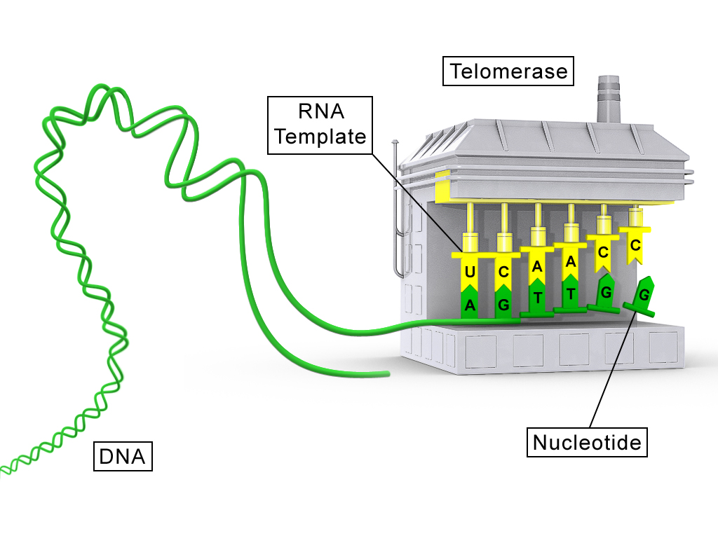
Telomerase is a reverse transcriptase (TERT) carrying its own RNA template (TERC). It base-pairs the RNA to the 3′ overhang, adds one repeat, translocates and repeats. Pol α–primase then fills in the complementary strand. Germ cells, stem cells and most cancers express telomerase; most somatic cells do not, so their telomeres shorten until they stop dividing (replicative senescence). Bacteria and most archaea avoid the problem with circular chromosomes.
DNA damage and repair
| Agent | Main lesion | Main fix |
|---|---|---|
| UVB (280–315 nm) | Absorbed directly by DNA: cyclobutane pyrimidine dimers and 6-4 photoproducts that kink the helix | Nucleotide excision repair |
| UVA (315–400 nm) | Mostly indirect: reactive oxygen species oxidize bases (8-oxoguanine); fewer dimers | Dimers by NER; oxidative lesions by other pathways |
| Ionizing radiation (X-rays, γ-rays) | Hydroxyl radicals and direct hits break the backbone; double-strand breaks are the dangerous lesion | NHEJ or HDR |
| Intercalating agents (ethidium bromide, acridines, doxorubicin) | Flat molecules slip between base pairs, stretch the helix and cause insertions or deletions during replication; some trap topoisomerase II, creating breaks | Frameshift mutations if unrepaired |
Nucleotide excision repair (NER) removes bulky, helix-distorting lesions by cutting out a short stretch of the damaged strand and resynthesizing it from the intact strand. In bacteria, UvrA and UvrB find the lesion, UvrC cuts on both sides (a 12–13 nucleotide piece; because it cuts on both sides, the UvrABC enzyme is called an excinuclease), UvrD helicase removes it, and Pol I and ligase fill in. Eukaryotes do the same job with more proteins: XPC (or a stalled RNA polymerase, in transcription-coupled repair) detects damage, the TFIIH helicases open the DNA, and XPG and XPF–ERCC1 cut out about 24–32 nucleotides. Defects cause xeroderma pigmentosum, extreme UV sensitivity.
Double-strand breaks have two fates. Non-homologous end joining (NHEJ) ligates the ends directly. In eukaryotes, Ku70/80 binds the ends, DNA-PKcs and Artemis trim them, and XRCC4–ligase IV seals them. It works in any cell-cycle phase but often adds or deletes a few bases. Many bacteria (such as mycobacteria) have a minimal NHEJ of Ku plus ligase D; E. coli lacks it. Homology-directed repair (HDR) copies an intact homologous template, usually the sister chromatid, so it is accurate but limited to S and G2. The ends are resected to 3′ single-stranded tails (RecBCD in bacteria; MRN and CtIP in eukaryotes). A recombinase filament (RecA; Rad51, loaded by BRCA2) invades the homolog, polymerase copies across the gap, and Holliday junctions are resolved (RuvABC in bacteria).
Spontaneous and chemical damage. Not all damage comes from radiation. Every cell loses thousands of purine bases a day by depurination (hydrolysis of the N-glycosidic bond, leaving an abasic or AP site), the most common spontaneous lesion. Deamination turns cytosine into uracil (and 5-methylcytosine into thymine, which is why methylated CpG sites are mutation hotspots, giving C→T transitions). Uracil in DNA is clearly foreign and is removed by uracil-DNA glycosylase (UNG), but thymine from 5mC deamination is a normal base, so the resulting G·T mismatch needs special glycosylases (TDG and MBD4) that remove the T opposite G; if they miss it, replication fixes a C·G → T·A transition. Reactive oxygen species make 8-oxoguanine, which pairs with A and causes G→T transversions. Replication itself makes errors, and tautomeric shifts (rare enol or imino forms of bases) cause mispairing. Strand slippage in repeats adds or removes repeat units, the mechanism of trinucleotide-repeat expansion.
| Mutagen class | Examples | Effect |
|---|---|---|
| Base analogs | 5-bromouracil (resembles T), 2-aminopurine (resembles A) | Incorporated, then mispair → transitions |
| Alkylating agents | EMS, nitrogen mustards, MMS | Add alkyl groups (O⁶-methylguanine pairs with T) → G·C→A·T transitions |
| Deaminating agents | Nitrous acid | C→U, A→hypoxanthine → transitions |
| Hydroxylamine | — | Modifies C to pair with A → C→T only |
| Intercalating agents | Ethidium bromide, acridine orange, proflavine | Insertions or deletions → frameshifts |
| UV light | UVB, UVC | Pyrimidine dimers → C→T transitions at dipyrimidine sites, the "UV signature" |
| Ionizing radiation | X-rays, γ-rays | Strand breaks, especially double-strand breaks → deletions, rearrangements |
The Ames test screens chemicals for mutagenicity using Salmonella strains with a his⁻ mutation that cannot make histidine. Bacteria are plated on medium lacking histidine with the test chemical (often plus rat-liver extract, which converts some chemicals into their active forms as the body would). Only revertants (back-mutations to his⁺) form colonies, so a mutagen gives many more colonies than the control. Because most carcinogens are mutagens, it is a cheap first screen for carcinogens.
Every repair pathway in one table.
| Pathway | Fixes | Key steps and proteins (bacteria; eukaryotes) |
|---|---|---|
| Direct reversal | Pyrimidine dimers; O⁶-methylguanine | Photolyase uses visible light to split dimers (bacteria, plants, many animals, but not placental mammals). MGMT (O⁶-methylguanine methyltransferase) moves the methyl group onto its own cysteine, a suicide reaction that uses up one protein per lesion |
| Base excision repair (BER) | Small, non-distorting base damage: uracil, 8-oxoG, alkylated bases, AP sites | A DNA glycosylase scans the DNA, flips out the damaged base and removes it, leaving an AP site; AP endonuclease (APE1) cuts the backbone 5′ of it; Pol β (eukaryotes) or Pol I (bacteria) fills one nucleotide and removes the sugar remnant; ligase seals. Single-strand breaks build up if BER fails |
| Nucleotide excision repair (NER) | Bulky, helix-distorting lesions: pyrimidine dimers, chemical adducts | Recognition of distortion (UvrA–UvrB; XPC or a stalled RNA polymerase), dual incision (UvrC; XPF and XPG), removal of an oligonucleotide, gap filling and ligation. Transcription-coupled repair (Mfd/TRCF in bacteria, CSB in humans) preferentially fixes the template strand of active genes; defects cause Cockayne syndrome |
| Mismatch repair (MMR) | Replication errors that escaped proofreading: mismatches and small loops | E. coli MutS finds the mismatch, MutL joins, MutH nicks the unmethylated new strand at a hemimethylated GATC site (Dam methylation marks the old strand); exonuclease removes the stretch; Pol III resynthesizes; ligase seals. Eukaryotes use MutS and MutL homologs (MSH2, MLH1) and recognize the new strand by its nicks. Defects cause Lynch syndrome (hereditary colon cancer) and microsatellite instability |
| NHEJ | Double-strand breaks, any time in the cycle | Ku binds ends, trimming, ligase IV. Fast but error-prone (small indels); occasionally joins the wrong ends, causing translocations |
| Alternative end joining (MMEJ / TMEJ) | Double-strand breaks when NHEJ fails or HDR is unavailable | After resection, short microhomologies (a few bases) on the two ends anneal, and DNA polymerase θ (Pol theta, a polymerase fused to a helicase-like domain) extends from them; leaves deletions flanked by microhomology and sometimes templated insertions. Highly error-prone. HR-deficient (BRCA-mutant) cancers depend on it, so Pol θ inhibitors are synthetic-lethal with BRCA loss |
| HDR / homologous recombination | Double-strand breaks and collapsed forks, in S and G2 | Resection to 3′ tails, RecA/Rad51 strand invasion, copy from the sister chromatid, Holliday junction resolution. Accurate. After strand invasion forms a D-loop, two routes are possible: in synthesis-dependent strand annealing (SDSA) the invading strand is extended, then released and annealed back to the other end of the break, so no Holliday junction forms and the product is always non-crossover (the main route in mitotic cells); in the double Holliday junction (dHJ) route the second end is captured, two junctions form, and their resolution gives crossover or non-crossover products (dissolution by BLM helicase–topoisomerase gives non-crossovers). In meiosis the dHJ route, cut by resolvases, makes the crossovers |
| Translesion synthesis / SOS | Lesions that block the replicative polymerase | Specialized low-fidelity polymerases (Pol IV and V, induced in the bacterial SOS response when RecA activates cleavage of the LexA repressor; Pol η, ζ in eukaryotes) copy past damage. It is damage tolerance, not repair, and it is mutagenic |
The 2015 Nobel Prize in Chemistry went to Tomas Lindahl (base excision repair), Aziz Sancar (nucleotide excision repair) and Paul Modrich (mismatch repair).
Repair diseases show why each pathway matters: xeroderma pigmentosum (NER; extreme UV sensitivity, skin cancer), Lynch syndrome (MMR), BRCA1/2 hereditary breast and ovarian cancer (HDR), ataxia-telangiectasia (ATM kinase, damage signaling), Bloom and Werner syndromes (RecQ helicases), Fanconi anemia (cross-link repair). Many chemotherapies exploit repair defects: PARP inhibitors kill BRCA-deficient tumor cells (synthetic lethality): blocking PARP-dependent single-strand-break repair turns nicks into double-strand breaks at forks, which cells without homologous recombination cannot fix; X-rays and radiotherapy kill rapidly dividing cancer cells, which are more vulnerable because they replicate damaged DNA and often have broken checkpoints.
Mutations
Substitutions swap one base: a transition swaps purine for purine or pyrimidine for pyrimidine, and a transversion swaps between classes. Insertions and deletions add or remove bases. Chromosomal rearrangements move large segments (deletions, duplications, inversions, translocations); they are covered under "Nondisjunction and karyotypes".
| Mutation | Effect on the protein |
|---|---|
| Silent | Codon changes but the amino acid does not (the code is redundant) |
| Missense | One amino acid changes; effect ranges from none to severe |
| Nonsense | A premature stop codon truncates the protein |
| Frameshift | Insertion or deletion not a multiple of three shifts every downstream codon |
| In-frame indel | Whole codons added or removed; frame preserved |
Classifying a mutation from sequences. Line up the two sequences and ask: did one base change (substitution), or did the length change (insertion or deletion)? For a substitution, translate both codons. Then name the class.
| Mutation term | Meaning |
|---|---|
| Germline vs somatic | In cells that make gametes (heritable) vs in body cells (not passed on; causes cancer and mosaicism) |
| Spontaneous vs induced | From normal processes (replication errors, depurination, deamination, tautomers) vs caused by a mutagen |
| Forward vs reverse (reversion) | Wild type → mutant vs mutant → wild type at the same site |
| Suppressor mutation | A second mutation elsewhere that restores the phenotype: intragenic (for example a +1 frameshift restored by a nearby −1) or intergenic (for example a tRNA whose anticodon now reads a stop codon) |
| Loss-of-function vs gain-of-function | Reduces or abolishes activity (usually recessive) vs increases activity or adds a new one (usually dominant) |
| Conditional (temperature-sensitive) | Phenotype only under restrictive conditions; useful for studying essential genes |
| Neutral | Changes the sequence without changing fitness |
| Regulatory mutation | In a promoter, enhancer or other cis element: the protein is normal but made in the wrong amount, place or time |
| Up-promoter / down-promoter mutation | A promoter change that makes the sequence more like the consensus (for example a TATA box closer to TATAAA, or a bacterial −10 box closer to TATAAT) binds the transcription machinery better and raises transcription; a change away from the consensus lowers it |
Where in the gene matters. A mutation in the coding sequence can change the protein; a mutation deep in an intron usually does nothing, because introns are removed, unless it creates a new splice site; a mutation at a splice site (the GU at the intron's 5′ end, the AG at its 3′ end, the branch-point A, or the last bases of an exon) can cause exon skipping, intron retention or use of a hidden cryptic splice site, often changing the reading frame. A mutation in the 5′ or 3′ UTR can change mRNA stability or translation; a promoter mutation changes how much mRNA is made; a mutation that destroys the start codon prevents translation. Deletions of a whole region (such as the 5p deletion in cri-du-chat) remove many genes at once and are usually more harmful than point mutations. Trinucleotide repeat expansions happen when the polymerase slips on a repetitive template and a hairpin forms in the new strand, adding repeats; the repeat grows across generations (anticipation): CAG in Huntington disease (polyglutamine in the protein), CGG in fragile X (in the 5′ UTR, causing silencing by methylation), CTG in myotonic dystrophy (3′ UTR) and GAA in Friedreich ataxia (intron).
Mutagenesis as a tool. Random mutagenesis (chemicals, UV, transposons, error-prone PCR) scatters mutations for forward genetic screens (phenotype first, then find the gene). Targeted mutagenesis changes a chosen site (site-directed mutagenesis with mutagenic primers, CRISPR editing) and needs the sequence first (reverse genetics: gene first, then the phenotype).
Eukaryotic gene expression and regulation
Gene expression is the conversion of the information in a gene into a functional product, usually a protein but sometimes a functional RNA. In eukaryotes, transcription and RNA processing happen in the nucleus and translation happens in the cytoplasm, so each step (chromatin opening, transcription, processing, export, mRNA decay, translation, protein modification and degradation) is a separate control point. Most regulation still happens at the start of transcription, because that is the cheapest place to stop making a product the cell does not need.
The central dogma and the parts of a gene
Francis Crick's central dogma (1958) states that sequence information flows from nucleic acid to protein and never back from protein. The general transfers are DNA → DNA (replication), DNA → RNA (transcription) and RNA → protein (translation). The special transfers are RNA → DNA (reverse transcription, by retroviruses such as HIV, retrotransposons and telomerase) and RNA → RNA (RNA replication by RNA viruses). A question asking for an "exception to the central dogma" almost always wants reverse transcription. Prions are not an exception: they pass on a protein shape, not a sequence.
Why use an RNA intermediate at all? Each mRNA can be translated many times (amplification), the DNA stays protected in the nucleus, and the cell can control expression at every RNA step. Every somatic cell carries essentially the same genome; a liver cell and a neuron differ because they express different sets of genes (differential gene expression). Housekeeping genes (glycolysis enzymes, ribosomal proteins) are expressed in nearly every cell; tissue-specific genes are switched on by particular combinations of transcription factors.
A eukaryotic protein-coding gene has these parts, from upstream to downstream:

| Part | Transcribed? | Translated? | Job |
|---|---|---|---|
| Enhancer / silencer | Usually no | No | Binds activators or repressors; can be far away, in either direction or inside an intron |
| Promoter (core + proximal) | No (sits upstream of +1) | No | Positions RNA polymerase and sets the start site (+1) |
| 5′ UTR | Yes | No | Ribosome loading and scanning; carries regulatory elements such as IREs and uORFs |
| Start codon to stop codon (exons) | Yes | Yes | Coding sequence (CDS) |
| Introns | Yes | No (spliced out) | Allow alternative splicing; can hold enhancers and small RNA genes |
| 3′ UTR | Yes | No | Stability and localization signals, miRNA sites, AAUAAA polyadenylation signal |
| Poly(A) tail | No (added after cleavage) | No | Not encoded in the gene, so it never appears in genomic DNA |
Because the promoter is not transcribed, a cDNA made from mRNA contains no TATA box and no introns, but it does carry the UTRs and the poly(A) stretch. About 1–2% of the human genome codes for protein (exons of roughly 20,000 protein-coding genes); some test keys round this to "about 3%". A gene's transcription unit runs from +1 to the end of transcription, so the primary transcript is the same length as the transcription unit, including introns.
Conservation as evidence of function. Sequences that matter are under purifying selection, so they change slowly between species. Exons, splice sites, promoters and many enhancers are far more conserved than the bulk of intron and intergenic DNA, and within exons the third codon position (often synonymous) changes fastest. Comparative genomics uses this to find enhancers and new genes: a non-coding stretch conserved from human to fish is probably a regulatory element. A ratio of nonsynonymous to synonymous change (dN/dS) below 1 signals purifying selection; above 1 signals positive selection.
RNA types and who makes them
Most of a cell's RNA is rRNA (about 80%), then tRNA (about 15%); mRNA is only a few percent. The table lists the RNAs that tests name, with the polymerase that makes each in eukaryotes.
| RNA | Size | Made by (eukaryotes) | Job |
|---|---|---|---|
| mRNA | Hundreds to thousands of nt | Pol II | Carries the coding sequence to the ribosome |
| tRNA | ~76 nt (73–93) | Pol III | Adapter: anticodon reads the codon, 3′ end carries the amino acid |
| 28S, 18S, 5.8S rRNA | 28S ~5 kb, 18S ~1.9 kb, 5.8S ~160 nt | Pol I, as one 45S precursor in the nucleolus | Ribosome structure; 28S is the catalytic peptidyl transferase |
| 5S rRNA | ~120 nt | Pol III (separate genes, outside the nucleolus) | Large-subunit structure |
| Bacterial 23S, 16S, 5S rRNA | — | The single RNA polymerase | 16S in the 30S subunit pairs with Shine–Dalgarno; 23S is catalytic |
| snRNA (U1, U2, U4, U5, U6) | 100–200 nt | Pol II, except U6 by Pol III | Spliceosome; base-pair with splice sites and branch point |
| snoRNA | 60–300 nt | Mostly from Pol II introns | Guide rRNA modification: C/D box → 2′-O-methylation; H/ACA box → pseudouridine |
| miRNA | ~22 nt mature | Mostly Pol II (a few Pol III) | Imperfect pairing to 3′ UTRs; represses translation and speeds decay of many targets |
| siRNA | ~21 nt | No dedicated polymerase: cut by Dicer from long dsRNA (viral, transposon, experimental) | Perfect pairing; Argonaute slices one target |
| piRNA | 24–31 nt | Pol II, from piRNA clusters | Germline; with PIWI proteins silences transposons |
| lncRNA | > 200 nt | Mostly Pol II | Chromatin regulation (XIST, HOTAIR), scaffolds, decoys |
| 7SL RNA | ~300 nt | Pol III | RNA core of the signal recognition particle (SRP) |
| Telomerase RNA (TERC) | ~450 nt | Pol II | Template for telomere repeats |
| RNase P RNA | ~340 nt | Pol III | Ribozyme that cuts the 5′ leader off pre-tRNA |
Ribozymes are catalytic RNAs: the large-subunit rRNA (peptide bond formation), RNase P, group I and group II self-splicing introns, and the U2–U6 snRNA core of the spliceosome. Their existence supports the RNA world hypothesis, in which RNA served as both genetic material and catalyst before DNA and proteins.
Transcription
Transcription is DNA-directed RNA synthesis. RNA polymerase unwinds about 12–14 bp to form a transcription bubble, reads the template (antisense, non-coding) strand 3′→5′ and adds ribonucleotides (rNTPs) to the 3′-OH of the growing RNA, so RNA grows 5′→3′. Each addition releases pyrophosphate. Unlike DNA polymerase, RNA polymerase needs no primer, uses rNTPs with U instead of T, and has no separate 3′→5′ proofreading exonuclease; it corrects a few errors by backtracking and cleaving the 3′ end (helped by TFIIS in eukaryotes, GreA/GreB in bacteria). Its error rate is about 1 in 10⁴–10⁵, far higher than replication, which is tolerable because RNAs are short-lived copies. Different genes on the same chromosome can use different strands as template.
RNA polymerases compared
Bacteria have one RNA polymerase. The core enzyme (α₂ββ′ω) can elongate but cannot find promoters; adding a σ (sigma) factor makes the holoenzyme, which can.
| Subunit (bacteria) | Job |
|---|---|
| α (two copies) | Assembles the enzyme; the α C-terminal domain binds UP elements and some activators (such as CAP) |
| β | Forms the active site with β′; binds NTPs; target of the antibiotic rifampicin |
| β′ | Binds the DNA template; holds the catalytic Mg²⁺ |
| ω | Helps fold and assemble β′ |
| σ (σ⁷⁰ for most genes) | Recognizes the −35 and −10 promoter boxes (helix-turn-helix in region 4 binds −35), helps melt the −10 region, then is released early in elongation |
Alternative sigma factors switch whole gene sets on together (σ³² heat shock, σ⁵⁴ nitrogen limitation, σˢ stationary phase). Eukaryotes divide the work among specialized enzymes, all of them multi-subunit enzymes related to the bacterial core.
| Polymerase | Location | Products | Promoter / factors | α-amanitin |
|---|---|---|---|---|
| Pol I | Nucleolus | 45S pre-rRNA (→ 28S, 18S, 5.8S) | Upstream control element + core; SL1 (contains TBP) and UBF | Insensitive |
| Pol II | Nucleoplasm | mRNA, most snRNAs, most miRNAs, lncRNAs, snoRNA host transcripts, TERC | TATA/Inr/DPE core; TFIID, A, B, E, F, H plus Mediator | Very sensitive |
| Pol III | Nucleoplasm | tRNA, 5S rRNA, U6 snRNA, 7SL, 7SK, RNase P RNA, Alu elements | Often internal promoters (A and B boxes inside tRNA genes); TFIIIA, B (contains TBP), C | Moderately sensitive |
| Bacterial RNAP | Cytoplasm (nucleoid) | All RNA | −35/−10 boxes via σ | — |
| Archaeal RNAP | Cytoplasm | All RNA | TATA box + BRE; TBP and TFB (TFIIB homolog), TFE | — |
Plants add Pol IV and Pol V, which work in RNA-directed DNA methylation (Pol IV makes the precursors of 24-nt siRNAs; Pol V makes scaffold transcripts that those siRNAs pair with). Mitochondria use a single-subunit, phage-like polymerase. Only Pol II has the long C-terminal domain (CTD), tandem repeats of the heptad YSPTSPS (52 in humans, 26 in yeast), whose phosphorylation pattern coordinates initiation, capping, splicing and polyadenylation.
The archaeal polymerase is the key comparison: archaea have one polymerase like bacteria, but it resembles Pol II in subunit structure, and its promoter (a TATA box bound by TBP plus a BRE bound by TFB) is a simplified version of the eukaryotic Pol II system.
Promoter elements and general transcription factors
A promoter is the DNA sequence where RNA polymerase and its factors assemble; it is upstream (5′ on the coding strand) of the transcription start site (+1). Positions upstream are negative and there is no position 0. The Pol II core promoter spans about −40 to +40 and is built from a mix of elements; no element is found in every promoter.
| Element | Position | Consensus (coding strand) | Bound by | Notes |
|---|---|---|---|---|
| TATA box (Goldberg–Hogness box) | About −25 to −30 | TATAAA (TATAWAWR) | TBP, the TATA-binding protein subunit of TFIID | Present in only a minority of human promoters (roughly 10–25% depending on definition); common in strongly regulated genes |
| BRE (TFIIB recognition element) | Just upstream or downstream of TATA | G/C-rich | TFIIB | Helps orient the complex |
| Inr (initiator) | Spans +1 | YYANWYY (A at +1) | TAFs of TFIID | Can work without TATA |
| DPE (downstream promoter element) | About +28 to +32 | RGWYV | TAFs of TFIID | Mainly in TATA-less promoters, with an Inr |
| CAAT box (proximal) | About −75 to −80 | GGCCAATCT | NF-Y (CBF) | Proximal element; raises efficiency |
| GC box (proximal) | Often −40 to −110, often several copies | GGGCGG | Sp1 | Typical of housekeeping (CpG-island) promoters |
TATA-less, GC-rich promoters often start transcription at several nearby sites rather than one. The TATA box is the eukaryotic counterpart of the bacterial −10 (Pribnow) box: both are AT-rich, which makes them easy to melt because A·T pairs have two hydrogen bonds instead of three.
Pol II cannot recognize a promoter by itself. General transcription factors (GTFs) assemble with it into the preinitiation complex (PIC) in a standard order (D, A, B, F + Pol II, E, H):
| GTF | Job |
|---|---|
| TFIID | First to bind. TBP binds the TATA minor groove and bends the DNA about 80°; about 13 TAFs (TBP-associated factors) bind Inr and DPE and contact activators. The largest GTF (most subunits) |
| TFIIA | Stabilizes TBP on DNA and blocks inhibitors |
| TFIIB | Bridges TBP and Pol II, binds the BRE, helps choose the start site |
| TFIIF | Escorts Pol II to the promoter; prevents nonspecific DNA binding |
| TFIIE | Recruits and stimulates TFIIH |
| TFIIH | ATP-dependent helicase (XPB) opens the DNA; kinase (CDK7) phosphorylates CTD serine 5, releasing Pol II from the promoter (promoter escape). Also works in nucleotide excision repair |
| Mediator | Large coactivator bridge that carries signals from activators bound at enhancers to Pol II and stimulates TFIIH's CTD kinase; repressors can act through it to lower initiation |
There is no Pol II "TFIIC"; TFIIIC is a Pol III factor. TBP is shared by all three eukaryotic polymerases (in SL1, TFIID and TFIIIB) and by archaea.
Initiation, elongation and termination
Initiation. The PIC forms on the core promoter (closed complex). TFIIH melts the DNA (open complex) and Pol II starts making RNA. The first few nucleotides are often released and restarted (abortive initiation) while the polymerase stays put and pulls downstream DNA into itself (scrunching); the stress stored in the scrunched DNA helps the polymerase break its promoter contacts. TFIIH phosphorylates CTD serine 5, and Pol II escapes. In bacteria the same steps occur with σ, which is released once the RNA is about 10 nt long.
Elongation. Pol II moves about 1–4 kb per minute, keeping an RNA–DNA hybrid of about 8–9 bp in the bubble. Many genes have Pol II paused 20–60 nt downstream of the start until the kinase P-TEFb phosphorylates CTD serine 2 and pausing factors; serine 2 phosphorylation then recruits splicing and 3′-end factors. Nucleosomes ahead of the polymerase are displaced and rebuilt behind it by histone chaperones (such as FACT).
Termination. Pol II passes the AAUAAA polyadenylation signal; the RNA is cleaved and the polyadenylation machinery takes over the released 3′ end. The exonuclease Xrn2 enters at the new 5′ end of the RNA still coming out of the polymerase, degrades it, catches up and knocks Pol II off the DNA (the torpedo model). Pol I uses a terminator protein; Pol III stops at a run of T's in the coding strand without needing a hairpin.
Bacteria use two mechanisms, which tests ask about often:
- Intrinsic (rho-independent): the RNA has a GC-rich inverted repeat that folds into a hairpin (stem-loop), followed by a run of about 7–8 U's. The hairpin stalls the polymerase and the weak rU·dA pairs let the RNA slip off. No protein and no ATP needed.
- Rho-dependent: the Rho protein, a hexameric ATP-dependent 5′→3′ RNA helicase, loads onto a C-rich, G-poor rut site on the RNA, chases the polymerase and unwinds the RNA–DNA hybrid when the polymerase pauses. Uses ATP.
To recognize an intrinsic terminator in a sequence question, look for an inverted repeat (a sequence followed a few bases later by its reverse complement) followed by A's in the template (U's in the RNA). That inverted repeat is what lets the RNA form a hairpin.
Bacteria, archaea and eukaryotes compared
The 2027 rules center on eukaryotes, but older questions compare all three domains. An operon is a bacterial cluster of genes under one promoter and operator, transcribed into one polycistronic mRNA. Operon regulation (lac inducible, trp repressible) is no longer in the rules.
| Feature | Bacteria | Archaea | Eukaryotes |
|---|---|---|---|
| RNA polymerases | One (α₂ββ′ω + σ) | One, Pol II-like (~11–13 subunits) | Three nuclear (I, II, III) |
| Promoter | −35 (TTGACA) and −10 Pribnow box (TATAAT), 17 ± 1 bp apart | TATA box (~−26) + BRE | TATA (~−25 to −30), Inr, DPE, CAAT, GC boxes, enhancers |
| Promoter recognition | σ factor | TBP + TFB (+ TFE) | TBP/TFIID + other GTFs + Mediator |
| Termination | Intrinsic hairpin + U run, or Rho-dependent | U-rich stretches; a CPSF-like factor | Pol II: cleavage at poly(A) site and torpedo; Pol III: T run |
| Location | Cytoplasm; coupled with translation | Cytoplasm; coupled | Nucleus; translation in cytoplasm |
| mRNA | Often polycistronic | Often polycistronic | Monocistronic |
| Processing | None for mRNA (no cap, no splicing); poly(A) marks RNA for degradation | Few introns, mainly in tRNA and rRNA, removed by a protein endonuclease | Cap, poly(A) tail, splicing, editing |
| mRNA lifetime | Minutes | Minutes | Minutes to days (median hours) |
| Ribosome | 70S = 30S + 50S | 70S | 80S = 40S + 60S (cytoplasm); 70S-like in mitochondria and chloroplasts |
| Initiator amino acid | N-formylmethionine (fMet) | Methionine | Methionine |
| Ribosome finds start by | Shine–Dalgarno (AGGAGG, ~8 nt before AUG) pairing with the 3′ end of 16S rRNA | Shine–Dalgarno or leaderless mRNAs | 5′ cap binding and scanning to the first AUG in a Kozak context |
| Initiation / elongation / release factors | IF1–3; EF-Tu, EF-Ts, EF-G; RF1 (UAA, UAG), RF2 (UAA, UGA), RF3 | aIF (eukaryote-like); aEF1α, aEF2; aRF1 | eIFs (eIF2, eIF4F …); eEF1A, eEF2; eRF1 (all three stops), eRF3 |
| Histones | No | Many have histone-like proteins | Yes |
Coupling is possible in bacteria because there is no nuclear envelope: ribosomes start translating the 5′ end of an mRNA while its 3′ end is still being made. It is impossible in eukaryotes because the pre-mRNA must be processed and exported first.
Reading strands: from DNA to mRNA
The coding strand (sense, non-template) has the same sequence as the mRNA, with T for U, and is the strand written in databases. The template strand (antisense) is complementary and antiparallel to the mRNA.
RNA maturation

The primary transcript of a protein-coding gene is pre-mRNA (part of heterogeneous nuclear RNA, hnRNA). All three processing steps happen co-transcriptionally, while the RNA is still being made, with the factors riding on the Pol II CTD. Bacterial mRNA gets none of them. rRNA and tRNA are processed too: the 45S pre-rRNA is cut into 18S, 5.8S and 28S and chemically modified under snoRNA guidance, and pre-tRNAs have their 5′ leader removed by RNase P, their 3′ trailer trimmed, CCA added to the 3′ end and many bases modified.
Capping and polyadenylation
5′ capping comes first, once the RNA is about 20–30 nt long. Three enzymes act: an RNA triphosphatase removes the terminal phosphate, guanylyltransferase adds GMP in an unusual 5′–5′ triphosphate linkage, and a methyltransferase adds a methyl (from S-adenosylmethionine) to N7 of the guanine, giving m⁷GpppN. The cap protects the 5′ end from exonucleases, is bound by the nuclear cap-binding complex for export and splicing of the first intron, and is recognized in the cytoplasm by eIF4E to start translation.
Polyadenylation sets the 3′ end. CPSF binds the AAUAAA signal (written 5′→3′ in the RNA) and CstF binds a GU- or U-rich element downstream; the RNA is cut between them, about 10–30 nt past AAUAAA, and poly(A) polymerase adds about 200–250 adenines in mammals (fewer, ~70–90, in yeast) without a template. Poly(A)-binding protein (PABP) coats the tail, protecting the mRNA and helping translation. The tail is why mature mRNA can be purified with oligo-dT and why cDNA ends in a poly(A)/poly(T) stretch that is absent from the genomic sequence. Some genes have several poly(A) sites (alternative polyadenylation), producing mRNAs with longer or shorter 3′ UTRs and different numbers of miRNA sites. In bacteria, a short poly(A) tail is a degradation signal, not a stabilizer.
Splicing and the spliceosome
Introns are transcribed sequences removed from the RNA; exons are the sequences joined together and kept (including the UTR parts). Nearly all nuclear pre-mRNA introns follow the GU–AG rule:
- 5′ splice site: exon…AG | GURAGU… (the intron starts with GU)
- Branch point: an adenine about 18–40 nt upstream of the 3′ end of the intron
- Polypyrimidine tract: a run of C's and U's between branch point and 3′ splice site
- 3′ splice site: …YAG | G…exon (the intron ends with AG)
The spliceosome is a large complex of five snRNPs (U1, U2, U4, U5 and U6, each an snRNA plus proteins) and over 100 other proteins. U1 snRNA base-pairs with the 5′ splice site; SF1 (BBP in yeast) and U2AF bind the branch point, polypyrimidine tract and AG; U2 snRNP then replaces SF1 and base-pairs around the branch point so the branch A bulges out; the U4/U6·U5 tri-snRNP joins; U1 and U4 leave, and U6 pairs with both the 5′ splice site and U2, forming the catalytic RNA core. Splicing is two transesterifications, with no net change in the number of phosphodiester bonds and no ATP used in the chemistry itself (ATP is used by helicases that rearrange the snRNPs):
- The 2′-OH of the branch-point A attacks the phosphate at the 5′ splice site. The 5′ exon is freed, and the intron's 5′ G is joined to the A by a 2′–5′ bond, forming a lariat.
- The freed 3′-OH of the 5′ exon attacks the 3′ splice site. The exons are joined and the lariat is released, then debranched and degraded.
The spliceosome leaves an exon junction complex (EJC) about 20–24 nt upstream of each exon–exon junction, which helps export and marks the mRNA for nonsense-mediated decay checks. Other intron types: group I self-splicing introns (Tetrahymena rRNA, using a free guanosine), group II self-splicing introns (organelles; they form a lariat and are thought to be ancestors of the spliceosome), a minor U12-type spliceosome for rare introns (<0.5% of human introns; first found as AU–AC introns, though most U12-type introns actually have GU–AG ends), and tRNA introns removed by protein enzymes. Most bacterial genes and human mitochondrial genes have no introns, which is why a human nuclear gene must be supplied as cDNA to be expressed correctly in bacteria.
Alternative splicing
Alternative splicing joins different combinations of exons from one pre-mRNA, so one gene can make several protein isoforms. About 95% of human multi-exon genes are alternatively spliced, which is how about 20,000 protein-coding genes produce far more distinct proteins.
| Type | What happens |
|---|---|
| Exon skipping (cassette exon) | An exon is included in some mRNAs and skipped in others; the most common type in animals |
| Mutually exclusive exons | Exactly one of two (or more) exons is kept |
| Alternative 5′ splice site | The donor site moves, changing the 3′ end of the upstream exon |
| Alternative 3′ splice site | The acceptor site moves, changing the 5′ start of the downstream exon |
| Intron retention | An intron stays in the mRNA; most common in plants and fungi |
| Alternative promoters / first exons and alternative poly(A) sites | Different 5′ or 3′ ends (not splicing strictly, but often grouped with it) |
The choice is made by RNA-binding proteins: SR proteins bind exonic splicing enhancers (ESEs) and promote inclusion; hnRNP proteins bind splicing silencers and promote skipping. Their levels differ between tissues, so splicing patterns are tissue-specific. Classic examples: Drosophila sex determination (Sex-lethal → transformer → doublesex), calcitonin vs CGRP from one gene in thyroid vs neurons, and Drosophila Dscam, with a theoretical 38,016 isoforms. If an included or skipped exon is not a multiple of 3 nt, everything downstream shifts frame.
RNA editing
RNA editing changes the sequence of an RNA after transcription, so the protein differs from what the gene predicts.
- A-to-I editing by ADAR enzymes (adenosine deaminases acting on RNA) in double-stranded RNA regions. Inosine pairs like G, so the ribosome and splicing machinery read I as G. It can change codons (the GluA2 glutamate receptor Q/R site, CAG → CIG read as Arg, which makes the channel impermeable to Ca²⁺), create or destroy splice sites, and alter miRNA targeting. It is most common in Alu-rich regions of human transcripts.
- C-to-U editing by APOBEC1 of apolipoprotein B mRNA. In the small intestine, codon 2153 CAA (Gln) becomes UAA (stop), producing apoB-48 (48% of full length, used in chylomicrons); the liver makes unedited apoB-100 from the same gene. Relatives in the APOBEC3 family deaminate C to U in single-stranded DNA, mutating HIV's newly made DNA (G→A hypermutation); HIV's Vif protein targets APOBEC3 for degradation.
- U insertion and deletion guided by guide RNAs in trypanosome mitochondria, where some mRNAs are rewritten extensively.
mRNA export, decay and nonsense-mediated decay
Only properly capped, spliced and polyadenylated mRNA is exported through nuclear pores (by the NXF1/TAP receptor); unspliced RNA is retained and degraded. In the cytoplasm, mRNA lifetime is a major control point. The usual decay route is deadenylation (CCR4–NOT shortens the tail) → decapping (Dcp2) → 5′→3′ degradation by Xrn1, or 3′→5′ degradation by the exosome. AU-rich elements (AREs) in the 3′ UTR of short-lived mRNAs (cytokines, proto-oncogenes such as c-fos) speed deadenylation. Protein and mRNA turnover is fast compared with DNA, which is not turned over at all except by repair.
Quality-control pathways destroy faulty mRNAs:
- Nonsense-mediated decay (NMD): if a ribosome stops at a premature stop codon more than about 50–55 nt upstream of the last exon–exon junction, an EJC remains downstream; UPF1 detects this and triggers decay. NMD protects cells from truncated, possibly dominant-negative proteins. A premature stop in the last exon, or within ~50 nt of the last junction, escapes NMD.
- Non-stop decay: an mRNA with no stop codon (ribosome reads into the poly(A) tail).
- No-go decay: ribosomes stalled on a damaged or strongly structured mRNA.
Regulatory elements
Regulation is mostly about deciding when, where and how much a gene is transcribed. The vocabulary that tests check first is cis versus trans.
| Feature | Cis-acting element | Trans-acting factor |
|---|---|---|
| What it is | A DNA (or RNA) sequence | A diffusible molecule, usually a protein (or an RNA such as a miRNA) |
| Acts on | Only the molecule it sits on | Any copy of its target, on any chromosome |
| Examples | Promoter, enhancer, silencer, insulator, response element, splice site, 3′ UTR element, riboswitch | Transcription factors, activators, repressors, hormone receptors, miRNAs, IRP |
| Mutation test in a diploid | Affects only the allele on the same chromosome (allele-specific) | Affects both alleles; a wild-type copy elsewhere can rescue it |
Cis-acting elements
The core promoter sets the start site (see the promoter table above). Proximal promoter elements (CAAT and GC boxes) lie within about 250 bp upstream. Enhancers are clusters of binding sites for activators. They can lie thousands to a million base pairs away, upstream, downstream or inside an intron, and work in either orientation, because the DNA loops (helped by Mediator and cohesin) to bring bound activators into contact with the promoter. Enhancers give tissue specificity: a gene can have separate enhancers for different tissues. Silencers bind repressors and lower transcription. Insulators (boundary elements) block an enhancer from acting on a promoter when placed between them, and act as barriers against the spread of heterochromatin.
In vertebrates the main insulator protein is CTCF. With cohesin it anchors chromatin loops, and convergent CTCF sites mark the borders of topologically associating domains (TADs): regions of roughly 0.1–1 Mb within which DNA contacts are frequent and across which they are rare, so enhancers mostly act on genes in their own TAD. Deleting a TAD boundary can let an enhancer switch on the wrong gene (causing, for example, limb malformations). TADs and loops are measured with Hi-C (see "Genomics assays (State and National)").
Trans-acting factors and response elements
Transcription factors (TFs) are modular proteins. A DNA-binding domain reads a specific sequence, usually in the major groove, and a separate activation domain (acidic, glutamine-rich or proline-rich) or repression domain recruits coactivators, Mediator, histone acetyltransferases or corepressors. Many TFs work as dimers, which lengthens the recognized site and raises binding affinity and specificity.
| DNA-binding motif | Structure | Examples |
|---|---|---|
| Helix-turn-helix / homeodomain | Recognition helix lies in the major groove; homeodomain is 60 amino acids | Hox proteins, bacterial repressors, σ factor region 4 |
| Zinc finger | Zn²⁺ held by Cys and His (C₂H₂) or four Cys (C₄) | Sp1, TFIIIA, steroid hormone receptors, CTCF |
| Leucine zipper (bZIP) | Coiled-coil dimer of leucine-rich helices; basic region binds DNA | CREB, Fos/Jun (AP-1) |
| Helix-loop-helix (bHLH) | Two helices joined by a loop; dimerizes | MyoD, Myc/Max |
Activators increase PIC assembly by recruiting Mediator, TFIID and HATs; repressors compete with activators for sites, mask their activation domains, or recruit histone deacetylases and methyltransferases. Control is combinatorial: a handful of TFs in different combinations specify hundreds of cell types. Master regulators can switch cell fate on their own: MyoD turns fibroblasts into muscle cells, and the four Yamanaka factors (OCT4, SOX2, KLF4, c-MYC) reprogram adult cells into induced pluripotent stem cells. Pioneer factors (such as FOXA) can bind DNA inside closed nucleosomal chromatin and open it for others.
Response elements are how eukaryotes coordinate many genes without operons: a common short sequence near many genes lets one signal-activated TF switch all of them on together.
| Signal | Trans-acting factor | Cis response element |
|---|---|---|
| cAMP (e.g., after glucagon or adrenaline) | CREB, phosphorylated by protein kinase A, then recruits the HAT CBP/p300 | CRE, TGACGTCA |
| Glucocorticoid (cortisol) | Glucocorticoid receptor: held in the cytoplasm by Hsp90, binds hormone, dimerizes, moves into the nucleus | GRE (palindromic; nuclear receptors bind as dimers) |
| Estrogen | Estrogen receptor | ERE |
| Heat and misfolded proteins | HSF1 | Heat shock element (HSE) |
| Growth factors / serum | Serum response factor | Serum response element (SRE) |
Steroid hormones are lipid-soluble, so they cross the membrane and bind intracellular (nuclear) receptors, which are themselves zinc-finger TFs. The thyroid hormone receptor binds its element even without hormone and represses; hormone binding converts it into an activator. Peptide hormones act through surface receptors and second messengers such as cAMP.
Riboswitches
A riboswitch is a cis-acting structure in an mRNA, usually in the 5′ UTR, that binds a small metabolite directly, with no protein involved. It has an aptamer (binding pocket) and an expression platform that changes shape when the ligand binds. In bacteria the switch usually forms either a transcription terminator or an antiterminator hairpin, or hides or exposes the Shine–Dalgarno sequence. In the thiamine pyrophosphate (TPP) riboswitch, for example, low TPP allows an antiterminator and transcription continues; high TPP stabilizes the terminator, ending transcription of thiamine-synthesis genes. That is negative feedback by the end product. The only known eukaryotic riboswitches are TPP riboswitches in fungi, algae and plants, which act through splicing or 3′-end processing (in Neurospora, TPP binding changes which splice site is used). In a reporter assay where the riboswitch turns a gene off, more luciferase signal means less riboswitch action.
Small regulatory RNAs: miRNA, siRNA and piRNA

RNA interference is sequence-specific silencing guided by a short RNA loaded into an Argonaute protein, forming the RNA-induced silencing complex (RISC). The three classes differ in origin and in how they pair with targets.
| Feature | miRNA | siRNA | piRNA |
|---|---|---|---|
| Origin | Cell's own genes (often in introns), transcribed mostly by Pol II as a pri-miRNA hairpin | Long double-stranded RNA: viruses, transposons, or introduced by an experimenter | Pol II transcripts of piRNA clusters full of transposon fragments |
| Processing | Drosha + DGCR8 (the Microprocessor) cut pri-miRNA to a ~60–70 nt pre-miRNA hairpin → Exportin-5 (Ran-GTP) → Dicer → ~22 nt duplex | Dicer cuts dsRNA into ~21 nt duplexes | Dicer-independent; amplified by the "ping-pong" cycle |
| Protein partner | Argonaute (AGO1–4 in humans) | Argonaute 2 (has slicer activity) | PIWI proteins |
| Pairing | Imperfect; the seed (nucleotides 2–8) pairs with 3′ UTR sites | Perfect over its length | Near-perfect to transposon RNA |
| Result | Translational repression plus deadenylation and decay; one miRNA regulates hundreds of mRNAs | Cleavage ("slicing") of one target mRNA | Transposon RNA cleaved; DNA methylation and H3K9me3 directed to transposon copies |
| Where | Most cells | Antiviral defense (plants, invertebrates); lab knockdowns | Germline |
Only one strand of the duplex (the guide) stays in RISC; the other (passenger, or miRNA*) is discarded. miRNAs repress translation and speed mRNA decay, so they lower protein levels and often mRNA levels too. Losing a miRNA that restrains an oncogene, or overexpressing one that restrains a tumor suppressor, can both promote cancer. Antisense RNA is a related idea: a single-stranded RNA complementary to an mRNA blocks its translation. How RNAi is used to make knockdowns in the lab belongs to "Knockouts, knockdowns and complementation".
Epigenetic regulation
Epigenetic regulation changes gene expression without changing the DNA sequence, through marks that are copied through cell division. The main marks are DNA methylation and histone modifications, read together with nucleosome positioning and non-coding RNAs.
DNA methylation. In mammals a methyl group is added to carbon 5 of cytosine in CpG dinucleotides (5-methylcytosine, 5mC). About 70–80% of CpGs in the genome are methylated, mostly in repeats and transposons. CpG islands are stretches of about 0.5–2 kb that are rich in CpG; they overlap the promoters of about 60–70% of human genes, including nearly all housekeeping genes, and are normally unmethylated and active. Methylation of a promoter CpG island silences the gene, either by blocking TF binding or by recruiting methyl-CpG-binding proteins (MeCP2, MBD proteins) that bring in histone deacetylases. Hypermethylation of tumor-suppressor promoters is common in cancer. CpG is under-represented in the genome overall because 5mC deaminates spontaneously to thymine, a mutation hot spot (CpG → TpG).
| Enzyme | Role |
|---|---|
| DNMT3A, DNMT3B | De novo methylation: set new patterns, mainly in early embryo and germ cells |
| DNMT1 (with UHRF1) | Maintenance: after replication, recognizes hemimethylated CpG and methylates the new strand, so the pattern is inherited through mitosis |
| TET enzymes | Oxidize 5mC to 5-hydroxymethylcytosine and beyond, starting active demethylation |
Bacteria methylate DNA too, but for different reasons: Dam methylase marks GATC (used to tell the old strand in mismatch repair), and restriction–modification systems protect their own DNA from their own restriction enzymes. Measuring methylation by bisulfite sequencing or nanopore reads belongs to Techniques.
Histone modifications. Histone tails carry many reversible chemical marks; the combination, read by specific "reader" proteins, is called the histone code.
| Mark | Writer → reader | Meaning |
|---|---|---|
| Lysine acetylation (e.g., H3K9ac, H3K27ac) | HATs (CBP/p300, GCN5); removed by HDACs; read by bromodomains | Neutralizes the positive lysine, loosening DNA–histone contacts; active chromatin. H3K27ac marks active enhancers |
| H3K4me3 | Trithorax/COMPASS (SET-domain methyltransferases) | Active promoters |
| H3K36me3 | SETD2 | Bodies of transcribed genes |
| H3K9me3 | SUV39H1, SETDB1; read by HP1 (chromodomain) | Constitutive heterochromatin (centromeres, transposons) |
| H3K27me3 | Polycomb PRC2 (EZH2) | Facultative heterochromatin: developmentally silenced genes such as Hox clusters outside their domains; the inactive X |
| H4K16ac | MOF | Decompacts chromatin; the Drosophila male X |
Acetylation is not only a histone mark. Thousands of non-histone proteins are acetylated on lysines, in eukaryotes and in bacteria, and the effect depends on the protein: acetylating p53 stabilizes and activates it, while acetylating a metabolic enzyme's active-site lysine can switch it off (bacterial acetyl-CoA synthetase is inactivated this way and reactivated by the deacetylase CobB, a sirtuin). Because acetylation removes a positive charge, it can change DNA binding, protein–protein contacts, stability (it competes with ubiquitination for the same lysines) or catalysis, so it can raise or lower activity.
Methylation does not change charge, so its effect depends entirely on which lysine is marked and which reader binds. ATP-dependent chromatin remodelers (SWI/SNF, ISWI, CHD, INO80 families) slide, evict or swap nucleosomes to expose or hide promoters; histone variants (H2A.Z at promoters, H3.3 in active genes, CENP-A at centromeres) add another layer. Euchromatin and heterochromatin themselves are introduced in "DNA structure and organization".
Classic epigenetic phenomena.
- X inactivation: the lncRNA XIST coats one X in cis in female mammals, which becomes H3K27me3-rich and methylated, forming the Barr body. Number of Barr bodies = number of X chromosomes − 1. See "Sex determination and sex linkage".
- Genomic imprinting: parent-of-origin expression set by methylation of imprinting control regions in the germline (the H19/IGF2 example above). See "Inheritance outside the nucleus and parent-of-origin effects".
- Position-effect variegation: in Drosophila, a white⁺ gene moved next to heterochromatin is silenced in some cells and not others, giving mottled eyes, which shows that silencing can spread along a chromosome.
- Agouti viable yellow mice: a retrotransposon (IAP) inserted upstream of the agouti gene drives ectopic expression unless it is methylated. A mother's diet rich in methyl donors increases methylation and shifts pups from yellow and obese toward brown and lean. Genetically identical, epigenetically different.
Dosage compensation outside mammals uses the same kinds of chromatin marks: MSL/MOF writing H4K16ac on the Drosophila male X, and a condensin-like complex damping both C. elegans X's. See "Sex determination and sex linkage".
Yeast two-hybrid screening
The yeast two-hybrid (Y2H) screen finds proteins that physically bind each other, using the modularity of transcription factors. The yeast activator Gal4 has a DNA-binding domain (BD) and an activation domain (AD) that work only when they are close together; they do not need to be covalently joined.
- The protein of interest (bait) is fused to the Gal4 BD. A library of candidate partners (prey, often from a cDNA library) is fused to the AD.
- Both plasmids go into a yeast strain whose reporter genes sit downstream of Gal4 binding sites (UAS). Each plasmid carries a nutritional marker (commonly TRP1 on the bait and LEU2 on the prey), so growth without tryptophan and leucine shows that a cell has both plasmids.
- If bait and prey bind each other in the nucleus, the BD (on the DNA) is tethered to the AD, which recruits the transcription machinery and switches on the reporters: HIS3 (growth without histidine), ADE2 (growth without adenine) or lacZ (blue colonies with X-gal). The inhibitor 3-AT is often added to suppress leaky HIS3 expression.
- Prey plasmids from growing colonies are isolated and sequenced to identify the partner.
Limitations. The interaction must happen in the yeast nucleus, so membrane proteins and secreted proteins often fail (variants such as the split-ubiquitin system handle membrane proteins). Fusions can block a binding surface (false negatives). Yeast may lack the modifications (some phosphorylations) or third partners an interaction needs. False positives come from auto-activating baits, "sticky" prey and overexpression. Hits must be confirmed by another method such as co-immunoprecipitation. Related screens: the one-hybrid screen finds proteins that bind a DNA sequence, and the three-hybrid screen finds RNA–protein interactions.
Translation
Translation is mRNA-directed protein synthesis on ribosomes. The mRNA is read 5′→3′, one codon at a time, and the polypeptide grows from its N-terminus to its C-terminus. Three RNAs are directly involved: mRNA (the message), tRNA (the adapters) and rRNA (structure and catalysis). DNA is not directly involved.
The genetic code
The code was cracked in the 1960s. Crick and Brenner's frameshift experiments with proflavin-induced insertions and deletions in phage T4 (1961) showed the code is read in triplets from a fixed start: one or two added bases ruined the gene, but three insertions restored function. Nirenberg and Matthaei showed that poly(U) RNA made polyphenylalanine (UUU = Phe). Khorana's repeating copolymers and Nirenberg and Leder's triplet-binding assay assigned the rest.
With four bases, a doublet code gives only combinations, too few for 20 amino acids; a triplet code gives . (A hypothetical four-base codon would give .) Of the 64 codons, 61 specify amino acids and 3 are stops (UAA "ochre", UAG "amber", UGA "opal"). AUG codes for methionine and is the start codon in all domains; bacteria occasionally start at GUG or UUG, still inserting fMet.
| Property | Meaning |
|---|---|
| Triplet | Three nucleotides per codon |
| Non-overlapping | Each base belongs to one codon in a given frame |
| Comma-free | No spacers or punctuation between codons; the start codon sets the frame |
| Degenerate (redundant) | Most amino acids have more than one codon (synonymous codons); only Met (AUG) and Trp (UGG) have one |
| Unambiguous | Each codon specifies one amino acid only |
| Nearly universal | The same in almost all organisms, which is why a human gene can be expressed in bacteria |
| Colinear | Codon order matches amino acid order |
Degeneracy is mostly in the third position: for example all four GCN codons are Ala. Leu, Ser and Arg have six codons each; Ile has three. Organisms use synonymous codons at different frequencies (codon usage bias), so genes moved between species are often "codon-optimized".
Exceptions to universality: in vertebrate mitochondria UGA = Trp, AUA = Met, and AGA and AGG are stops; in yeast mitochondria CUN = Thr; Mycoplasma reads UGA as Trp; some ciliates read UAA and UAG as Gln. Selenocysteine, the 21st amino acid, is inserted at certain UGA codons when a SECIS stem-loop is present (in the 3′ UTR in eukaryotes); it occurs in all three domains, including about 25 human selenoproteins. Pyrrolysine, the 22nd, is inserted at UAG in some methanogenic archaea and a few bacteria.

The full table, arranged the usual way: find the row by the first (5′) base and third (3′) base, then the column by the middle base. Codons are written 5′→3′ in mRNA.
ReferenceStandard genetic code (mRNA codons)· 16 rows
Rows: first base and third base. Columns: second base. Three-letter and one-letter amino acid codes in each cell.
No rows match.
| 1st base | 3rd base | 2nd = U | 2nd = C | 2nd = A | 2nd = G |
|---|---|---|---|---|---|
| U | U | UUU Phe (F) | UCU Ser (S) | UAU Tyr (Y) | UGU Cys (C) |
| U | C | UUC Phe (F) | UCC Ser (S) | UAC Tyr (Y) | UGC Cys (C) |
| U | A | UUA Leu (L) | UCA Ser (S) | UAA Stop | UGA Stop |
| U | G | UUG Leu (L) | UCG Ser (S) | UAG Stop | UGG Trp (W) |
| C | U | CUU Leu (L) | CCU Pro (P) | CAU His (H) | CGU Arg (R) |
| C | C | CUC Leu (L) | CCC Pro (P) | CAC His (H) | CGC Arg (R) |
| C | A | CUA Leu (L) | CCA Pro (P) | CAA Gln (Q) | CGA Arg (R) |
| C | G | CUG Leu (L) | CCG Pro (P) | CAG Gln (Q) | CGG Arg (R) |
| A | U | AUU Ile (I) | ACU Thr (T) | AAU Asn (N) | AGU Ser (S) |
| A | C | AUC Ile (I) | ACC Thr (T) | AAC Asn (N) | AGC Ser (S) |
| A | A | AUA Ile (I) | ACA Thr (T) | AAA Lys (K) | AGA Arg (R) |
| A | G | AUG Met (M), start | ACG Thr (T) | AAG Lys (K) | AGG Arg (R) |
| G | U | GUU Val (V) | GCU Ala (A) | GAU Asp (D) | GGU Gly (G) |
| G | C | GUC Val (V) | GCC Ala (A) | GAC Asp (D) | GGC Gly (G) |
| G | A | GUA Val (V) | GCA Ala (A) | GAA Glu (E) | GGA Gly (G) |
| G | G | GUG Val (V) | GCG Ala (A) | GAG Glu (E) | GGG Gly (G) |
Every amino acid has the same backbone, an amino group, an α-carbon with H and a carboxyl group (so one nitrogen in the backbone of each residue), and differs only in its side chain (R group). The side chain's chemistry predicts where a residue sits in a folded protein and how damaging a missense change is likely to be.
| Class | Amino acids (codes) | Notes |
|---|---|---|
| Nonpolar (hydrophobic) | Gly (G), Ala (A), Val (V), Leu (L), Ile (I), Met (M), Pro (P), Phe (F), Trp (W) | Buried in the protein core or in membranes. Gly is the smallest and most flexible; Pro is rigid and breaks helices |
| Polar, uncharged | Ser (S), Thr (T), Cys (C), Asn (N), Gln (Q), Tyr (Y) | Hydrogen bonding. Cys forms disulfide bonds; Ser, Thr and Tyr are phosphorylated; Asn carries N-linked sugars |
| Acidic (negative at pH 7) | Asp (D), Glu (E) | — |
| Basic (positive at pH 7) | Lys (K), Arg (R), His (H) | His is only partly charged at pH 7. Lys and Arg rich sequences bind DNA and form nuclear localization signals |
Classifications differ slightly between textbooks (Tyr, Trp and Gly are sometimes moved between classes), so accept the test's own table if it gives one. A missense change within a class (Leu → Ile) is "conservative"; a change across classes (Glu → Val in sickle-cell hemoglobin) is more likely to disrupt function. Mutation notation such as Pro250Arg means proline at residue 250 replaced by arginine.
tRNA and aminoacyl-tRNA synthetases

tRNA is the adapter Crick predicted. Each is about 76 nt and folds by intramolecular base pairing into a cloverleaf (secondary structure) with four stems, which folds further into an L shape (tertiary structure) about 7 nm long, with the anticodon at one end and the amino acid at the other.
- Acceptor stem with the 3′ CCA end; the amino acid is attached by an ester bond to the 3′ (2′ or 3′ OH) of the terminal A. CCA is added after transcription in eukaryotes.
- D arm, named for dihydrouridine.
- Anticodon arm: its loop carries the three-base anticodon that pairs antiparallel with the codon.
- Variable loop, which differs in length between tRNAs.
- TψC arm (T = ribothymidine, ψ = pseudouridine), which contacts the ribosome.
tRNAs carry many modified bases (inosine, pseudouridine, dihydrouridine, methylated bases), made after transcription. Not all tRNAs are identical outside the anticodon: they share the overall shape but differ in sequence, which lets synthetases tell them apart.
Aminoacyl-tRNA synthetases attach the correct amino acid to each tRNA ("charging"). There is typically one synthetase per amino acid (20 in total), each recognizing all the tRNAs for its amino acid (isoacceptors) by identity elements in the acceptor stem and anticodon. The reaction has two steps:
Hydrolysis of the released pyrophosphate drives the reaction, so charging costs two high-energy phosphate bonds per amino acid (ATP → AMP). Synthetases proofread: isoleucyl-tRNA synthetase, for example, hydrolyzes valine mistakenly attached to tRNA^Ile at a separate editing site. A charged tRNA is called an aminoacyl-tRNA. The ribosome checks only the codon–anticodon match, not the amino acid: in a classic experiment, Cys already attached to tRNA^Cys was chemically converted to Ala, and Ala was then inserted at Cys codons. So a synthetase error (say, Lys attached to the tRNA that reads UUU) puts the wrong amino acid at every position specified by that codon.
Wobble. Crick's wobble hypothesis: the first two codon positions pair strictly, but the third codon position (paired with the anticodon's 5′ base, position 34) tolerates some non-Watson–Crick pairs. This is why one tRNA can read several synonymous codons, and why cells need far fewer than 61 tRNA types (about 32 minimum; bacteria have about 40–45 anticodon species).
| Anticodon 5′ base (position 34) | Pairs with codon 3′ base |
|---|---|
| C | G only |
| A | U only (rare; usually modified to I) |
| G | C or U |
| U | A or G |
| I (inosine) | U, C or A |
Ribosomes
Ribosomes are made of rRNA (about two-thirds of the mass) and proteins. Their subunits are assembled in the nucleolus from rRNA and imported ribosomal proteins, then exported. Svedberg (S) units measure sedimentation rate, which depends on shape as well as mass, so they do not add: 40S + 60S = 80S, and 30S + 50S = 70S.
| Ribosome type | Small subunit | Large subunit | Whole ribosome |
|---|---|---|---|
| Bacteria (archaea: same rRNAs and sizes, more proteins) | 30S: 16S rRNA + ~21 proteins | 50S: 23S + 5S rRNA + ~33 proteins | 70S |
| Eukaryotic cytoplasm | 40S: 18S rRNA + ~33 proteins | 60S: 28S + 5.8S + 5S rRNA + ~47 proteins | 80S |
| Mitochondria and chloroplasts | — | — | 70S-like (bacterial ancestry) |
The small subunit binds mRNA and checks codon–anticodon pairing (the decoding center). The large subunit holds the peptidyl transferase center, where the rRNA, not protein, catalyzes peptide bonds, so the ribosome is a ribozyme. Three tRNA binding sites span both subunits and are formed mostly by rRNA:
- A site (aminoacyl): accepts each incoming charged tRNA.
- P site (peptidyl): holds the tRNA carrying the growing chain. The initiator tRNA is the only one that enters the P site directly.
- E site (exit): holds the uncharged tRNA on its way out.
A polysome (polyribosome) is one mRNA being translated by many ribosomes at once, spaced as close as about one per 80 nt; it multiplies the protein output of each mRNA. Bacterial ribosome-targeting antibiotics (streptomycin on 30S; chloramphenicol, erythromycin on 50S; tetracycline blocking the A site) exploit the differences between 70S and 80S ribosomes.
Initiation, elongation and termination of translation

Initiation (eukaryotes). The small subunit always binds before the large one.
- eIF2·GTP carries the initiator Met-tRNAi (a ternary complex). It joins the 40S subunit with eIF1, eIF1A, eIF3 and eIF5 to form the 43S preinitiation complex.
- eIF4F binds the 5′ cap: eIF4E (cap-binding), eIF4G (scaffold) and eIF4A (an ATP-dependent RNA helicase that unwinds 5′ UTR structure). eIF4G also binds PABP on the poly(A) tail, circularizing the mRNA (the "closed loop"), which boosts initiation and ensures only intact mRNAs are translated well.
- The 43S complex loads at the cap and scans 5′→3′ (using ATP) to the first AUG in a good Kozak context, gccRccAUGG, where the purine (R, usually A) at −3 and G at +4 matter most. A weak context lets some ribosomes skip the first AUG (leaky scanning), which can produce two proteins from one mRNA.
- Base pairing of Met-tRNAi with AUG triggers eIF2's GTP hydrolysis; factors leave; eIF5B·GTP joins the 60S subunit, forming the 80S initiation complex with Met-tRNAi in the P site and the A site empty. eIF2·GDP is recycled to eIF2·GTP by the exchange factor eIF2B.
In bacteria, the 30S subunit with IF1, IF3 and IF2·GTP binds the Shine–Dalgarno sequence, which pairs with the 3′ end of 16S rRNA and positions the AUG; the initiator is fMet-tRNA^fMet; the 50S joins when IF2 hydrolyzes GTP. Because ribosomes bind internal Shine–Dalgarno sites, each gene in a polycistronic mRNA has its own start. The formyl group and often the Met are later removed.
Elongation repeats three steps for each codon:
- Decoding: EF-Tu·GTP (eukaryotes eEF1A) brings an aminoacyl-tRNA to the A site. A correct codon–anticodon match triggers GTP hydrolysis and release of the factor (recycled by EF-Ts / eEF1B). One GTP.
- Peptide bond formation: the amino group of the A-site amino acid attacks the ester bond holding the chain on the P-site tRNA. The whole chain moves onto the A-site tRNA. Catalyzed by rRNA; no GTP.
- Translocation: EF-G·GTP (eukaryotes eEF2) moves the ribosome 3 nt toward the 3′ end. The peptidyl-tRNA moves from A to P, the empty tRNA from P to E (and then leaves), and the A site is free for the next codon. One GTP.
Bacterial ribosomes add about 15–20 amino acids per second, eukaryotic ones about 5–6, with about one error per 10⁴ codons.
Termination. When a stop codon enters the A site, no tRNA matches it. A release factor binds instead (it mimics the shape of a tRNA): eRF1 reads all three stops in eukaryotes, assisted by eRF3·GTP; bacteria use RF1 (UAA, UAG) or RF2 (UAA, UGA), plus RF3. The release factor positions a water molecule in the peptidyl transferase center, which hydrolyzes the bond between the polypeptide and the P-site tRNA, releasing the protein. The subunits are then separated and recycled (ABCE1 in eukaryotes; RRF + EF-G in bacteria). If translation is frozen just as the stop codon reaches the A site, you would isolate a complete ribosome with the finished polypeptide still attached to the tRNA in the P site.
Translation calculations
Use 4 high-energy bonds per amino acid (2 to charge the tRNA, 2 per elongation cycle) and say so on your paper; the exact bookkeeping of initiation and recycling differs between textbooks. The breakdown behind the formula: each amino acid costs 2 phosphate bonds to charge (ATP → AMP + PPᵢ), and each of the elongation cycles costs 2 GTP (EF-Tu/eEF1A and EF-G/eEF2). The first amino acid is delivered by eIF2 (1 GTP) rather than eEF1A, and termination uses 1 GTP (eRF3), giving exactly . Some textbooks add a second initiation GTP (eIF5B), ATP for scanning, or GTP/ATP for ribosome recycling; graders mainly check the 2 + 2 per residue logic. GTP is the direct energy source for the ribosome's movements; ATP is spent on charging.
Why a protein sequence cannot give back the gene: the code is degenerate (each amino acid may have up to 6 codons), the protein says nothing about introns, UTRs or promoters, and processing may have removed parts (signal peptide, initiator Met). Back-translation gives only a set of possible coding sequences.
Protein targeting, folding and modification
All cytoplasmic translation starts on free ribosomes. Where the protein goes is decided by sorting signals in its own sequence.
- Secretory pathway (bound ribosomes on rough ER): an N-terminal signal peptide (about 15–30 residues with a hydrophobic core) emerges from the ribosome and is bound by the signal recognition particle (SRP, 7SL RNA + proteins). SRP pauses elongation and docks the ribosome on the SRP receptor in the ER membrane; the chain is threaded through the Sec61 translocon as it is made (co-translational translocation), and signal peptidase cuts off the signal. Hydrophobic stop-transfer sequences anchor membrane proteins. Products: secreted proteins (insulin, antibodies, collagen, digestive enzymes), plasma-membrane proteins (receptors), and proteins of the ER, Golgi and lysosomes. ER proteins carry a C-terminal KDEL retention signal; lysosomal enzymes are tagged with mannose-6-phosphate in the Golgi.
- Free ribosomes (cytosol): cytosolic proteins (glycolytic enzymes, hemoglobin, actin) and proteins imported after translation into the nucleus (with a nuclear localization signal rich in Lys and Arg, such as PKKKRKV), mitochondria and chloroplasts (N-terminal amphipathic transit peptides) and peroxisomes (C-terminal SKL).
Protein structure levels.
| Level | What it is | Held by |
|---|---|---|
| Primary | Amino acid sequence, N to C | Covalent peptide bonds |
| Secondary | α-helices and β-pleated sheets of the backbone | Hydrogen bonds between backbone C=O and N–H |
| Tertiary | 3D fold of one chain | Side-chain interactions: hydrophobic effect, hydrogen bonds, ionic bonds (salt bridges), van der Waals forces, covalent disulfide bonds (Cys–Cys) |
| Quaternary | Assembly of two or more chains (hemoglobin α₂β₂) | The same forces as tertiary |
The primary sequence determines the fold (Anfinsen's experiment with ribonuclease), but many proteins need chaperones to fold efficiently and avoid aggregation: Hsp70 binds exposed hydrophobic stretches of nascent chains, chaperonins (Hsp60/GroEL–GroES) provide an enclosed folding chamber, and Hsp90 matures signaling proteins such as steroid receptors. Protein structure is more conserved in evolution than sequence, because many sequences fold the same way.
Post-translational modifications (PTMs) change activity, location or lifetime.
| Modification | Residue / site | Example and purpose |
|---|---|---|
| Phosphorylation | Ser, Thr, Tyr (by kinases; removed by phosphatases) | Switches enzymes and signaling proteins on or off (CREB, eIF2α) |
| Glycosylation | N-linked on Asn (starts in ER); O-linked on Ser/Thr (Golgi) | Folding, sorting, cell-surface recognition (ABO antigens) |
| Proteolytic cleavage | Specific peptide bonds | Removal of signal peptide; proinsulin → insulin; zymogens → active proteases |
| Disulfide bonds | Cys pairs (in ER, by protein disulfide isomerase) | Stabilize secreted proteins (antibodies, insulin) |
| Acetylation, methylation | Lys, Arg, N-terminus | Histone code; protein stability |
| Ubiquitination, SUMOylation | Lys | Degradation, signaling, nuclear transport |
| Lipidation | Cys, Gly, C-terminus | Membrane anchoring (Ras farnesylation) |
| Hydroxylation | Pro, Lys in collagen | Collagen triple-helix stability; needs vitamin C |
Bacteria do few of these modifications (no N-linked glycosylation like eukaryotes), one reason eukaryotic proteins are often expressed in yeast or mammalian cells instead of E. coli.
Ubiquitin–proteasome system. Proteins are marked for destruction by chains of the 76-residue protein ubiquitin. An E1 activating enzyme (uses ATP) passes ubiquitin to an E2 conjugating enzyme, and an E3 ligase (which supplies substrate specificity; there are hundreds) transfers it to a lysine on the target. A chain of at least four ubiquitins linked through Lys48 directs the protein to the 26S proteasome (a 20S proteolytic core with 19S caps), which unfolds it using ATP and cuts it into short peptides; the ubiquitin is recycled. This system destroys misfolded proteins and short-lived regulators such as cyclins (via the anaphase-promoting complex) and p53. Lys63-linked chains signal rather than destroy.
Translational control
Regulating translation lets a cell change protein levels in minutes without making new mRNA. Most control acts on initiation.
- eIF2α phosphorylation (global). Four stress kinases phosphorylate eIF2α: PKR (double-stranded RNA from viruses), PERK (unfolded proteins in the ER), GCN2 (amino acid starvation, sensed as uncharged tRNA) and HRI (heme deficiency in red cell precursors). Phosphorylated eIF2·GDP binds eIF2B tightly and sequesters it, so little eIF2·GTP·Met-tRNAi is regenerated and most translation stops. A few mRNAs with upstream ORFs, such as ATF4 (GCN4 in yeast), are translated more when ternary complex is scarce, because reinitiating ribosomes skip the inhibitory uORF and reach the real start codon. This is the integrated stress response.
- mTOR and 4E-BPs (global). Unphosphorylated 4E-binding proteins bind eIF4E and block its binding to eIF4G. When nutrients, growth factors and energy are plentiful, mTORC1 phosphorylates 4E-BPs, releasing eIF4E and raising cap-dependent translation (it also activates S6 kinase). Rapamycin inhibits mTORC1. mRNAs with 5′ terminal oligopyrimidine (TOP) tracts, including those for ribosomal proteins, respond most strongly.
- IRE/IRP (specific). Iron-responsive elements are stem-loops bound by iron regulatory proteins when iron is low. An IRE in the 5′ UTR of ferritin mRNA (iron storage) blocks 43S recruitment, so ferritin is not made. IREs in the 3′ UTR of transferrin receptor mRNA (iron uptake) protect it from decay, so more receptor is made. When iron is high, IRPs release (IRP1 becomes cytosolic aconitase with an Fe-S cluster; IRP2 is degraded): ferritin is translated and transferrin receptor mRNA decays. One trans factor has opposite effects depending on where its cis element sits.
- Upstream ORFs (uORFs) in the 5′ UTR divert or slow scanning ribosomes and usually reduce translation of the main ORF.
- IRES (internal ribosome entry sites) are structured RNA elements that recruit ribosomes without the cap. Poliovirus and hepatitis C virus use them; poliovirus also shuts down host cap-dependent translation by cleaving eIF4G; some cellular mRNAs use IRESs during stress, mitosis or apoptosis.
- miRNAs bound to 3′ UTRs repress initiation and recruit deadenylases (see above); they do not slow tail removal, they speed it.
- Poly(A) tail control: in oocytes, stored mRNAs with short tails are activated by cytoplasmic polyadenylation (CPEB proteins) at maturation.
- Localized translation: mRNAs carry 3′ UTR "zip codes" that send them to one part of the cell before translation (bicoid mRNA at the anterior of the Drosophila egg; β-actin mRNA at the leading edge of moving cells).
- 5′ UTR structure: stable hairpins near the cap hinder scanning and lower translation; in bacteria, structure that hides the Shine–Dalgarno sequence blocks initiation.
Repeat-associated non-AUG (RAN) translation is an abnormal case that recent tests mention: expanded repeats (CAG, CGG, GGGGCC) form hairpins that let ribosomes start without an AUG, in several frames, making toxic homopolymeric or dipeptide-repeat proteins. A CGG repeat read in the GGC frame gives polyglycine; GCN repeats encode polyalanine; CAG encodes polyglutamine.
Mobile genetic elements (State and National)
Transposable elements (TEs), or transposons ("jumping genes"), are DNA sequences that move from one site in a genome to another, usually encoding the enzyme that moves them. Barbara McClintock discovered them in maize in the 1940s and received the 1983 Nobel Prize. She found that the Ds (Dissociation) element caused chromosome breaks and could insert into a pigment gene, making colorless kernels, but moved only when Ac (Activator) was present elsewhere in the genome. When Ds jumped back out of the pigment gene in a cell during kernel development, that cell's descendants made pigment, producing spotted (variegated) kernels. Ac is autonomous (it encodes the transposase); Ds is a non-autonomous deletion derivative that keeps the terminal sequences and borrows Ac's transposase in trans.
TEs are classified by their intermediate. Class I elements (retroelements) move through RNA by copy-and-paste; Class II elements (DNA transposons) move as DNA, usually by cut-and-paste.
| Element | Class / intermediate | Enzymes | Hallmarks | Human genome share |
|---|---|---|---|---|
| DNA transposons (Ac/Ds, P elements, mariner, bacterial IS and Tn) | II; DNA, cut-and-paste (some replicative) | Transposase (+ resolvase in Tn3) | Terminal inverted repeats; short target-site duplication | ~3% (all inactive in humans) |
| LTR retrotransposons (Ty1 yeast, copia flies, endogenous retroviruses) | I; RNA, copy-and-paste | Reverse transcriptase, RNase H, integrase (pol); gag | Long terminal repeats; gag and pol but no env; reverse transcription in virus-like particles | ~8% |
| LINEs (LINE-1) | I; RNA (non-LTR) | ORF1p (RNA-binding) + ORF2p (endonuclease and reverse transcriptase) | Up to ~6 kb, poly(A) 3′ end; target-primed reverse transcription in the nucleus; most copies 5′-truncated and dead | ~17% (all LINEs ~20%) |
| SINEs (Alu) | I; RNA (non-LTR) | None of their own; borrow LINE-1 ORF2p | ~300 bp, derived from 7SL RNA, Pol III promoter inside, A-rich tail; >1 million copies | Alu ~10% (all SINEs ~13%) |
| Retroviruses (HIV) | I; RNA genome → DNA provirus | Reverse transcriptase, integrase, protease | LTRs + gag, pol and env; infect new cells | — |
Altogether about 45% of the human genome is TE-derived, against about 1.5% that codes for protein. Plant genomes can be even richer: most of the maize genome is LTR retrotransposons.
DNA transposons
A transposase binds the element's terminal inverted repeats, cuts it out and inserts it into a staggered cut in the target DNA. The host fills the single-stranded gaps on each side, creating a short target-site duplication (TSD): a direct repeat of host sequence (for example 8 bp for Ac, 9 bp for Tn5 and Tn10, the dinucleotide TA for mariner elements) flanking the insertion. The TSD is made by insertion and stays behind as a "footprint" if the element later leaves, often leaving a small mutation.
Cut-and-paste does not by itself increase copy number, but copies still accumulate when an element jumps from a newly replicated region to an unreplicated one, or when the gap it leaves is repaired using the sister chromatid that still carries a copy.
Bacterial transposons carry antibiotic resistance between plasmids and chromosomes:
- Insertion sequences (IS elements): the simplest, about 0.7–2.5 kb, encoding only a transposase between inverted repeats.
- Composite transposons: a cargo gene flanked by two IS elements, which supply the transposase. Tn10 carries tetracycline resistance between two IS10 copies; Tn5 carries kanamycin/neomycin resistance between two IS50 copies.
- Replicative (complex) transposons such as Tn3 (ampicillin resistance, bla) copy themselves into a new site through a cointegrate that a resolvase separates, leaving a copy at both sites.
Transposons can hop from the chromosome onto an R plasmid, which conjugation then spreads between cells and even species; this is a major route to multidrug resistance. Mariner-family transposons such as Himar1 need no host factors and insert at TA sites, which makes them standard tools for genome-wide insertion mutagenesis (Tn-seq).
Retrotransposons
Retrotransposons are transcribed into RNA, reverse-transcribed into DNA and inserted at a new site, so the original copy stays and the number of copies rises with every event.
LTR retrotransposons follow a retrovirus-like cycle inside the cell: transcription from the LTR, translation of gag and pol, reverse transcription inside a virus-like particle in the cytoplasm, and integration of the double-stranded DNA by integrase. They lack env (or have a broken one), so they cannot leave the cell. Endogenous retroviruses are the remains of ancient retroviral infections of the germline.
LINE-1 works differently (target-primed reverse transcription, TPRT): the L1 RNA and its ORF2 protein return to the nucleus; the endonuclease nicks the target DNA, and the freed 3′-OH primes reverse transcription of the RNA directly at the insertion site. Reverse transcription often stops early, so most copies are 5′-truncated and immobile; only about 80–100 L1 copies per human genome remain able to move. Alu elements are transcribed by Pol III from their internal promoter and hijack L1's ORF2p. Individual Alu copies are not all transcribed at once; most are silent at any time.
Retroviruses
A retrovirus has an RNA genome (two copies of single-stranded, positive-sense RNA per particle) and copies it into DNA that becomes a permanent part of the host genome. HIV, a lentivirus, is the standard example.
- Entry: HIV's envelope protein gp120 binds CD4 and a co-receptor (CCR5 or CXCR4) on helper T cells and macrophages; gp41 fuses the membranes.
- Reverse transcription in the cytoplasm: reverse transcriptase (an RNA-dependent and DNA-dependent DNA polymerase with an RNase H domain that degrades the RNA template) uses a host tRNA as primer and makes double-stranded DNA with an LTR (U3-R-U5) at each end.
- Integration: the DNA enters the nucleus and integrase inserts it into host DNA, creating a short TSD (5 bp for HIV). The integrated copy is the provirus, inherited by all daughter cells.
- Expression: host RNA polymerase II transcribes the provirus from the 5′ LTR promoter (HIV's Tat protein boosts elongation); some RNAs are spliced and others exported unspliced (with Rev). Full-length RNA serves as both genome and mRNA for gag and gag-pol.
- Assembly, budding and maturation: gag, pol and genome RNA assemble at the membrane carrying Env; viral protease cuts the gag-pol polyprotein to make the particle infectious.
| Gene | Products |
|---|---|
| gag | Structural proteins: matrix, capsid, nucleocapsid |
| pol | Protease, reverse transcriptase (with RNase H), integrase |
| env | Envelope glycoproteins (gp120, gp41 in HIV) |
Why HIV evolves so fast. Reverse transcriptase has no 3′→5′ proofreading, so it makes roughly one error per 10⁴–10⁵ nucleotides (in-cell estimates about 3 × 10⁻⁵), on the order of 0.1–1 mutation per ~10 kb genome per cycle. Combined with huge virus numbers, this creates a diverse population in each patient that escapes the immune system and quickly evolves drug resistance, which is why HIV is treated with combinations of drugs. Drug targets map onto the life cycle: nucleoside analogs such as AZT and zalcitabine (ddC) lack a 3′-OH and terminate the growing DNA chain; non-nucleoside RT inhibitors bind RT elsewhere; integrase inhibitors; protease inhibitors; and the CCR5 blocker maraviroc. People homozygous for the CCR5-Δ32 deletion lack the co-receptor and resist most HIV strains.
Retroviruses in medicine and cancer. Integration near a proto-oncogene can switch it on (the LTR promoter or enhancer drives it: insertional activation), and some retroviruses carry captured oncogenes (v-src in Rous sarcoma virus). Retroviral and lentiviral vectors are used for gene therapy because they integrate stably. Simple (gamma)retroviral vectors need the nuclear envelope to break down in mitosis, so they cannot transduce non-dividing cells such as mature neurons; lentiviral vectors can import their DNA through nuclear pores. Integration can also cause insertional oncogenesis, as in early SCID-X1 gene therapy trials, where vector insertion near LMO2 caused leukemia.
Effects on genomes and silencing
Transposition is a major source of mutation and of genome evolution:
- Insertional mutation: inserting into an exon destroys the coding sequence; into an intron or UTR it can disrupt splicing, add a poly(A) signal or create cryptic splice sites. Mendel's wrinkled peas carry a transposon-like insertion in the starch-branching enzyme gene; human cases include LINE-1 insertions into the factor VIII gene (hemophilia A) and Alu insertions in NF1.
- Rearrangements: unequal crossing over between dispersed copies of the same element (two Alus) causes deletions and duplications.
- Regulatory rewiring: TEs carry promoters, enhancers and polyadenylation signals that can be co-opted by nearby genes.
- Domestication: host genes evolved from TEs, including the RAG1/RAG2 recombinase of V(D)J recombination (from a Transib DNA transposon), telomerase reverse transcriptase (related to retroelement RTs), and placental syncytin genes (from retroviral env).
- Diagnostics: a TE insertion unique to a tumor or lineage can be detected by PCR across the insertion site (a bigger band) or by Southern blotting.
Hosts keep TEs quiet, especially in the germline, where new insertions would be inherited. piRNAs with PIWI proteins cleave TE transcripts and direct DNA methylation and H3K9me3 to TE copies; DNA methylation of TE promoters (most methylated CpGs in the genome are in TEs) and heterochromatin (H3K9me3 written by SETDB1, recruited by KRAB zinc-finger proteins that recognize specific TE families) keep them silent in the soma; plants use siRNA-directed DNA methylation. When methylation is lost (in some cancers, or in global demethylation in early embryos), TE transcription rises.
Techniques
Most technique questions ask one of four things: what a method does and in what order, which reagent or enzyme does each job, which method fits a stated goal, or what a result (a gel, a trace, a Ct value, a colony count) means. The 2027 rules list PCR and its applications, Illumina and Nanopore sequencing, molecular cloning, and knockout/knockdown/complementation logic for every level, and add RNAi, homologous recombination with Cre-Lox, CRISPR, ChIP-seq, Hi-C and RNA-seq at State and National. Gels and blots are not named in the rules but sit underneath almost every technique question, so they come first.
Gel electrophoresis and blots
Gel electrophoresis separates molecules by size by pulling them through a porous gel with an electric field. DNA and RNA carry one negative charge per nucleotide on the phosphate backbone, so their charge-to-mass ratio is nearly constant and they all move toward the positive electrode (anode); the only thing that differs is how easily each fragment snakes through the pores. Short fragments slip through and travel far; long ones lag near the wells. "Run to red": the red lead is positive.
How a gel is run. Agarose (a polysaccharide purified from red seaweed) is melted in buffer (TAE or TBE, which carry current and hold pH), poured with a comb to form wells, and set. Samples are mixed with loading dye, which contains glycerol or Ficoll to make the sample dense enough to sink into the well and tracking dyes to show progress. In a 1% agarose gel, bromophenol blue runs with roughly 300 bp DNA and xylene cyanol with roughly 4 kb, so you stop the run before the faster dye leaves the gel. A DNA ladder (fragments of known size) runs in its own lane on the same gel; sizes are read only against a ladder run under identical conditions. DNA is invisible until stained.
| Stain | How it works | Notes |
|---|---|---|
| Ethidium bromide (EtBr) | Flat molecule that intercalates between base pairs; fluoresces orange under UV | Sensitive and cheap, but a mutagen (the same intercalation causes frameshifts); UV also damages the DNA you want to recover |
| SYBR Safe, GelRed, SYBR Green | Dyes that bind DNA and fluoresce far more when bound | Marketed as less mutagenic or less cell-permeable; SYBR Safe can be viewed with blue light, which spares DNA for cloning |
| Methylene blue | Binds the backbone electrostatically; visible without UV | Safe but much less sensitive |
Agarose percentage sets pore size. Low percentage (about 0.7%) resolves large fragments (roughly 0.8–12 kb); high percentage (2%) resolves roughly 50–2,000 bp. Polyacrylamide gels (PAGE) have much smaller pores and can resolve fragments differing by a single nucleotide, so they are used for small DNA, the original Sanger sequencing gels and proteins. Capillary electrophoresis runs the same separation in a thin polymer-filled capillary with a laser detector at the end, which is how modern Sanger sequencing and STR genotyping are read. Fragments above about 50 kb all co-migrate in an ordinary gel and need pulsed-field electrophoresis, which keeps switching the field direction.
Plasmid forms run differently from their size. An uncut plasmid prep usually shows two or three bands even though every molecule has the same length. Supercoiled DNA is compact and usually runs fastest; linear DNA (cut once) runs at its true size; nicked (open circular) DNA, with one strand broken, is floppy and runs slowest. So an uncut plasmid's brightest band often appears smaller than the plasmid really is. Only linearized DNA can be sized against a linear ladder.

Reading a gel. Band position gives size; band intensity gives amount (a band twice as bright holds about twice the mass of DNA). A smear means DNA of many lengths: degraded or sheared DNA, overloading, or simply a genomic DNA digest (millions of fragments). A bright band below about 50 bp in a PCR lane is primer dimer. Extra bands in a PCR lane mean non-specific priming. No bands in any lane, ladder included, means a staining or running problem; no band in the sample lane alone means the reaction failed. If the leads are reversed, the DNA runs backward out of the wells and is lost in the buffer. RNA can be run the same way (under denaturing conditions so secondary structure does not affect speed). Proteins need SDS-PAGE: SDS denatures them and coats them with negative charge roughly in proportion to length, and a reducing agent (β-mercaptoethanol or DTT) breaks disulfide bonds, so they separate by mass alone.
A gel is also a quantity check. Concentration of a pure DNA solution is measured by UV absorbance: an A₂₆₀ of 1.0 ≈ 50 µg/mL double-stranded DNA (40 µg/mL for RNA). An A₂₆₀/A₂₈₀ ratio of about 1.8 indicates clean DNA (about 2.0 for RNA); a lower ratio suggests protein or phenol contamination.
Blots and hybridization probes
A blot moves separated molecules out of a fragile gel onto a membrane, where a specific probe can find one target among thousands. A probe for DNA or RNA is a labeled single-stranded nucleic acid (radioactive ³²P, a fluorophore or digoxigenin) that base-pairs (hybridizes) with its complement. The names are a pun on Edwin Southern, who invented the DNA version in 1975.
| Method | Target | Separated on | Probe | Typical question it answers |
|---|---|---|---|---|
| Southern blot | DNA | Agarose gel after restriction digest | Labeled DNA (or RNA) | Is this sequence present, in how many copies, on what size fragment? (RFLPs, gene targeting checks, transgene copy number) |
| Northern blot | RNA | Denaturing agarose gel | Labeled DNA or RNA | Is this gene transcribed in this tissue, how much, and what size is the mRNA (splice forms)? |
| Western blot | Protein | SDS-PAGE | Antibodies (primary, then labeled secondary) | Is this protein made, how much, what size, is it modified? |
| Microarray | Labeled cDNA (or DNA) from the sample | Not separated; hybridized to probes fixed in spots | The array spots are the probes | Expression (or copy number) of thousands of known genes at once |
| FISH | DNA or RNA in fixed cells or chromosomes | Not separated | Fluorescent DNA probe | Where a sequence is in the nucleus, chromosome or tissue |
| Colony or plaque hybridization | DNA in bacterial colonies or phage plaques lifted onto a membrane | Not separated | Labeled DNA | Which clone in a library carries the gene |
Southern blot procedure. (1) Digest genomic DNA with a restriction enzyme. (2) Run it on an agarose gel. (3) Denature the DNA in the gel with alkali (NaOH) so it is single-stranded and can pair with a probe. (4) Transfer (blot) it to a nitrocellulose or nylon membrane, classically by capillary action of buffer wicking through the gel into paper towels, keeping the band pattern. (5) Fix it to the membrane by baking or UV crosslinking. (6) Block, then hybridize with the labeled probe. (7) Wash away unbound or loosely bound probe; higher temperature and lower salt make the washes more stringent. (8) Detect by autoradiography (X-ray film) or chemiluminescence. Only fragments carrying the probe's sequence light up. A Northern follows the same steps with RNA but skips the alkali step, because alkali hydrolyzes RNA; the gel itself is run denaturing (with formaldehyde). A Western transfers proteins electrically, blocks the membrane (milk or BSA), adds a primary antibody against the protein, then an enzyme-linked secondary antibody that produces light or color; a loading control such as actin, tubulin or GAPDH shows that every lane had equal protein.
The electrophoretic mobility shift assay (EMSA, gel shift) runs a labeled DNA fragment with and without a protein: a bound protein slows the DNA, shifting its band upward, which shows binding in vitro.
PCR
The polymerase chain reaction (Kary Mullis, 1983; Nobel Prize in Chemistry 1993) copies one chosen region of DNA exponentially. Two primers define the region: each binds one strand at one end of the target, pointing toward the other, and every product made in one cycle becomes a template in the next. That is the "chain reaction."
| Step | Temperature | What happens |
|---|---|---|
| Initial denaturation | 94–98 °C, 1–3 min | Fully separates long genomic DNA; activates hot-start enzymes |
| Denaturation | 94–98 °C, 15–30 s | Heat breaks the hydrogen bonds between strands (replaces helicase) |
| Annealing | about 50–65 °C (about 3–5 °C below the primers' Tm), 15–30 s | Primers base-pair to their complementary sites; this step sets specificity |
| Extension | 72 °C (Taq's optimum), about 1 min per kb | Polymerase adds dNTPs to each primer's 3′ OH, 5′→3′ |
| Repeat | 25–35 cycles | Each cycle roughly doubles the target |
| Final extension | 72 °C, 5–10 min | Finishes incomplete products |

Ingredients: template DNA, two DNA primers (single-stranded oligonucleotides, usually 18–25 nt), the four dNTPs (building blocks and energy source; pyrophosphate is released as each is added), a thermostable DNA polymerase, and buffer with Mg²⁺ (the polymerase's essential cofactor). PCR needs no helicase, primase, SSB, ligase, RNA primers or restriction enzymes: heat separates the strands, synthetic DNA primers replace primase, and the products are not ligated. Leaving out primers, dNTPs, polymerase or Mg²⁺ gives no product.
Why Taq? Taq polymerase comes from Thermus aquaticus, a bacterium from Yellowstone hot springs. It survives repeated 95 °C steps, so it need not be re-added each cycle as the original E. coli enzyme had to be. Taq has no 3′→5′ proofreading exonuclease, so it makes roughly one error per 10⁴–10⁵ bases, and it adds an extra untemplated A to 3′ ends (exploited in TA cloning). High-fidelity enzymes such as Pfu (from the archaeon Pyrococcus furiosus), Phusion and Q5 proofread and are used when the product will be cloned or expressed.
Why PCR plateaus. After about 30 cycles, primers and dNTPs run low, the polymerase loses activity, and abundant product strands re-anneal to each other instead of to primers, so doubling stops.
Primer design
| Rule | Reason |
|---|---|
| 18–25 nt | Long enough to be unique in a genome (4¹⁸ ≈ 7 × 10¹⁰), short enough to anneal quickly; longer adds little specificity |
| 40–60% GC | Gives a Tm in the useful 55–65 °C range |
| Forward and reverse Tm within about 5 °C | One annealing temperature must suit both |
| GC clamp: a G or C in the last 1–2 bases at the 3′ end (not more than 3 G/C in the last 5) | G·C pairs have three hydrogen bonds and stack strongly, so the 3′ end the polymerase extends is held firmly |
| No self-complementarity or palindromes | Prevents hairpins (a primer folding on itself) |
| No complementarity between the two primers, especially at 3′ ends | Prevents primer dimers, short products made of two primers |
| Avoid runs of 4+ of one base and repeats | Prevents slipping and mispriming |
| Flank, do not sit inside, repeats (STRs) | Product size then reports repeat number cleanly |
Writing primers from a sequence. The forward primer is identical to the top (coding) strand at the left end, written 5′→3′. The reverse primer is the reverse complement of the top strand at the right end, also written 5′→3′. Restriction sites or Gibson overlaps can be added to the 5′ ends of primers, since only the 3′ portion must match the template.
PCR math
Count what the question counts. "Copies" normally means double-stranded molecules, starting from one double-stranded template: 2ⁿ after n cycles. If a question starts from "one strand" and asks for strands, it usually still expects 2ⁿ; state your assumption in a free-response answer.
Exact-length products. The original template strands are long. In cycle 1, each primer makes a strand with a defined 5′ end but a ragged 3′ end. Strands with both ends set by the primers first appear in cycle 3 (two of them). After that they dominate.
PCR variants and troubleshooting
The table lists the variants tests name; pick by the goal in the question.
| Variant | How it differs | Use |
|---|---|---|
| RT-PCR | Reverse transcriptase first copies RNA into cDNA | Detect an mRNA or RNA virus; products lack introns |
| qPCR (real-time PCR) | Fluorescence measured every cycle | Quantify starting template |
| RT-qPCR | RT, then qPCR | Measure gene expression (mRNA level) |
| Nested PCR | A second round with primers inside the first product | Very high specificity and sensitivity from scarce or messy samples |
| Multiplex PCR | Several primer pairs in one tube, giving different-sized products | Test for several targets (pathogens, STR loci) at once |
| Hot-start PCR | Polymerase blocked (antibody, chemical or wax) until the first heating | Prevents mispriming and primer dimers formed while the tube sits at room temperature |
| Touchdown PCR | Annealing temperature starts high and drops each cycle | Early cycles amplify only the perfect match, which then outcompetes off-targets |
| Colony PCR | Template is a bacterial colony | Screen clones for an insert |
| RAPD | Single short (about 10 nt) arbitrary primer | Fingerprint genomes without sequence knowledge |
| Error-prone PCR | Fidelity deliberately lowered | Random mutagenesis (below) |
| Digital PCR | Sample split into thousands of tiny reactions, each positive or negative | Absolute copy counting without a standard curve |
| Observation | Likely cause | Fix |
|---|---|---|
| Extra bands of the wrong size | Annealing too cool, too much Mg²⁺ or primer, primers not unique | Raise annealing temperature, touchdown, hot start, redesign primers |
| Bright band below 50 bp | Primer dimers | Redesign primers (3′ complementarity), hot start |
| No product | Missing reagent, annealing too hot, inhibitors in the sample, GC-rich template, degraded template | Check reagents, lower annealing temperature, dilute template, add DMSO or betaine for GC-rich templates (they disrupt secondary structure); BSA relieves some inhibitors |
| Smear | Too many cycles, too much template, nuclease contamination | Fewer cycles, less template, fresh reagents |
| Product in the no-template control | Contamination with template or previous product | New reagents, separate pre- and post-PCR areas |
Sanger sequencing
Sanger (chain-termination) sequencing (Frederick Sanger, 1977) reads the sequence of a single DNA region. It is a primer-extension reaction with one primer, template, DNA polymerase, all four dNTPs and a small amount of dideoxynucleotides (ddNTPs). A ddNTP lacks the 3′ OH (it has H at both 2′ and 3′), so once the polymerase adds one, no phosphodiester bond can form to the next nucleotide and that strand stops. Because ddNTPs are rare relative to dNTPs (on the order of 1:100), each strand stops at a random matching position, giving a nested set of fragments ending at every base. Modern dye-terminator sequencing labels each of the four ddNTPs with a different color and runs all four in one tube; capillary electrophoresis separates the fragments by single-base length differences and a laser reads the color of each terminal base. The original method used four tubes (one ddNTP each), radioactive label, and four lanes of a polyacrylamide gel.

Compared with PCR, Sanger uses one primer (so product accumulates linearly, not exponentially), adds fluorescent ddNTPs, makes fragments of every length instead of one defined product, and reads about 700–1,000 bases at about 99.99% accuracy.
Ratio problems. Too much ddNTP → strands stop early → only short fragments, no long reads. Too little → few terminations → weak signal and only long products. Leaving out normal dNTPs → only the first ddNTP is added → a single fragment size. A weak signal for one base means too little of that ddNTP. The template must be single-stranded during the reaction (denatured double-stranded DNA is fine) and you must already know enough sequence to design the primer; sequencing an unknown insert uses primers in the flanking vector.
Interpreting traces. Clean single peaks give the sequence. Two overlapping peaks at one position mean a heterozygous SNP. Clean sequence that turns into overlapping peaks from one point onward means two templates that differ by an insertion or deletion there, such as a heterozygous indel after CRISPR editing or a mixed colony. Sanger is the choice for checking a cloned insert, confirming one variant, or sequencing one PCR product; it was the method of the Human Genome Project (finished 2003) but is too slow and costly per base for whole genomes now.
Error-prone PCR and mutagenesis
Error-prone PCR deliberately lowers fidelity to scatter random point mutations through a gene: use non-proofreading Taq, add Mn²⁺, raise Mg²⁺, and unbalance the dNTP concentrations. The mutant library is cloned and screened or selected for an improved property. Repeating mutation and selection is directed evolution (Frances Arnold, Nobel Prize in Chemistry 2018), which can improve enzymes, including Cas9 variants with broader PAMs or fewer off-target cuts. Site-directed mutagenesis makes one planned change: primers carrying the mismatch amplify the whole plasmid with a high-fidelity polymerase, then DpnI digests the parental plasmid (which is Dam-methylated because it came from E. coli) while the new, unmethylated PCR product survives. Random mutagenesis (error-prone PCR, chemicals such as EMS, UV, transposons) serves forward genetics (find the gene behind a phenotype); targeted changes serve reverse genetics (change a known gene, observe the phenotype).
RT-PCR and RT-qPCR
Reverse transcriptase (from retroviruses) copies RNA into complementary DNA (cDNA) using a primer: oligo(dT) (binds poly(A) tails, so mRNA only), random hexamers (all RNA), or a gene-specific primer. Its RNase H activity then degrades the RNA in the RNA–DNA hybrid. PCR on the cDNA amplifies an RNA's sequence; because the template was mature mRNA, introns are missing, so a primer pair spanning an intron gives a shorter product from cDNA than from genomic DNA. A no-RT control (all reagents except reverse transcriptase) should be blank; a product there means genomic DNA contamination. A no-template control checks for contamination of reagents.
qPCR measures product as it forms. SYBR Green fluoresces when bound to any double-stranded DNA: cheap, but it also reports primer dimers and off-target products, so a melt curve follows (heat the product slowly; one specific product gives one sharp melting peak, while primer dimers melt at a lower temperature). A TaqMan probe is an oligo between the primers carrying a reporter dye and a quencher; Taq's 5′→3′ exonuclease degrades the probe as it extends, separating dye from quencher, so signal arises only from the intended target (and several colors allow multiplexing).
The Ct (threshold cycle, also Cq) is the cycle at which fluorescence crosses a threshold set in the exponential phase. More starting template → lower Ct. At 100% efficiency, one cycle is a twofold difference and 3.32 cycles is tenfold. Ct values above about 35 are unreliable.
Illumina and Nanopore sequencing
Next-generation (second-generation) sequencing reads millions to billions of short fragments in parallel, so it can sequence whole genomes and count molecules. Third-generation platforms (Oxford Nanopore and PacBio) read single long molecules without amplification.
Illumina workflow. (1) Library prep: fragment DNA (about 200–500 bp), repair the ends, add an A overhang, and ligate adapters (sequences that bind the flow cell and the sequencing primers, plus an index or barcode so many samples can be pooled); a few PCR cycles enrich adapter-carrying fragments. (2) Denature and flow the single strands over a flow cell coated with oligos complementary to the adapters. (3) Bridge amplification: each bound strand bends over to pair with a nearby oligo and is copied; repeated cycles build a cluster of about a thousand identical copies, enough signal for a camera. (4) Sequencing by synthesis: add all four nucleotides, each with a different fluorescent dye and a reversible terminator blocking the 3′ OH, so each strand adds exactly one base; image the color of every cluster; cleave off dye and blocker; repeat, one base per cycle. Errors or phasing within a cluster limit reads to a few hundred bases.

Paired-end sequencing reads each fragment from both ends (for example 2 × 150 bp). Because the fragment length is roughly known, the two reads' spacing helps place reads in repeats and reveals deletions, insertions and rearrangements. Illumina instruments trade output for read length: the MiSeq has the lowest output (gigabases per run) and longest reads (up to 2 × 300 bp on the classic MiSeq; the newer MiSeq i100 offers 2 × 500 bp on one flow cell), suited to small genomes, amplicons and plasmids; the NextSeq is a mid-range benchtop machine (hundreds of gigabases); the NovaSeq gives the highest output (terabases per run, usually 2 × 150 bp) for human genomes and large RNA-seq projects.
Nanopore workflow. A motor protein (a helicase) unwinds DNA and ratchets one strand through a protein nanopore in a membrane at a controlled speed. A voltage drives ions through the pore; the bases occupying the pore at any moment (a few nucleotides) partially block the current in a characteristic way, and software decodes the current trace into sequence. Without the motor, DNA would pass too fast to read. There is no amplification and no fluorescence, so it reads native DNA or RNA directly, detects modified bases (5-methylcytosine), produces data in real time, and runs on a palm-sized MinION in the field or an operating room.

| Feature | Sanger | Illumina (next-generation) | Nanopore (third-generation) |
|---|---|---|---|
| Principle | Chain termination, capillary electrophoresis | Bridge amplification, sequencing by synthesis with reversible fluorescent terminators | Ionic current through a protein pore |
| Read length | About 700–1,000 bases | Short, about 50–300 bases (often paired-end) | Long: typically tens of kb, record reads over a megabase |
| Accuracy | Very high (about 99.99%) | Very high per base (about 99.9%) | Lower per read, improved to about 99% with newer chemistry; errors cluster in homopolymers |
| Throughput | One sequence per reaction | Millions to billions of reads per run | Variable; a few to hundreds of gigabases |
| Amplification needed | PCR or cloning of the template | Yes (library PCR and clusters) | No |
| Best for | Checking one insert or variant | Counting (RNA-seq, ChIP-seq), SNVs and small indels, population and clinical sequencing | De novo assembly across repeats, structural variants, full-length transcripts, methylation, field and real-time work |
Other platforms appear as distractors: PacBio SMRT (single polymerase in a zero-mode waveguide, long reads; "HiFi" mode re-reads a circular molecule to reach high accuracy), Ion Torrent (detects the H⁺ released when a base is added, as a pH change), 454 pyrosequencing (detects light from released pyrophosphate; discontinued) and SOLiD (sequencing by ligation; discontinued). Bisulfite sequencing maps methylation: bisulfite converts unmethylated C to U (read as T) but leaves 5-methylcytosine as C, so Cs that remain mark methylated sites.
Choose the platform from the goal. Short, accurate, cheap reads for counting and small variants; long reads for repeats, rearrangements, tandem-repeat sizes, phasing, isoforms and methylation; Sanger for a single short region. A new genome is often assembled from long reads and then polished with Illumina reads (a hybrid approach). Whole-exome sequencing reads only protein-coding exons (about 1–2% of the genome), a cheaper first test for a suspected Mendelian disease.
Restriction enzymes and DNA fingerprinting
Restriction endonucleases are bacterial enzymes that cut double-stranded DNA at specific short recognition sequences. Their natural job is defense against phage: the bacterium's own DNA is protected because a partner methyltransferase methylates the same sites in the host genome (the restriction–modification system), while unmethylated phage DNA is cut. A bacterium without them is easily lysed by phages. The type II enzymes used in labs cut inside or right next to their site. Most sites are palindromic: the sequence reads the same 5′→3′ on both strands (EcoRI: 5′-GAATTC-3′, complement 3′-CTTAAG-5′). Enzymes are named for their source: EcoRI = Escherichia coli, strain RY13, enzyme I (the first found in that strain; the first restriction enzyme characterized was HindII, from Haemophilus influenzae, in 1970); HindIII = Haemophilus influenzae Rd.
Sticky (cohesive) ends come from a staggered cut that leaves short single-stranded overhangs; any two ends with complementary overhangs base-pair and are easy to ligate. Blunt ends come from a cut straight across; any blunt end joins any other, so ligation is less efficient and orientation is random.
| Enzyme | Site (^ = cut on top strand) | Ends |
|---|---|---|
| EcoRI | G^AATTC | 5′ overhang AATT |
| BamHI | G^GATCC | 5′ overhang GATC |
| HindIII | A^AGCTT | 5′ overhang AGCT |
| PstI | CTGCA^G | 3′ overhang TGCA |
| SmaI | CCC^GGG | Blunt |
| EcoRV | GAT^ATC | Blunt |
| NotI | GC^GGCCGC | 5′ overhang; 8-bp site, rare cutter |
| BsaI (type IIS) | GGTCTC(N)1^ | Cuts outside its site; 4-nt overhang of any sequence |
Counting fragments. n cuts in a linear molecule give n + 1 fragments; n cuts in a circular plasmid give n fragments. One cut in a plasmid gives one linear band at full plasmid length. The total of the fragment sizes always equals the molecule's size.
A mutation that destroys a restriction site merges two fragments (0.8 + 1.0 kb → 1.8 kb); one that creates a site splits a fragment. This is the basis of RFLPs.
DNA fingerprinting compares the highly variable, non-coding parts of genomes.
| Marker | What varies | Detected by | Notes |
|---|---|---|---|
| RFLP (restriction fragment length polymorphism) | A SNP or indel that creates or destroys a restriction site, or changes fragment length | Digest → gel → Southern blot with a probe (or PCR then digest) | The first DNA markers; needs a lot of intact DNA |
| VNTR / minisatellite | Number of copies of a 10–100 bp repeat | Southern blot (Alec Jeffreys's original fingerprinting, 1984–85) or PCR | Highly polymorphic, long alleles |
| STR / microsatellite | Number of copies of a 2–6 bp repeat | PCR with fluorescent primers flanking the repeat, then capillary electrophoresis | Today's forensic standard (the US CODIS database uses 20 core STR loci); works on tiny or degraded samples |
| SNP | One base | Sequencing or arrays | Biallelic, so many are needed; used in ancestry and GWAS |
Why PCR replaced RFLP typing. RFLP and VNTR typing by Southern blot needs micrograms of intact, high-quality DNA, a radioactive or chemiluminescent probe, and days to weeks of digestion, blotting, hybridization and film exposure, and it is labor-intensive and costly per sample. PCR-based STR typing works from nanograms of DNA (even partly degraded samples such as old stains), amplifies many loci at once (multiplex), is automated on capillary instruments, gives exact allele sizes that can be stored in databases, and takes hours. The trade-off is that PCR is very sensitive to contamination.
Paternity and identity. Each person shows one or two bands per locus (one allele from each parent; one band if homozygous). In a paternity test, every band in the child must match a band in the mother or the alleged father; at every locus where the child carries a band absent from the mother, the father must carry it. Mismatches at two or more loci exclude a man (a single mismatch can be a rare STR mutation); matching many independent loci makes a random match vanishingly unlikely (multiply the genotype frequencies across loci, the product rule). For a crime scene, the suspect must match the evidence at every locus.
Molecular cloning
Cloning inserts a DNA fragment into a vector, a DNA molecule that replicates in a host, so the fragment is copied (and often expressed). The result is recombinant DNA: DNA joined from different sources. The vector is the taxi; the cloned gene is the passenger.
Features of a plasmid cloning vector. An origin of replication (ori) lets it replicate in the host independently of the chromosome; high-copy origins (such as pUC, hundreds per cell, "relaxed") give lots of DNA, low-copy ones ("stringent") are stable for large or toxic inserts. A selectable marker, usually an antibiotic-resistance gene (ampR/bla, kanR, tetR), keeps only cells that took up the plasmid. A multiple cloning site (MCS, polylinker) packs many unique restriction sites into a short stretch, so you can choose enzymes that do not cut your insert. An expression vector adds a promoter that works in the host (T7 or lac in E. coli; CMV or EF1α in mammalian cells), a ribosome-binding site (Shine–Dalgarno) or Kozak sequence, often a tag (His-tag, GFP fusion) and a terminator or poly(A) signal. Inducible promoters (lac with IPTG, araBAD with arabinose, Tet with doxycycline) switch expression on when wanted. Plasmids that can integrate into the bacterial chromosome are episomes.
Choose a vector by insert size:
| Vector | Typical insert size | Host |
|---|---|---|
| Plasmid | Up to about 10–15 kb | Bacteria |
| Bacteriophage λ (replacement vector) | Up to about 20–25 kb | E. coli, packaged into phage heads |
| Cosmid (plasmid with λ cos sites) / fosmid | About 35–45 kb | E. coli |
| BAC (bacterial artificial chromosome, F-plasmid based) | About 100–300 kb | E. coli, one copy per cell, very stable |
| YAC (yeast artificial chromosome) | About 100 kb to 1–2 Mb | Yeast; needs a centromere, two telomeres and an origin |
Order from smallest to largest insert: plasmid < λ < cosmid < BAC < YAC. BACs were the backbone of the Human Genome Project's clone-by-clone sequencing.
Cloning workflow
- Get the insert. Usually PCR from cDNA. To express a eukaryotic gene in bacteria, use cDNA (made from mature mRNA by reverse transcriptase), not genomic DNA: bacteria cannot splice introns, so a genomic copy would be translated into a long, wrong protein. Genomic DNA from any cell (a cheek swab) carries the gene; mRNA must come from a tissue that expresses it.
- Cut or prepare ends. Digest insert and vector with the same enzyme(s), or design Gibson/Golden Gate overlaps. Two different enzymes give non-compatible ends and force directional cloning (the insert can go in only one way, and the vector cannot close on itself). Check that the enzymes do not cut inside the insert, or it will be chopped up.
- Ligate. DNA ligase (T4 ligase, which uses ATP) forms phosphodiester bonds between a 3′ OH and a 5′ phosphate. Sticky ends anneal first and ligate efficiently; blunt ends need more ligase and give both orientations.
- Transform competent E. coli and allow recovery in rich medium (about an hour, so the resistance protein is made).
- Select on antibiotic plates: only cells with a plasmid grow.
- Screen colonies for the right construct: colony PCR, a diagnostic restriction digest of miniprep DNA, then Sanger sequencing to confirm the sequence and junctions.
- Grow and isolate the plasmid (miniprep, midiprep or maxiprep, by scale: alkaline lysis then column purification) and move it into the expression host.
Preventing empty vectors. A vector cut with one enzyme (or blunt) can re-ligate to itself and give colonies with no insert. Treating the cut vector with alkaline phosphatase (CIP, SAP, rSAP) removes its 5′ phosphates, so it cannot close on itself; the insert still has phosphates and supplies one for each junction, and the leftover nicks are repaired inside the cell. Directional cloning with two enzymes avoids the problem too.
Selection vs screening. Selection kills or prevents growth of everything without the wanted feature (antibiotic plates, so untransformed cells die). Screening lets everything grow and you inspect each one (colony PCR, a color or fluorescence reporter). Selection handles millions of cells; screening is limited by how many you can check.
Reporter genes make expression visible: GFP (green fluorescent protein from the jellyfish Aequorea victoria; glows under blue/UV light, needs no substrate), luciferase (firefly enzyme that produces light from luciferin and ATP; very quantitative), lacZ (β-galactosidase turns X-gal blue), and GUS in plants. A promoter fused to a reporter shows when and where the promoter is active; a protein fused to GFP shows where the protein goes.
pGLO, the classroom plasmid, carries the GFP gene under the arabinose-inducible araBAD promoter (with its regulator araC) and the bla gene for ampicillin resistance. Transformed cells grow on ampicillin plates; they glow green under UV only when arabinose is present. Untransformed cells do not grow on ampicillin at all.
Satellite colonies are tiny colonies of untransformed cells around a real ampicillin-resistant colony: β-lactamase secreted by the resistant colony destroys ampicillin nearby.
Assembly methods
Restriction cloning depends on convenient sites. Two newer methods join fragments without relying on sites already in the sequence.
| Method | How it works | Strengths |
|---|---|---|
| Gibson assembly | Fragments share 20–40 bp overlapping ends (added by PCR primers). In one 50 °C reaction, a 5′ exonuclease chews back 5′ ends to expose single-stranded 3′ overhangs, the overlaps anneal, a polymerase fills the gaps and a thermostable ligase seals the nicks | Seamless (no scar), any sequence, several fragments at once in a set order, no restriction enzymes needed in the assembly |
| Golden Gate cloning | Type IIS enzymes (BsaI, BsmBI) cut outside their recognition site, leaving custom 4-nt overhangs. Digestion and ligation run together; the recognition sites are lost from the correct product, so it can no longer be recut and accumulates | Many fragments in a set order in one pot, standardized parts, high efficiency |

Badly designed overlaps (too short, mismatched Tm, or repeated sequence shared by two junctions) let fragments join in the wrong order. Golden Gate fails if a BsaI site lies inside a part, so such sites are removed first ("domesticated").
cDNA and libraries
cDNA is DNA copied from RNA by reverse transcriptase: first strand primed by oligo(dT) on the poly(A) tail (or random primers), RNA removed by RNase H, second strand made by DNA polymerase. A cDNA library clones the cDNA from one tissue at one time: it contains only expressed genes, in proportion to their expression, without introns or regulatory regions, so it is small and suits expression in bacteria. A genomic library clones fragments of the whole genome (restriction-cut or sheared) into plasmids, BACs or phage: every gene is present, with introns, promoters and non-coding DNA, regardless of expression. A clone is found in a library by colony hybridization with a labeled probe, by PCR, or by expression of a selectable function. RACE (rapid amplification of cDNA ends) recovers the unknown 5′ or 3′ end of a cDNA.
Getting DNA into cells
| Cell type | Method | How it works |
|---|---|---|
| Bacteria | Heat shock of chemically competent cells | Cold CaCl₂ neutralizes repulsion between DNA and the negatively charged membrane; a brief 42 °C pulse (about 30–60 s) then ice drives DNA in |
| Bacteria, yeast, mammalian cells | Electroporation | A high-voltage pulse opens transient pores in the membrane; usually the most efficient for bacteria |
| Some bacteria (Streptococcus pneumoniae, Bacillus subtilis) | Natural competence (transformation) | Cells take up free DNA from the environment (Griffith's experiment); lab strains such as E. coli are made artificially competent instead (heat shock, electroporation) |
| Bacteria | Conjugation | A conjugative plasmid (F factor) transfers through a pilus from donor to recipient; an Hfr ("high frequency of recombination") strain, whose F factor has integrated into the chromosome, drags chromosomal genes along with it. One-way gene transfer, not sexual reproduction (no gametes fuse, no meiosis) |
| Bacteria | Transduction | A phage accidentally packages bacterial DNA and injects it into the next host |
| Mammalian cells | Lipofection, calcium phosphate, PEI | Lipid or polymer particles carry DNA across the membrane by endocytosis |
| Mammalian cells, tissues | Viral transduction | Lentivirus, AAV, adenovirus deliver genes efficiently, even to hard-to-transfect cells |
| Embryos, oocytes | Microinjection | DNA, mRNA or Cas9 protein injected with a fine needle into the pronucleus or cytoplasm |
| Plants | Agrobacterium tumefaciens, gene gun | The Ti plasmid's T-DNA (which naturally causes crown gall tumors) is replaced by the gene of interest and transferred into the plant genome; or DNA-coated gold particles are shot into cells |
"Transformation" means DNA uptake by bacteria or yeast; "transfection" means non-viral DNA delivery into animal cells; "transduction" means delivery by a virus. Electroporation (pores in membranes) is not electrophoresis (separation in a gel).
Bacteriophages as tools. A phage in the lytic cycle injects its genome, takes over the cell, makes progeny and lyses the host. A temperate phage such as λ can instead enter the lysogenic cycle: its genome integrates into the host chromosome as a prophage and is copied with it until stress induces it to excise and turn lytic. Prophages can carry genes that change the host, as the cholera toxin gene in Vibrio cholerae comes from a phage. In generalized transduction, a phage in its lytic cycle packages a random piece of host DNA by mistake; in specialized transduction, an imprecisely excised prophage carries the host genes next to its insertion site. Phage λ was adapted as a cloning vector, phage enzymes (T4 ligase, T7 RNA polymerase) are lab staples, and Cre recombinase comes from phage P1.
Expression and troubleshooting
Bacterial vs eukaryotic expression. E. coli is fast, cheap and gives high yields, but it does not splice introns, add most eukaryotic post-translational modifications (such as glycosylation), or always fold complex proteins (they can aggregate into inclusion bodies). Yeast, insect or mammalian cells give proper folding, modifications and secretion at higher cost. Recombinant insulin, human growth hormone and interferon were early products made in microbes.
| Problem | Likely cause | Fix |
|---|---|---|
| No colonies at all | Dead competent cells, wrong antibiotic or too much, ligation failed, no recovery step before kanamycin | Run a positive control plasmid; check plates; repeat ligation |
| Many colonies on the vector-only control | Uncut vector or vector self-ligation | Digest to completion, gel-purify the vector, dephosphorylate it |
| Colonies with plasmid but no insert | Vector re-ligated without insert | Alkaline phosphatase, two-enzyme directional cloning |
| Insert backwards | Single enzyme or blunt ends allow both orientations | Directional cloning; screen by digest or sequencing |
| Insert cut into pieces | Enzyme site inside the insert | Choose other enzymes, or use Gibson/Golden Gate |
| Plasmid correct, no protein | Genomic DNA with introns, insert out of frame, missing promoter or RBS, premature stop, rare codons, toxic protein | Use cDNA, check frame by sequencing, use an expression vector |
| Small colonies around large ones | Satellite colonies on ampicillin | Pick isolated colonies; do not over-incubate; use carbenicillin, which is more stable |
Transient and stable transfection
Expression in mammalian cells can be transient or stable. In transient transfection (lipid reagents, electroporation), the plasmid stays outside the chromosomes, expression peaks within 1–3 days, and the plasmid is diluted and lost as cells divide. It is fast and good for short experiments. A stable cell line carries the gene integrated into its genome: cells are transfected (or transduced with a lentivirus), selected for weeks with a drug such as puromycin or G418 (neomycin analog), and single clones are expanded. It takes longer but gives uniform, lasting expression for long-term or large-scale work.
Knockouts, knockdowns and complementation
These experiments test what a gene does. A knockout permanently removes or disrupts the gene at the DNA level; a knockdown lowers its expression, usually at the RNA level, and is often partial and reversible. Complementation (rescue) adds a working copy back to the mutant to show the defect was caused by that gene, not an off-target change. (For the genetic complementation test between two recessive mutants, see "Complementation and epistasis" under Mendelian genetics.)
The logic is about necessity and sufficiency. A loss-of-function experiment (knockout or knockdown) tests necessity: if removing the gene removes the phenotype, the gene is needed for it. A gain-of-function experiment (expressing the gene where it is normally absent, overexpressing it, or transplanting the cells that make it) tests sufficiency: if adding the gene produces the phenotype, it is enough on its own.
| Feature | Knockout | Knockdown |
|---|---|---|
| Acts on | DNA (the gene itself) | mRNA (or translation) |
| Tools | CRISPR-Cas9 with NHEJ, homologous recombination in ES cells, Cre-Lox deletion | siRNA, shRNA, antisense oligos, morpholinos, CRISPRi |
| Completeness | Usually complete (null) | Partial; residual protein remains |
| Duration | Permanent and heritable | Transient (siRNA lasts days) or as long as an shRNA or CRISPRi construct is expressed |
| Speed | Weeks to months to make a line | Days |
| Best when | You want a clean null or a heritable model | The knockout is lethal, partial loss mimics disease, timing matters, or many genes must be screened fast |
| Caveats | Lethality of essential genes; genetic compensation by related genes can hide a phenotype; in-frame indels may leave function | Off-target silencing, incomplete loss can hide phenotypes; often milder than the knockout |
Always confirm the perturbation at the right level: genotype by PCR and sequencing (knockout), mRNA by RT-qPCR, protein by Western blot. A knockout allele can be confirmed by a Southern blot (a fragment of new size) or PCR across the deletion. Controls: wild type, a non-targeting guide or scrambled siRNA, and the mock-treated sample.
RNAi, antisense and morpholinos
RNA interference (RNAi) silences a gene after transcription. Discovered by Fire and Mello in C. elegans (1998; Nobel 2006), it is triggered by double-stranded RNA. Dicer cuts long dsRNA (or an shRNA hairpin) into 21–23 nt siRNA duplexes with 2-nt 3′ overhangs. The duplex loads into RISC; the passenger strand is discarded and the guide strand directs Argonaute (Ago2, the "slicer") to cleave any mRNA that matches it perfectly, so the mRNA is degraded and less protein is made. (How endogenous miRNAs regulate genes, with partial pairing in 3′ UTRs, is covered under "Eukaryotic gene expression and regulation.")
RNAi outside the lab. Because dsRNA triggers silencing in many plants and invertebrates, it is used in agriculture: crops engineered to make dsRNA against an insect gene (SmartStax PRO maize targets the western corn rootworm Snf7 gene), and dsRNA sprays ("spray-induced gene silencing") or baits that pests or fungal pathogens take up. Targeting a sequence unique to the pest gives species specificity, and naked dsRNA is quickly broken down by RNases, UV and microbes in soil and water, which limits how long non-target organisms are exposed. The same instability makes field delivery the main technical hurdle, and vertebrates (whose gut and long-dsRNA interferon response differ) are not silenced by eating it.
In the lab: synthetic siRNAs are transfected for transient knockdown (a few days). Long dsRNA works in worms, flies and plants but triggers an antiviral interferon response in mammalian cells, which is why 21-nt siRNAs are used there. shRNAs (short hairpin RNAs) are expressed from a vector, usually from an RNA polymerase III promoter (U6 or H1), and can be delivered by lentivirus for stable, long-term knockdown. Validate with RT-qPCR and Western blot; use a non-targeting control and at least two different siRNAs, because seed-region matches cause off-target silencing. RNAi drugs exist: patisiran (2018) was the first approved siRNA drug.
Antisense oligonucleotides (ASOs) are short, chemically stabilized single-stranded DNA-like oligos that pair with a target RNA. "Gapmer" ASOs recruit RNase H, which cuts the RNA in the RNA–DNA hybrid (tofersen lowers SOD1 in a familial form of ALS); splice-switching ASOs block a splice site to include or skip an exon (nusinersen restores SMN2 exon 7 inclusion in spinal muscular atrophy; eteplirsen skips a DMD exon in Duchenne muscular dystrophy to restore the reading frame). Morpholinos are uncharged antisense analogs that work by steric blocking, not degradation: a translation-blocking morpholino covers the start codon, a splice-blocking one covers a splice junction. They are standard knockdown tools in zebrafish and Xenopus embryos, but off-target toxicity means morphant phenotypes should be confirmed with a genetic mutant.
Gene targeting and Cre-Lox (State and National)
Homologous recombination gene targeting in mouse embryonic stem (ES) cells (Capecchi, Evans and Smithies; Nobel 2007) was the first way to make a targeted knockout. The targeting vector carries a selectable marker (neo, conferring resistance to G418) inserted into or replacing an essential exon, flanked by homology arms (several kb matching the gene on each side). Homologous recombination swaps the disrupted copy into the chromosome. Because random integration is far more common than targeting, positive–negative selection is used: neo inside the arms is kept by both events (positive selection with G418 kills cells with no integration), while a thymidine kinase (HSV-tk) gene placed outside the arms is lost by true homologous recombination but kept by random integration of the whole vector; ganciclovir kills tk-expressing cells (negative selection). Survivors are checked by PCR and Southern blot.
Making a knockout or transgenic mouse line. The change must reach the germline, or it will not be inherited. ES-cell route: targeted ES cells → injected into a host blastocyst → implanted in a foster mother → chimeric pups (patches of ES-derived coat color) → bred; pups carrying ES-derived coat color got the allele through the germline → heterozygotes → intercross heterozygotes → one quarter homozygous knockouts (if the knockout is viable). CRISPR route: inject Cas9 and guide RNA (as RNA or protein) into the zygote → founders, often mosaic (different cells carry different edits, because cutting continues after the first divisions) → cross founders to wild type to isolate one allele per F1 animal → sequence → intercross. Embryonic lethality of a homozygous knockout (heterozygotes normal) is the usual signal to switch to a conditional knockout.

In Cre-Lox, the 34 bp loxP site (two 13 bp inverted repeats around an 8 bp asymmetric spacer that gives the site a direction) is placed around an essential exon (a "floxed" allele). The recombinase Cre (from bacteriophage P1) recombines two loxP sites without any other cofactor: same orientation excises the DNA between them as a circle, opposite orientation inverts it, and sites on two different molecules can integrate one into the other. Crossing a floxed mouse with one expressing Cre from a tissue-specific promoter deletes the gene only in that tissue (a conditional knockout), which avoids embryonic lethality and isolates the tissue's role. An inducible CreER (Cre fused to a modified estrogen receptor ligand-binding domain) stays in the cytoplasm until the animal receives tamoxifen, adding timing control. A "lox-STOP-lox" cassette in front of a reporter or oncogene turns expression on wherever Cre acts, which is used for lineage tracing. FLP-FRT is the analogous system from the yeast 2-micron plasmid and is often used alongside Cre-Lox (for example to remove a selection marker). Tet-On/Tet-Off systems switch transgenes on or off with doxycycline.
CRISPR-Cas biology (State and National)
CRISPR stands for Clustered Regularly Interspaced Short Palindromic Repeats. It is an adaptive immune system in bacteria and archaea (about 40% of sequenced bacteria and most archaea carry one) that remembers past infections by phages and plasmids. A CRISPR locus has Cas genes (CRISPR-associated) and, next to them, an array: a leader sequence, then identical repeats (about 20–50 bp, partly palindromic, so their RNA forms hairpins) alternating with unique spacers (about 30–40 bp), each copied from a past invader. Repeats were first noticed in E. coli by Ishino in 1987; the spacers' phage origin was recognized in 2005; and in 2007 Barrangou and colleagues showed in the yogurt bacterium Streptococcus thermophilus that acquiring a phage spacer makes the cell resistant to that phage.
Immunity runs in three stages:
- Adaptation (spacer acquisition). Cas1 and Cas2 (the integrase complex; Csn2 also participates in type II-A) capture a fragment of invading DNA, chosen next to a PAM (protospacer adjacent motif), and insert it as a new spacer at the leader end of the array, duplicating a repeat. The array is therefore a dated record: the newest spacers sit nearest the leader. Knocking out Cas1 or Cas2 stops new learning but leaves existing immunity intact.
- Expression (crRNA biogenesis). The array is transcribed into one long pre-crRNA, which is cut into mature crRNAs, each carrying one spacer. In type II systems, a tracrRNA (trans-activating crRNA, discovered by Charpentier's group in 2011) base-pairs with the repeat portions and RNase III cleaves the duplex, with Cas9 holding it. Types I and III use Cas6-family endonucleases; Cas12a processes its own pre-crRNA.
- Interference. The crRNA guides the effector to a matching protospacer in invading DNA; if a PAM is present and the guide pairs, the invader is cut and destroyed. If Cas9 is disabled, or no spacer matches, the cell is infected.
Self vs non-self. The spacer in the cell's own array is identical to the protospacer but is flanked by repeats, not by a PAM, so the system does not cut its own CRISPR locus. A cell that acquires a spacer matching its own genome next to a PAM cuts its own chromosome and usually dies. Phages fight back with anti-CRISPR (Acr) proteins; AcrIIA4, from Listeria monocytogenes prophages, is a DNA mimic that binds SpCas9 (only after the guide RNA is loaded) in the PAM-interacting pocket, blocking DNA recognition and covering the RuvC active site. Labs use it as an "off switch" to limit editing in time or tissue.
| Class | Type | Effector | Target | Distinctive features |
|---|---|---|---|---|
| 1 (multi-protein effector) | I | Cascade complex + Cas3 (helicase-nuclease) | DNA, needs PAM | Most common type; Cas3 degrades the target progressively |
| 1 | III | Csm/Cmr complex with Cas10 | RNA, and DNA that is being transcribed | No PAM; self is recognized when the crRNA's repeat-derived tag pairs with the target |
| 1 | IV | Minimal complexes | Mostly plasmids | Often lacks Cas1/Cas2 and a CRISPR array |
| 2 (single-protein effector) | II | Cas9 + crRNA + tracrRNA | dsDNA, 3′ PAM (NGG for SpCas9) | HNH and RuvC domains, blunt cut |
| 2 | V | Cas12 (Cas12a = Cpf1) | dsDNA, 5′ T-rich PAM (TTTV for Cas12a) | One RuvC domain, staggered cut, no tracrRNA (V-A); collateral ssDNA cleavage |
| 2 | VI | Cas13 | RNA | HEPN RNase domains; collateral RNA cleavage once activated |
Class 2 systems use one protein, which is why Cas9 and Cas12 became genome-editing tools.
CRISPR-Cas9 genome editing (State and National)
In 2012, Jinek, Charpentier, Doudna and colleagues showed that Streptococcus pyogenes Cas9 (SpCas9) can be programmed in vitro to cut any DNA matching its guide, and fused crRNA and tracrRNA into one single guide RNA (sgRNA) of about 100 nt: a 20-nt spacer at the 5′ end that matches the target, plus a scaffold that binds Cas9. In 2013, Zhang's and Church's groups edited human cells. Doudna and Charpentier shared the 2020 Nobel Prize in Chemistry.
Mechanism. Cas9 is a bilobed protein: the REC (recognition) lobe holds the guide and the guide–DNA duplex, and the NUC (nuclease) lobe holds the HNH and RuvC nuclease domains and the PAM-interacting domain. (1) Guide binding switches Cas9 into a DNA-searching shape. (2) Cas9 scans DNA for its PAM, 5′-NGG-3′ (any base then two Gs) on the non-target strand. (3) At a PAM, it unwinds the adjacent DNA and tests whether the guide pairs with the target strand, starting from the PAM-proximal "seed" (about 10–12 nt); if not, it lets go and the DNA re-zips. (4) A full match forms an R-loop, the nuclease domains swing into place, and, with Mg²⁺, Cas9 cuts: HNH cleaves the target strand (the strand complementary to the guide) and RuvC cleaves the non-target strand (the one carrying the NGG). Both cuts are at the same position, 3 bp upstream (5′) of the PAM, giving a blunt double-strand break. For eukaryotes, Cas9 is fused to a nuclear localization signal (a short stretch of basic lysine and arginine residues) and the sgRNA is usually expressed from a Pol III (U6) promoter, or the two are delivered pre-assembled as a ribonucleoprotein (RNP).

Designing a guide. Find NGG in the target region on either strand. The 20 nt immediately 5′ of the NGG, on the same strand, is the protospacer; the sgRNA spacer has that same sequence (with U for T). The PAM is in the DNA, not in the guide. On the top strand, a CCN reads as NGG on the bottom strand, so it is also a PAM. Target an early coding exon (or an essential domain) so a frameshift destroys the protein. There are 4²⁰ ≈ 1.1 × 10¹² possible 20-nt guides, so a 20-nt match is usually unique in a genome.
Repair decides the outcome. (Mechanisms of NHEJ and HDR are in "DNA damage and repair.")
| Goal | Supply | Pathway | Result |
|---|---|---|---|
| Knockout | sgRNA + Cas9 | NHEJ (fast, any cell-cycle phase, error-prone) | Random small insertions or deletions (indels) at the cut; a non-multiple-of-3 indel shifts the frame, creates a premature stop and usually triggers nonsense-mediated decay. About one third of indels are in-frame and may leave a working protein, so clones must be sequenced |
| Precise edit or knock-in | sgRNA + Cas9 + donor template with homology arms (single-stranded oligo for small changes, plasmid for large inserts), with the PAM mutated in the donor to prevent re-cutting | HDR (accurate, only in S/G2, less efficient, poor in non-dividing cells) | Point mutation corrected, tag or gene inserted |
| Deletion | Two sgRNAs | NHEJ joins the outer ends | The region between the cuts is removed |
Different clones from one experiment carry different indels (−1, −2, +1, +6, sometimes deletions of hundreds of bases), because NHEJ repair is stochastic; a clone with two different alleles gives clean Sanger sequence up to the cut and overlapping peaks after it. Limitations: off-target cuts at sites with a few mismatches (especially PAM-distal ones), mosaicism in embryos, in-frame indels, low HDR efficiency, large unintended deletions, immune responses to Cas9, delivery (SpCas9's gene is about 4.1 kb, close to AAV's roughly 4.7 kb packaging limit, so the smaller Staphylococcus aureus Cas9 or split systems are used), and PAM availability. Specificity improves with high-fidelity Cas9 variants, paired nickases, slightly truncated guides (17–18 nt), short-lived RNP delivery and careful guide choice. Engineered PAM variants (such as xCas9 and SpRY, made by directed evolution or structure-guided design) and Cas9s from other species broaden the sites that can be targeted. A longer PAM increases specificity but reduces the number of targetable sites.
Engineered Cas tools reuse the targeting without (or with less) cutting:
| Tool | What is changed | What it does |
|---|---|---|
| Nickase (nCas9) | One nuclease domain inactivated: D10A (RuvC dead, so HNH nicks the target strand) or H840A (HNH dead, so RuvC nicks the non-target strand) | Single-strand nicks; two nickases on opposite strands make a DSB only where both guides bind, cutting off-target breaks |
| dCas9 | Both domains dead (D10A + H840A) | Binds without cutting; a programmable DNA-binding platform |
| CRISPRi | dCas9 alone or dCas9-KRAB | Blocks RNA polymerase or represses the promoter (KRAB recruits silencing chromatin): reversible knockdown without DNA change |
| CRISPRa | dCas9-VP64, dCas9-VPR (VP64-p65-Rta) | Recruits activators to a promoter: switches genes on (gain-of-function screens) |
| Cytosine base editor (CBE) | Cytidine deaminase (APOBEC1) + nCas9 (D10A) + uracil glycosylase inhibitor (UGI), joined by flexible peptide linkers (glycine–serine-rich such as the XTEN linker) whose length sets how far the deaminase can reach, and so the editing window | Deaminates C → U in a small window (about positions 4–8 of the protospacer) on the displaced single strand; replication or repair turns C·G into T·A. UGI stops the cell removing the U; nicking the other strand makes the cell repair it using the edited strand as template |
| Adenine base editor (ABE) | Evolved TadA deaminase + nCas9 | A → inosine (read as G): A·T → G·C |
| Prime editor | nCas9 (H840A) fused to reverse transcriptase + pegRNA (guide + RT template + primer-binding site) | Nicks the PAM strand; the freed 3′ end primes reverse transcription of the edit from the pegRNA; all 12 base substitutions and small insertions/deletions, with no DSB or donor DNA |
| Cas12a (Cpf1) | Different enzyme | TTTV PAM 5′ of the target, staggered cut with 5′ overhangs distal to the PAM, processes its own crRNA array (easy multiplexing), no tracrRNA, smaller than SpCas9, generally fewer off-targets |
| Cas13 | Different enzyme | Cuts RNA: knockdown at the RNA level, RNA editing, and (with collateral cleavage) diagnostics such as SHERLOCK; Cas12 collateral activity underlies DETECTR |
Base and prime editors avoid double-strand breaks, so they give fewer indels and work in non-dividing cells. Older programmable nucleases, zinc-finger nucleases (ZFNs) and TALENs, recognize DNA with engineered protein domains fused to the FokI nuclease (which must dimerize), so each new target needs a new protein; CRISPR needs only a new 20-nt guide, which makes it cheaper, faster and easy to multiplex. (TALENs are no longer listed in the 2027 rules; they appear here only as the contrast that explains why CRISPR took over.)
Screens. A pooled genome-wide CRISPR knockout screen gives each cell one guide; after growth, guides against essential genes are depleted from the population (counted by sequencing the guides), while guides against genes whose loss helps survival under a drug are enriched.
Gene therapy and editing in patients
Gene therapy treats disease by adding, replacing, silencing or editing genes in a patient's cells. Viral vectors are made replication-defective: the viral genes needed to replicate and package new virus (such as gag, pol and env in a lentivirus) are removed from the vector and supplied only in the packaging cells, on separate plasmids. The vector particle can enter a cell and deliver (and, for lentivirus, integrate) the therapeutic gene, but the resulting provirus cannot make new infectious virions. Ex vivo therapy removes cells (usually blood stem cells or T cells), modifies them in culture and returns them; in vivo therapy delivers the vector into the body. Somatic editing affects only the patient; germline editing (embryos, gametes) is inherited by descendants and is not permitted clinically in most countries.
| Vector | Integrates? | Capacity | Notes |
|---|---|---|---|
| Gammaretrovirus | Yes | About 8 kb | Infects only dividing cells (poor for neurons); insertional activation of oncogenes caused leukemia in early X-SCID trials |
| Lentivirus (HIV-derived) | Yes | About 8–10 kb | Infects dividing and non-dividing cells; used for ex vivo therapies and CAR-T cells; long-term expression |
| AAV (adeno-associated virus) | Mostly no (episomal) | About 4.7 kb | Low immunogenicity, long expression in non-dividing tissue; serotypes target different tissues (Luxturna for RPE65 blindness, 2017; Zolgensma for SMA, 2019) |
| Adenovirus | No | Large (up to about 36 kb in gutless vectors) | Strong immune response; the 1999 death of Jesse Gelsinger in an adenoviral trial |
| Lipid nanoparticles | No | mRNA, siRNA or Cas9 RNP | Transient; accumulate in the liver; also used for mRNA vaccines |
Milestones: the first gene therapy trial (1990) gave ADA-deficient SCID patients their own T cells carrying a working ADA gene; the first CRISPR treatments were ex vivo PD-1 knockouts in T cells for cancer (China, 2016); EDIT-101 (2020) was the first CRISPR editor delivered directly into the body, injected under the retina for CEP290 blindness (LCA10); Casgevy (exa-cel), approved in late 2023 as the first CRISPR therapy, disrupts the BCL11A erythroid enhancer in patients' blood stem cells ex vivo so they make fetal hemoglobin, treating sickle cell disease and β-thalassemia.
Tn-seq and random insertion screens (State and National)
Tn-seq (transposon sequencing) finds which bacterial genes matter for growth in a condition. A transposon (commonly the Mariner element Himar1, which inserts at TA dinucleotides) is delivered to make a library of millions of mutants, each with one random insertion. The library is grown under a test condition; genomic DNA is cut so that each fragment contains the transposon end plus a little flanking genome (Himar1 versions carry MmeI sites, and MmeI cuts about 20 bp outside its site), and the junctions are sequenced. Genes with no insertions in the starting library are essential; genes whose insertions drop out during growth are needed for fitness in that condition. Tn-seq answers "which genes are required," not "which genes are expressed" (that is RNA-seq).
Genomics assays (State and National)
Each assay turns a biological question into DNA fragments that can be counted by sequencing. The key is what the fragments represent.
ChIP-seq (chromatin immunoprecipitation followed by sequencing) maps where a protein binds the genome in living cells. (1) Crosslink proteins to DNA in intact cells with formaldehyde, freezing the interactions. (2) Lyse cells and shear chromatin to about 200–500 bp by sonication or nuclease. (3) Immunoprecipitate with an antibody against the transcription factor or histone modification (on protein A/G beads). (4) Wash, reverse the crosslinks with heat, digest protein and purify the DNA. (5) Make a library and sequence. (6) Align reads and call peaks, regions with more reads than the input control (sheared chromatin before IP). An IgG (non-specific antibody) control checks background. ChIP-qPCR tests one known site instead of the whole genome. Histone marks read by ChIP-seq include H3K4me3 (active promoters), H3K27ac (active enhancers and promoters), H3K4me1 (enhancers), H3K36me3 (transcribed gene bodies), H3K27me3 (Polycomb repression) and H3K9me3 (heterochromatin). Paired-end reads help but single-end reads are standard for peak calling. Limitations: depends on antibody quality, needs many cells, and binding does not prove the protein regulates the nearby gene (combine with RNA-seq after knockdown).
Hi-C maps 3D contacts across the whole genome. (1) Crosslink chromatin. (2) Digest with a restriction enzyme (often a 4-cutter such as DpnII/MboI, or HindIII). (3) Fill in the sticky ends with biotin-labeled nucleotides, making blunt ends. (4) Proximity ligation: in dilute conditions, ends held near each other by the crosslinked chromatin ligate, so each junction joins two pieces of DNA that were close in 3D space. (5) Reverse crosslinks, shear, pull down biotin junctions with streptavidin. (6) Paired-end sequence: each read pair names two loci that touched. The result is a contact matrix. Its strong diagonal shows that nearby sequences touch most. Squares along the diagonal are TADs (topologically associating domains, roughly hundreds of kb to about 1 Mb, inside which sequences contact each other more than outside). Bright dots at square corners are loops, usually anchored by convergent CTCF sites held by cohesin (loop extrusion). A checkerboard pattern far from the diagonal shows A (active) and B (inactive) compartments. Related methods differ in scope: 3C tests one locus against one other by PCR, 4C one against all, 5C many against many, Hi-C all against all. To ask whether a particular enhancer touches a particular promoter 2 Mb away, use 3C or Hi-C, not ChIP-seq. Hi-C is a population average and its resolution is limited by sequencing depth.
RNA-seq measures the transcriptome. Library prep: (1) extract total RNA (most of it, about 80%, is rRNA); (2) enrich the RNA of interest, either by poly(A) selection with oligo(dT) beads (keeps mature mRNA) or by rRNA depletion (keeps non-polyadenylated RNAs such as many lncRNAs and histone mRNAs, and is required for bacteria, whose mRNA lacks poly(A) tails, and for degraded samples); (3) fragment; (4) reverse transcribe to cDNA (RNA is unstable and sequencers and PCR need DNA); (5) second strand, adapters, PCR; (6) sequence; (7) align to the genome, count reads per gene, normalize, and test for differential expression (fold change with adjusted p-values, often shown as a volcano plot). Normalization corrects for sequencing depth and gene length, since longer genes yield more fragments: CPM corrects depth only; RPKM/FPKM and TPM correct both. RNA-seq also reveals splice isoforms, fusion genes, novel transcripts and allele-specific expression; long-read (Nanopore, PacBio) RNA-seq reads whole isoforms end to end.
| Feature | Bulk RNA-seq | Single-cell RNA-seq (scRNA-seq) |
|---|---|---|
| Input | RNA pooled from thousands to millions of cells | Dissociated individual cells |
| How cells are kept separate | Not separated | Each cell is captured (often in a droplet with a gel bead) and its cDNA tagged with a cell barcode; unique molecular identifiers (UMIs) tag each original molecule so PCR duplicates are not counted twice |
| Output | Average expression of every gene | Expression per cell; clusters of cell types and states (UMAP plots), rare cells, developmental trajectories |
| Strengths | Deep coverage per gene, cheap per sample, good for differential expression and isoforms | Resolves heterogeneity; finds new cell types |
| Limits | Averages hide cell types; a change could be a shift in cell mix rather than in expression | Sparse data with many missed transcripts ("dropouts"), doublets, dissociation stress, costly, loses spatial position (spatial transcriptomics keeps it) |
DNA microarrays measure expression by hybridization rather than sequencing. Thousands of known DNA probes are fixed in spots on a chip. mRNA from the sample is reverse-transcribed into fluorescently labeled cDNA and hybridized; spot brightness reports how much of each transcript was present. In a two-color array, control cDNA is labeled green (Cy3) and treated cDNA red (Cy5) and both are hybridized to the same array: red spots mean the gene is higher in the treated sample, green lower, yellow equal, dark not expressed. The steps: isolate mRNA from each sample, reverse-transcribe it into cDNA carrying a fluorescent label, hybridize the labeled cDNA to the array, wash away unbound and loosely (nonspecifically) bound molecules so that only well-matched duplexes remain and background falls, scan the fluorescence of each spot, then normalize and compare. A bright spot strictly means something in the sample hybridized to that probe; the jump to "this gene is expressed this much" assumes the probe is specific, which is why cross-hybridization is the method's main weakness.
| Feature | Microarray | RNA-seq |
|---|---|---|
| Principle | Hybridization to known probes | Sequencing of cDNA |
| Prior sequence knowledge | Required | Not required; finds novel transcripts, isoforms, fusions, SNPs |
| Dynamic range | Limited (background at low end, saturation at high end) | Wide; counts can approach absolute quantification |
| Cross-hybridization | A problem for similar genes | Reads map to exact sequence |
| Both | Compare expression between conditions | Compare expression between conditions |
Choosing the assay. The table matches common goals to the technique tests expect.
| Question | Technique |
|---|---|
| Is a gene or pathogen DNA present in a sample? | PCR (or Southern blot) |
| How much of one mRNA, across a few samples? | RT-qPCR (or Northern blot) |
| How does the whole transcriptome change after a drug? | RNA-seq |
| Which cell types in a tissue express a gene? | scRNA-seq (or in situ hybridization) |
| Is the protein made, and how much? | Western blot |
| Where does a transcription factor bind, genome-wide? | ChIP-seq |
| Which chromatin is open? | ATAC-seq (Tn5 transposase inserts adapters into accessible DNA) or DNase-seq |
| Which cytosines are methylated? | Bisulfite sequencing or Nanopore |
| Does an enhancer physically contact a promoter? | 3C or Hi-C |
| Do two proteins interact? | Yeast two-hybrid or co-immunoprecipitation (see "Yeast two-hybrid screening") |
| Which genes are required for growth? | Tn-seq or a CRISPR knockout screen |
| Where on a chromosome is a sequence? | FISH |
Common pitfalls
Ligase joins, polymerase builds, helicase unwinds
DNA ligase is one of the most frequent correct answers in the archive (33 questions), alongside DNA polymerase (27) and helicase (21). Ask what each enzyme does to the DNA: ligase seals nicks, polymerase adds bases, helicase breaks hydrogen bonds between strands.
Incomplete dominance is not codominance
In incomplete dominance the heterozygote is intermediate (pink from red × white). In codominance both alleles show fully (AB blood type). Incomplete dominance is the correct answer on 30 archived questions.
Mitochondrial vs Y-linked
An affected father never passes a mitochondrial trait on; Y-linked traits go from an affected father to every son. Check the sex of the transmitting parent.
Carriers come from 2pq, not q²
q² is the affected frequency. For a rare disease, 2pq ≈ 2q, about 2/q times larger than q²: a factor of roughly 200 at q = 0.01.
Do not answer eukaryotic questions with bacterial machinery
Gene expression in the 2027 rules is eukaryotic. Sigma factors, Shine–Dalgarno sequences and operons are bacterial; the eukaryotic answers are general transcription factors, the 5′ cap with scanning to a Kozak AUG, and one gene per transcript with splicing. Replication, by contrast, asks for all three domains.
ΔΔCt sign
A lower Ct means more template. A negative ΔΔCt is upregulation (2^3 = 8-fold up when ΔΔCt = −3). Students often flip the sign and report a decrease.
Necessity and sufficiency are different experiments
A knockout that loses the phenotype shows necessity only. Sufficiency needs a gain-of-function experiment. Without a rescue, a knockout result may be off-target.
How tournaments test this
The question archive holds 5,036 Designer Genes questions, 4,962 of them Division C. The practice bank holds 12,821 Designer Genes questions, all Division C: about 4,000 exported from these archive tests, about 1,000 from older imports of questions the archive also has, and most of the rest (about 7,700) from older invitational tests, mostly 2019-21, that the archive does not have. About 3,100 archived questions are from the 2026 season, so recent tests dominate. The most represented tournaments are the Captains Summer Exchange (424 questions tagged 2026), the MIT Invitational (192, 2026), the Duke Invitational (160, 2021) and the Princeton Invitational (131, 2026). Older tests reflect earlier rules: the 196 archived questions that mention the lac operon, and the Sec/Tat items, come from seasons when gene expression was prokaryotic, so use them only for general transcription and translation logic.
Where the questions fall. In the October 2026 practice bank, where a question can carry more than one topic tag, the tags fall roughly like this: Mendelian genetics about 16%; lab techniques and genetic engineering about 14% each; mitosis and meiosis about 11%; DNA structure and transcription with RNA processing about 9% each; translation and the genetic code, and gene regulation, about 8% each; mutations and DNA repair, and DNA replication, about 7% each; pedigrees and sex linkage, and population genetics, about 5% each; and evolution and phylogenetics, and linkage and gene mapping, under 2% each. The rules say no topic should be overemphasized, so newer tests spread points more evenly than older ones did.
Of the archived questions, about 46% are multiple choice, 44% free response, 5% multi-select, 3% numeric and 2% true/false. Read each multi-select option on its own. Task tags put "which of the following" at about 11%, "explain or why" at 10.1%, "calculate" at 4.3%, "compare or contrast" at 2.2% and "interpret data" at 1.3%. The tags undercount reasoning, and the 2027 rules push further toward calculations and data interpretation, so expect more of both than past tests show.
Subtopic tags show where weight has fallen: molecular biology on 2,289 archived questions, genetics 2,104, DNA 1,372, heredity 1,222, biotechnology 968 and Mendelian genetics 708. The most common correct answers are DNA ligase (33), incomplete dominance (30), autosomal recessive (29), DNA polymerase (27), autosomal dominant (24), RNA polymerase (23), epistasis (22), helicase (21) and prophase I (21); 50% and 25% are frequent numeric answers. Recurring phrasings include "what is the probability," "which stage of meiosis," "which enzyme is required," and "a cell lacking ... would be expected to." For probability, identify the rule; for stages, name the event that separates chromosomes; for enzymes, name the function first.
Test yourself
These questions come from official keys in the archive and question bank, re-solved before inclusion. Questions on topics outside the 2027 rules (operons, Sec/Tat) were left out.
- 1
A pumpkin plant heterozygous for both orange pigment (orange is dominant, white is recessive) and warts (warts are dominant, no warts are recessive) is crossed with a pumpkin recessive for both traits. If the recombination frequency is 18%, how many of the 700 offspring will possess parental alleles?
Practice more Designer Genes questions →
- 1
Which model of DNA replication is generally accepted as correct?
From: University of Texas at Austin Invitational, 2019-20, Division C
Practice more Designer Genes questions →
- 1
Which of the following enzymes is NOT required for PCR?
2 questions from this set are no longer available.
Practice more Designer Genes questions →
- 1
This general transcription factor releases RNA polymerase from the promoter.
From: Captain's Summer Exchange 2019, Division C
Practice more Designer Genes questions →
Condensed facts
A print-ready summary of the formulas, ratios and tables most worth carrying on your one allowed sheet. Everything here is explained in the chapters above.
| Mode of inheritance | Fast test |
|---|---|
| Autosomal recessive | Unaffected parents → affected child; affected daughter of an unaffected father |
| Autosomal dominant | Every generation; affected parents → unaffected child; father → affected son rules out X-linked dominant |
| X-linked recessive | Mostly males; affected son ← carrier mother; never father → son |
| X-linked dominant | Affected father → all daughters, no sons |
| Y-linked | Father → all sons only |
| Mitochondrial | Mother → all children; father → none |
| Repair pathway | Lesion |
|---|---|
| Photolyase, MGMT | Dimers (light), O⁶-methylguanine (direct reversal) |
| BER (glycosylase, APE1) | Small base damage, uracil, 8-oxoG, AP sites |
| NER (UvrABC excinuclease in bacteria; XPC/TFIIH/XPF–XPG in humans) | Bulky distortions: UV dimers, adducts |
| MMR (MutS/L/H) | Replication mismatches |
| NHEJ | Double-strand breaks, any phase, error-prone |
| HDR | Double-strand breaks, S/G2, sister template, accurate |
| Enzyme (bacteria / eukaryotes) | Job |
|---|---|
| DnaA / ORC–Cdc6–Cdt1 | Initiator; load helicase (licensing in G1) |
| DnaB / CMG (MCM) | Helicase |
| SSB / RPA | Hold strands apart |
| Gyrase, topo I, topo IV / topo I, II | Relieve supercoils; unlink daughters |
| DnaG / Pol α–primase | RNA primers |
| Pol III / Pol ε (leading), Pol δ (lagging) | Main synthesis, proofreading 3′→5′ exo |
| β clamp / PCNA | Processivity |
| Pol I / RNase H + FEN1 | Primer removal |
| Ligase | Seals nicks |
| — / Telomerase (TERT + TERC) | Extends 3′ overhang (State/National) |
References and further reading
- Science Olympiad (soinc.org), for the 2027 rules manual and the event page.
- SciOly Wiki: Designer Genes, for event history. Use it as a pointer, not as a source for scope.
- OpenStax Biology 2e, for Mendelian genetics, meiosis, replication, eukaryotic transcription, RNA processing and translation.
- NCBI Bookshelf: Molecular Biology of the Cell and Molecular Cell Biology, for replication in the three domains, DNA repair, transposable elements and genome-editing methods.
- Addgene educational resources, for plasmid elements, Gibson and Golden Gate assembly, Cre-Lox and CRISPR.
- NHGRI Genetics Glossary, for definitions of linkage, epistasis, heritability and sequencing terms.