Forensics
2027 season
On this page(14 sections)
Forensics is a lab-and-logic event. A team of two receives a mock crime scene, a list of suspects and a bench of unknowns. In about 50 minutes you run tests on powders, plastics, fibers and chromatograms, examine physical evidence such as prints, glass and spatter, and then write an analysis that says which suspect the evidence points to and why the others are excluded. The points are spread across bench work and writing, so the habit that wins is simple: record what you actually observed, run the tests in a fixed order, and only then reason from the results in complete sentences.
Event format
| Rule | Detail |
|---|---|
| Team | Up to 2 students |
| Time | About 50 minutes |
| Eye protection | Class C goggles |
| Allowed | One 8.5" × 11" sheet per participant (two per team), both sides, any source; it may be sealed in a sheet protector with tape or laminated, with no affixed labels. No binder. One Class III calculator per participant. Teams may bring anything on the Division C chemistry lab equipment list from soinc.org; the Supervisor does not supply those items |
| Safety | Goggles, an apron or lab coat, skin covered from the neck to the wrists and toes, loose pants, and long hair tied back. Gloves are optional unless the host requires them |
| Supplied by the Supervisor | Iodine reagent (I₂ dissolved in KI solution), 1 M HCl, 1 M NaOH, Benedict's solution, a hot water bath, a Bunsen burner or equivalent heat source for flame tests, a waste container, chromatography materials and a wash bottle. Other equipment (a microscope, probes, candle and matches for fibers, density solutions for plastics, other reagents) may also be provided |
| Scoring | High score wins. Time is not scored |
| Penalties | Up to 10% for bringing prohibited lab equipment. A 10% penalty may be given if the station is not cleaned as directed. Removing safety gear or handling materials unsafely is penalized, up to disqualification |
| Build component | None. Nothing is built or impounded |
Check the lab equipment list early, since teams without those items may be at a disadvantage. The content outline (the substance list, the polymer lists, the nine physical-evidence topics, the level table and the score weights) is the same as last season.
What the rules cover
The competition has five parts. Parts 3.b through 3.e are evidence stations; part 3.f is the written analysis. The number of samples and topics depends on the level of the tournament.
| Part | Area | Regional | State | National |
|---|---|---|---|---|
| 3.b | Qualitative analysis of white salts and sugars | 3-8 samples | 6-10 samples | 10-14 samples |
| 3.c | Polymers: plastics, fibers and hair | 5-9 samples | 6-12 samples | 10-18 samples |
| 3.d | Chromatography and spectroscopy | 1 chromatogram type plus mass spectra | 1-2 types plus mass spectra | 1-3 types plus mass spectra |
| 3.e | Crime-scene physical evidence | 1-2 topics | 1-3 topics | 3-5 topics |
| 3.f | Written analysis of the crime | Required | Required | Required |
The rules list nine physical-evidence topics for part 3.e: fingerprints, DNA, glass, entomology, spatters, seeds and pollen, tracks and soil, blood, and bullet striations. A Regional uses only one or two of them, a National three to five, so you cannot predict which will appear and should prepare all nine. Mass spectra are part of 3.d at every level, including Regionals. The mock scene is built only from the topics a tournament uses, and questions and analysis can only draw on evidence that appeared in that competition. The approximate score weights are part 3.b about 20%, part 3.c about 20%, part 3.d about 15%, part 3.e about 15%, and part 3.f about 30%. Ties are broken by the score on the crime-scene analysis, and the analysis includes why suspects were eliminated or kept in the pool. The write-up outweighs any single bench station and decides close contests.
Rules map
Every item the rules name, and where this guide teaches it:
| Rule | What can be asked | Where in this guide |
|---|---|---|
| 3.b | Identify the 15 listed solids; their formulas, uses, reactions, flame colors, pH and solubility | Qualitative analysis of salts, the fifteen solids |
| 3.b | Iodine and Benedict's tests; carbohydrate chemistry | Benedict's and iodine tests |
| 3.c.i | Eight plastics (no burning by students; burn data may be given) | Plastics and density |
| 3.c.ii | Seven fibers, with burn tests | Fiber burn tests |
| 3.c.iii | Hair of human, bat, cow, squirrel and horse; medulla, cortex, cuticle, root, scale types | Hair structure |
| 3.d | Paper chromatography, TLC, Rf; mass spectra | Rf values, Mass spectra |
| 3.e.i | Eight fingerprint patterns, minutiae terms, skin layers, development methods and their chemistry | Fingerprints |
| 3.e.ii | Comparing DNA electropherograms; how DNA is copied (PCR) | DNA and PCR |
| 3.e.iii | Glass type from refractive index; order of fractures | Glass and refractive index |
| 3.e.iv | Time since death from insects | Time of death |
| 3.e.v | Angle, velocity and origin direction of spatters | Blood spatter |
| 3.e.vi | Matching seeds and pollen | Seeds and pollen |
| 3.e.vii | Tire tracks, footprints and soil composition | Tracks and soil |
| 3.e.viii | ABO typing with artificial blood; human, bird, mammal or reptile/amphibian blood cells | Blood typing |
| 3.e.ix | Matching bullet and casing striations | Bullet striations |
| 3.f | The written crime analysis and extra questions | Crime-scene analysis |
Strategy
Splitting the work and the clock
Give each partner a block of tests and share the write-up. Partner A takes salts and polymers (about 40% of the score), Partner B chromatography and physical evidence (about 30%), and both log results in one shared table.
A rough 50 minutes: about 5 reading the scenario and suspect list, 25 on the bench (write every observation at once, including negatives), 15 building the analysis from an evidence-by-suspect table, and 5 checking the write-up and cleaning the station, since the 10% cleanup penalty is easy to avoid.
What goes on the sheet
Use the sheet for what you cannot recall under pressure. The condensed facts at the end of this guide is a starting point: the fifteen-solids summary and net ionic equations, the plastics, fibers and hair tables, and the formulas for Rf, the medullary index, Snell's law, the angle of impact, the height of origin and degree hours, and short boxes on fingerprint patterns, development methods, ABO typing and blood cells. Add one worked example for each formula, the USDA texture triangle and any Supervisor-specific charts. With two sheets, give one to chemistry and polymers and one to physical evidence.
Qualitative analysis of salts (Part 3.b)
Part 3.b gives you white solids drawn from a fixed list of fifteen. They fall into natural groups: four sodium salts (the chloride, acetate, carbonate and hydrogen carbonate), the chlorides of lithium and potassium, three calcium salts (carbonate, nitrate, sulfate), magnesium sulfate, ammonium chloride, boric acid, and three carbohydrates (glucose, sucrose and cornstarch). No sample is a mixture, and every team gets the same set. Each identification is a sorting problem. One test places an unknown in a group, and a second, independent test picks the member. The written questions around the lab ask about the same fifteen solids: their formulas, common names, uses, equations, pH, solubility and the chemistry behind each test. This section covers both.
| Group | Members | Distinguishing test |
|---|---|---|
| Sodium salts | Sodium acetate, sodium chloride, sodium hydrogen carbonate, sodium carbonate | Strong yellow flame. pH, HCl and heating separate them |
| Carbonates | Sodium hydrogen carbonate, sodium carbonate, calcium carbonate | Fizz (CO₂) with dilute HCl |
| Lithium and potassium | Lithium chloride, potassium chloride | Crimson flame (lithium) or lilac flame seen through cobalt glass (potassium) |
| Calcium salts | Calcium nitrate, calcium sulfate, calcium carbonate | Orange-red flame. Carbonate fizzes, nitrate is very soluble, sulfate is barely soluble |
| Carbohydrates | Cornstarch, glucose, sucrose | Iodine (starch), Benedict's (reducing sugar), charring on heating |
| Other | Magnesium sulfate, boric acid, ammonium chloride | No metal flame color for Mg or NH₄⁺. Green flame for boric acid. Mg²⁺ precipitates and NH₄⁺ releases NH₃ with NaOH |
Test-day procedure
Each test is explained in the sections that follow; this is the order to run them in.
- Set up. Label a spot plate (or small test tubes) for each unknown and for each reagent. Start the hot-water bath first. Keep a dry, clean spatula for each sample, or wipe it between samples.
- Look and touch the container, not the sample. Record color, grain size and crystal shape with a hand lens. NaCl and KCl form cubes. Epsom salt forms needles. Boric acid forms pearly flakes. Cornstarch is a fine, squeaky powder. A damp or clumped sample suggests LiCl or Ca(NO₃)₂. Never taste, and waft rather than sniff.
- Water. Add a rice-grain amount to a few drops of distilled water. Note solubility and any warming or cooling, and test conductivity if a probe is provided.
- pH. Touch a drop of that solution to pH paper. Compare with the chart within a few seconds.
- HCl. Add 2–3 drops of 1 M HCl to fresh solid. Watch for bubbles (carbonates) and smell for vinegar (acetate).
- NaOH. Add 2–3 drops of 1 M NaOH to a dissolved portion. Watch for a white precipitate (Mg, Ca), and test the vapor with moist litmus (NH₄⁺).
- Iodine. Add 1 drop to the solid or its suspension. Blue-black means starch.
- Benedict's. Put a small amount in about 1–2 mL of reagent in a test tube and heat it in the boiling-water bath for 3–5 minutes. Run a reagent-only blank beside it for color comparison.
- Heat. For an unknown that is still unresolved (carbonate vs bicarbonate, hydrates, NH₄Cl, sugars), heat a small amount in a dry test tube and note condensation, mass loss, a subliming white smoke, or melting and charring.
- Flame test. It sorts the cations (Na, Li, K, Ca, B, none), so many teams run it early whenever the burner is free; it comes late here only because it uses up sample and needs a clean loop each time. Clean the wire with HCl until it gives no color between samples, and use cobalt glass for potassium.
- Controls and cleanup. If you have a sample you are sure of (for example the obvious starch), use it as a positive control for its test. Rinse wells with the wash bottle, put waste in the Supervisor's container, and leave the station clean, because cleanup can be scored.
The fifteen solids: master reference
The first table holds what you will look up during the lab. Solubility is given in grams per 100 mL of water near room temperature. pH is for a dilute (about 0.1 M or a few percent) solution, and your indicator paper will read to about ±0.5. Iodine results assume the I₂/KI reagent. Benedict's results assume heating in a boiling-water bath for 3–5 minutes.
ReferenceThe fifteen solids· 15 rows
M = molar mass of the form named first. Sol. = solubility, g per 100 mL water, about 20–25 °C. "—" = no visible change.
No rows match.
| Solid | Formula (common hydrates) | Common / trade names | M (g/mol) | Appearance | Sol. | pH | Flame | HCl | NaOH | Iodine | Benedict's | Heating |
|---|---|---|---|---|---|---|---|---|---|---|---|---|
| Sodium acetate | NaC₂H₃O₂ = CH₃COONa; trihydrate NaC₂H₃O₂·3H₂O (136.08) | "Hot ice" (trihydrate), E262 food additive | 82.03 | Colorless crystals (trihydrate) or white powder (anhydrous) | ~123 | ~8.5–9 | Yellow | No fizz; vinegar smell (acetic acid) | — | — | — (stays blue) | Trihydrate melts in its own water at 58 °C |
| Sodium chloride | NaCl | Table salt, rock salt, halite | 58.44 | Clear cubes | ~36 | ~7 | Yellow | — | — | — | — | Crackles (decrepitates); melts 801 °C |
| Sodium hydrogen carbonate | NaHCO₃ | Baking soda, bicarbonate of soda; mineral nahcolite | 84.01 | Fine white powder | ~9.6 | ~8.3 | Yellow | Vigorous fizz (CO₂) | — (forms CO₃²⁻, no visible change) | — | — | Decomposes to Na₂CO₃ + H₂O + CO₂; loses 36.9% of its mass |
| Sodium carbonate | Na₂CO₃; decahydrate Na₂CO₃·10H₂O (286.14); monohydrate | Soda ash (anhydrous), washing soda / sal soda (decahydrate) | 105.99 | White powder (anhydrous) | ~22–31 | ~11.5 | Yellow | Vigorous fizz (CO₂) | — | Iodine color may fade (strongly basic) | — | Stable at Bunsen heat (melts 851 °C); hydrate loses water |
| Lithium chloride | LiCl (also a monohydrate) | — (lithium salt) | 42.39 | White, deliquescent; clumps or goes wet in air | ~83 | ~7 | Crimson / carmine red | — | — | — | — | Melts ~605 °C |
| Potassium chloride | KCl | Potash / muriate of potash, salt substitute, mineral sylvite | 74.55 | Clear cubes | ~34 | ~7 | Lilac (pink-violet through cobalt glass) | — | — | — | — | Crackles; melts 770 °C |
| Calcium nitrate | Ca(NO₃)₂; tetrahydrate Ca(NO₃)₂·4H₂O (236.15) | Norwegian saltpeter, lime saltpeter, nitrocalcite | 164.09 | White granules or crystals, deliquescent | ~120+ | ~5.5–7 | Orange-red | — | White cloudiness / precipitate, Ca(OH)₂ | — | May cloud (CaCO₃ from reagent carbonate) | Hydrate melts; strong heat gives brown NO₂ fumes |
| Calcium sulfate | CaSO₄; dihydrate CaSO₄·2H₂O (172.17); hemihydrate CaSO₄·½H₂O (145.15) | Gypsum (dihydrate), plaster of Paris (hemihydrate), anhydrite, Drierite | 136.14 | Soft, chalky white powder | ~0.2–0.26 | ~7 | Orange-red (weak) | — | Slight cloudiness at most | — | — | Dihydrate loses water → plaster of Paris (~100–150 °C); anhydrous CaSO₄ melts only at about 1450 °C, far above a burner |
| Calcium carbonate | CaCO₃ | Limestone, chalk, marble, calcite, aragonite | 100.09 | Fine white powder, milky suspension | ~0.0013 | ~8–9.5 (suspension) | Orange-red (weak; insoluble) | Fizzes as it dissolves | — | — | — | Stable at Bunsen heat (→ CaO + CO₂ above ~840 °C) |
| Cornstarch | (C₆H₁₀O₅)ₙ | Corn flour (UK), starch | variable | Very fine, squeaky white powder | Insoluble cold; cloudy suspension, gels when hot | ~6–7 | Burns / chars (yellow) | — | — | Blue-black | — (blue) unless hydrolyzed | Browns, then chars; burnt smell |
| Glucose | C₆H₁₂O₆ | Dextrose, blood sugar, grape sugar | 180.16 | Fine white powder, sweet | ~91 | ~6–7 | Burns / chars (yellow) | — | — (yellows when hot) | — | Green → yellow → orange → brick red | Melts (~146 °C), caramelizes, chars |
| Sucrose | C₁₂H₂₂O₁₁ | Table sugar, cane / beet sugar, saccharose | 342.30 | Clear monoclinic grains | ~200 | ~6–7 | Burns / chars (yellow) | — | — | — | — (blue); positive after acid hydrolysis | Melts ~186 °C, caramelizes (~160 °C+), chars |
| Magnesium sulfate | MgSO₄; heptahydrate MgSO₄·7H₂O (246.47) | Epsom salt (heptahydrate), mineral epsomite | 120.37 | Clear needles/prisms (heptahydrate) | ~35 (anh.), ~113 (hepta) | ~6–7 | None (may show sodium yellow from impurity) | — | Heavy white gelatinous precipitate, Mg(OH)₂ | — | May cloud slightly | Hydrate melts and loses water, leaving white anhydrous solid |
| Boric acid | H₃BO₃ = B(OH)₃ | Boracic acid, orthoboric acid, mineral sassolite | 61.83 | Pearly white flakes, slippery / waxy feel | ~4.7 | ~5 (≈4 if saturated) | Green | — | — (neutralized, no visible change) | — | — | Loses water → HBO₂ → glassy B₂O₃ |
| Ammonium chloride | NH₄Cl | Sal ammoniac (salmiak) | 53.49 | White crystals | ~37 | ~5 | None | — | NH₃ smell; moist red litmus turns blue | — | Deep blue (Cu–ammine complex) | "Sublimes": white smoke that re-forms solid on the cool glass |
Tests ask about uses at least as often as about lab results. The second table collects uses and hazards, plus the one-line facts that recur.
ReferenceUses, hazards and notable facts· 15 rows
No rows match.
| Solid | Common uses | Hazards | Notable facts tests ask |
|---|---|---|---|
| Sodium acetate | Reusable hand warmers ("hot ice"); salt-and-vinegar chip flavoring (the acetate gives a sour, vinegary taste) and preservative; buffer with acetic acid; textile dyeing; de-icer | Low hazard; mild irritant | Conjugate base of acetic acid, so it is basic. A supersaturated trihydrate solution crystallizes on a click and releases heat |
| Sodium chloride | Seasoning and food preservation; road de-icing; 0.9% saline (isotonic with blood plasma, so red cells neither swell and burst, as they would in pure water, nor shrink); feedstock for the chlor-alkali process (Cl₂, NaOH) and the Solvay process | Excess intake raises blood pressure | Cubic crystals, about 39.3% Na and 60.7% Cl by mass. Salt of a strong acid and a strong base, so neutral |
| Sodium hydrogen carbonate | Leavening (baking soda; baking powder = NaHCO₃ + dry acid); antacid; grease-fire and dry-chemical extinguishers; cleaning; blood buffer (HCO₃⁻) | Low hazard | Amphiprotic HCO₃⁻. Made in the Solvay process. Heating converts it to Na₂CO₃ |
| Sodium carbonate | Glass making (largest use); water softening; laundry ("washing soda"); raising pool pH; paper and detergents | Irritant to eyes and skin (strongly basic) | Made by the Solvay process (Ernest Solvay, 1860s), which replaced the Leblanc process (Nicolas Leblanc, 1791). US supply is mined as trona. The decahydrate is efflorescent |
| Lithium chloride | Desiccant / dehumidifier; electrolysis feed (with KCl) to make lithium metal; brazing flux; red fireworks | Harmful if swallowed; lithium has a narrow therapeutic range | Lithium salts treat bipolar disorder (usually as Li₂CO₃). LiCl sold as a salt substitute in the late 1940s caused poisonings. The most deliquescent solid on the list |
| Potassium chloride | Salt substitute ("lite salt") for people who need to lower their sodium intake (it tastes salty but supplies K⁺ instead of Na⁺); fertilizer (potash); treatment of low potassium (hypokalemia); the heart-stopping drug in the three-drug lethal-injection protocol; IR optics | High doses cause cardiac arrest | In lethal injection it follows a sedative (e.g. midazolam or a barbiturate) and a paralytic (e.g. pancuronium bromide). Rock-salt (NaCl-type) crystal structure |
| Calcium nitrate | Fertilizer that supplies both N and Ca (for example against blossom-end rot); concrete set accelerator; wastewater odor control (prevents H₂S formation) | Oxidizer (intensifies fires); irritant | Deliquescent. Anhydrous solid dissolves exothermically, the tetrahydrate endothermically. Nitrates are always soluble |
| Calcium sulfate | Drywall and plaster; plaster of Paris casts; dental stone for casting shoe and tire impressions; Drierite desiccant (anhydrous, often with a CoCl₂ indicator); tofu coagulant; fire-resistant wallboard | Low hazard; dust irritant | Plaster sets by rehydrating hemihydrate to gypsum (exothermic). Solubility peaks near 40 °C and falls at higher temperature (anhydrite is "retrograde") |
| Calcium carbonate | Antacid (Tums) and calcium supplement; limestone for cement and quicklime (CaCO₃ → CaO + CO₂); chalk; filler in paper, paint and plastics; toothpaste abrasive | Low hazard | Main mineral of limestone, marble, chalk, shells and pearls. Dissolves in CO₂-rich (acidic) water as Ca(HCO₃)₂, which is how caves form. Its higher lattice energy (Ca²⁺ is +2) helps explain why it is insoluble while Na₂CO₃ dissolves |
| Cornstarch | Food thickener; "oobleck" (shear-thickening, non-Newtonian suspension); glove and dusting powder (dusted on rubber and plastic, such as gloves and balloons, to absorb moisture and keep surfaces from sticking together); bioplastics; adhesives | Fine dust is combustible | Extracted from the endosperm of the corn kernel. About 25% amylose and 75% amylopectin |
| Glucose | Energy source; IV dextrose; corn syrup; fermentation (yeast) | Low hazard | Monomer of starch, cellulose and glycogen. The only reducing sugar on the list |
| Sucrose | Table sugar; food sweetener and preservative | Low hazard | Glucose + fructose. Non-reducing. Caramelizes, then chars |
| Magnesium sulfate | Epsom-salt baths and soaks; laxative; IV treatment for eclampsia / pre-eclampsia seizures and for low magnesium; also severe asthma and some arrhythmias; Mg fertilizer (Mg is the central atom of chlorophyll); anhydrous form is a lab drying agent | Excess Mg²⁺ depresses nerve, muscle and heart function (Mg²⁺ competes with Ca²⁺) | Named for the mineral springs at Epsom, England. Heptahydrate is 51.2% water by mass. Anhydrous form is hygroscopic In the body Mg²⁺ lets muscles relax after contraction (it counters Ca²⁺) and is a cofactor for hundreds of enzymes, including every reaction that uses ATP. Given IV, Mg²⁺ dilates blood vessels and relaxes muscle. It is cleared almost entirely by the kidneys (renal excretion), so poor kidney function raises the risk of toxicity |
| Boric acid | Antiseptic and eyewash; ant and cockroach insecticide; flame retardant; borosilicate glass and fiberglass; neutron absorber in nuclear reactors; pH buffer | Toxic if swallowed in quantity; reproductive toxin | Weak, monoprotic Lewis acid (pKa ≈ 9.24), so a weak electrolyte. Trigonal-planar B(OH)₃. Green flame. Methyl borate test |
| Ammonium chloride | Dry-cell (Leclanché) battery electrolyte; soldering and galvanizing flux; expectorant in cough medicine; fertilizer; salmiak licorice flavoring; shampoo thickener | Irritant; releases NH₃ with bases, HCl fumes on heating | Formed in the Solvay process and recycled there to recover NH₃ (CaCl₂ is the waste); sold as a co-product, alongside soda ash, in Hou's process. Endothermic dissolution (solution feels cold). Acidic because NH₄⁺ is a weak acid |
Shape questions also come up. Carbonate, nitrate, the B(OH)₃ molecule and the carboxylate end of acetate are trigonal planar. Sulfate and ammonium are tetrahedral. Ionic solids such as NaCl and KCl have no "molecular geometry". Instead they form cubic rock-salt lattices.
Solubility, pH and heating
Start with the cheap tests. Put a small amount (about the size of a rice grain) in a well with a few drops of water and stir. Most of the list dissolves at once. Cornstarch forms a cloudy suspension that settles. Calcium carbonate gives a milky suspension. Calcium sulfate looks mostly undissolved, though about 0.2 g per 100 mL does dissolve. Boric acid and sodium hydrogen carbonate dissolve slowly in a small volume. Calcium nitrate, sodium acetate, lithium chloride and the sugars dissolve very freely. Among the three calcium salts, solubility alone puts them in order: nitrate (very soluble) > sulfate (slightly soluble) > carbonate (insoluble).
Why some solids dissolve. When an ionic solid dissolves, water molecules surround each ion (hydration): the partly negative oxygen ends point toward cations (Na⁺, Ca²⁺) and the partly positive hydrogen ends point toward anions (Cl⁻, SO₄²⁻). Water is polar, so it dissolves ionic and polar solids ("like dissolves like"). Because it dissolves more substances than any other liquid, water is often called the universal solvent. As for bond types, the salts on the list are ionic: cations and anions held together by ionic bonds (NaCl, KCl and LiCl have only ionic bonds). When an ion is polyatomic (SO₄²⁻, NO₃⁻, CO₃²⁻, HCO₃⁻, C₂H₃O₂⁻, NH₄⁺), its own atoms are joined by covalent bonds. So CaSO₄, Ca(NO₃)₂, Na₂CO₃, NaHCO₃, sodium acetate, MgSO₄ and NH₄Cl contain both ionic and covalent bonds. Glucose, sucrose, cornstarch and boric acid are molecular compounds, and all of their bonds are covalent. An ionic solid dissolves when the energy released by hydrating the ions is comparable to or greater than the lattice energy holding the crystal together. Doubly charged ions in a small, tight lattice hold together strongly. That is why CaCO₃ and CaSO₄ barely dissolve while the +1 salts dissolve. Sugars dissolve because their many –OH groups hydrogen-bond with water. Starch is a giant molecule whose chains hydrogen-bond to each other in packed granules, so it does not dissolve in cold water. A thick paste of cornstarch in water is oobleck, a shear-thickening (non-Newtonian) suspension of undissolved granules. Stirred slowly, water lubricates the grains and it pours. Struck or squeezed fast, the crowded grains are pushed into frictional contact and jam, so it briefly acts like a solid.
Solubility rules for these ions
The rules below predict every precipitate you can make by mixing the fifteen solids with each other or with the reagents. Ksp, the solubility product, is the equilibrium constant for a solid dissolving into its ions (for CaCO₃, Ksp = [Ca²⁺][CO₃²⁻]); the smaller it is, the less dissolves. Compare Ksp values directly only for salts with the same ion ratio.
| Ion | Rule | Relevance |
|---|---|---|
| Na⁺, K⁺, Li⁺, NH₄⁺ | Essentially all salts soluble | Every sodium, potassium, lithium and ammonium solid on the list dissolves |
| NO₃⁻, C₂H₃O₂⁻ | Always soluble | Calcium nitrate dissolves even though it contains Ca²⁺ |
| Cl⁻ | Soluble except Ag⁺, Pb²⁺, Hg₂²⁺ | All chlorides on the list dissolve |
| SO₄²⁻ | Soluble except Ca²⁺ (slightly), Sr²⁺, Ba²⁺, Pb²⁺ | CaSO₄ is the only sulfate on the list that barely dissolves (Ksp ≈ 5 × 10⁻⁵) |
| CO₃²⁻ | Insoluble except Group 1 and NH₄⁺ | CaCO₃ insoluble (Ksp ≈ 3 × 10⁻⁹); Na₂CO₃ soluble; MgCO₃ precipitates |
| OH⁻ | Insoluble except Group 1; Ca(OH)₂ slightly soluble | Mg(OH)₂ (Ksp ≈ 6 × 10⁻¹²) precipitates heavily; Ca(OH)₂ (Ksp ≈ 5 × 10⁻⁶) clouds |
So mixing sodium carbonate solution with calcium nitrate gives white CaCO₃, and mixing it with magnesium sulfate gives a white magnesium carbonate precipitate. In each case the spectator ions stay dissolved:
Spectator ions appear unchanged on both sides of the complete ionic equation (Na⁺ and NO₃⁻ above), so you cancel them to get the net ionic equation. Only strong electrolytes in solution are split into ions. Solids, gases, water and weak acids (CH₃COOH, H₂CO₃, H₃BO₃) stay written as formulas.
Electrolytes and conductivity
Ionic solids that dissolve are strong electrolytes: they dissociate completely, and the solution conducts electricity well. Examples are NaCl, KCl, LiCl, sodium acetate, Na₂CO₃, NaHCO₃, MgSO₄, Ca(NO₃)₂ and NH₄Cl. Boric acid is a weak electrolyte because only a tiny fraction ionizes. Glucose, sucrose and starch are nonelectrolytes: they dissolve as whole molecules, so the solution does not conduct. Insoluble CaCO₃ gives almost no conductivity, because conductivity needs ions in solution. If a probe is provided, conductivity separates the sugars (no reading) from look-alike salts. Use distilled water. Tap water conducts on its own because of dissolved minerals.
pH, hydrolysis and acid strength
Indicators. Litmus is the simplest: blue litmus turns red in acid, and red litmus turns blue in base; neutral solutions leave both unchanged. Litmus is a natural dye mixture extracted from lichens, mainly Roccella species. Universal indicator and pH paper show a color for each pH unit. Phenolphthalein is colorless in acid and pink above about pH 8.2.
pH is the negative log of the hydrogen-ion (hydronium) concentration: pH = −log[H⁺]. Each pH unit is a tenfold change in [H⁺]. A salt's pH comes from hydrolysis, in which an ion reacts with water. The conjugate base of a weak acid makes OH⁻, and the conjugate acid of a weak base makes H₃O⁺. Ions from strong acids and strong bases (Na⁺, K⁺, Li⁺, Ca²⁺, Cl⁻, NO₃⁻, SO₄²⁻) barely react with water, so their salts are close to neutral.
Ka, Kb and pKa. A weak acid's strength is its acid dissociation constant, Ka = [H⁺][A⁻]/[HA], and pKa = −log Ka, so a smaller pKa means a stronger acid. A base's Kb is defined the same way with OH⁻, and for a conjugate acid–base pair Ka × Kb = Kw = 1.0 × 10⁻¹⁴ (at 25 °C).
Mixing the most acidic and most basic solids. Ammonium chloride (pH ≈ 5) and sodium carbonate (pH ≈ 11.5) react when their solutions are mixed: the base takes a proton from ammonium and ammonia gas is released, NH₄⁺(aq) + CO₃²⁻(aq) → NH₃(g) + HCO₃⁻(aq) (with excess ammonium, the HCO₃⁻ can go on to give CO₂ and H₂O). The ammonia smell is the visible sign.
Buffers. A buffer is a weak acid plus its conjugate base (or a weak base plus its conjugate acid) in similar amounts; it resists pH change because the base soaks up added H⁺ and the acid soaks up added OH⁻. Its pH follows the Henderson–Hasselbalch equation, , so equal amounts give pH = pKa. Example: a NaHCO₃/Na₂CO₃ buffer with equal concentrations has pH ≈ 10.3 (pKa₂ of carbonic acid); with twice as much carbonate as bicarbonate, pH ≈ 10.3 + log 2 ≈ 10.6. Sodium acetate with acetic acid buffers near pH 4.76.
Why carbonate is more basic than bicarbonate. CO₃²⁻ is the conjugate base of HCO₃⁻, which is an extremely weak acid (pKa₂ ≈ 10.3). The weaker the acid, the stronger its conjugate base, so CO₃²⁻ has the larger Kb. HCO₃⁻ is the conjugate base of H₂CO₃ (pKa₁ ≈ 6.35), a stronger acid, so HCO₃⁻ is a weaker base. HCO₃⁻ can also give up H⁺, which keeps its pH near the average of the two pKa values (≈ 8.3). Both anions are Brønsted–Lowry bases because they accept protons.
Boric acid is a Lewis acid, not a proton donor. Boron in B(OH)₃ has only six valence electrons and an empty p orbital. It accepts an OH⁻ (an electron pair) from water to become tetrahedral B(OH)₄⁻, releasing the H⁺ that was left behind. It is monoprotic even though its formula shows three H atoms. Its pKa of 9.24 makes it a very weak acid and a weak electrolyte. With NaOH it forms sodium tetrahydroxyborate (or borax in concentrated solution):
Strong vs weak acids. A strong acid (HCl) ionizes completely, so 1 M HCl has [H⁺] = 1 M and pH = 0. A weak acid (acetic, carbonic, boric, NH₄⁺) ionizes only partly. "Weak" describes the fraction ionized, not the pH. Concentrated vinegar can read pH 2–3 and still be a weak acid.
Heat of solution: exothermic and endothermic dissolving
Dissolving breaks the lattice (costs energy) and hydrates the ions (releases energy). The sign of the net change depends on the form you dissolve, and hydrates usually feel cold because their ions arrive already partly hydrated.
| Feels warm (exothermic) | Feels cold (endothermic) |
|---|---|
| LiCl (about −37 kJ/mol), anhydrous MgSO₄ (strongly), anhydrous Na₂CO₃, anhydrous sodium acetate (about −17 kJ/mol), anhydrous Ca(NO₃)₂ | NH₄Cl (about +15 kJ/mol), KCl (about +17 kJ/mol), NaHCO₃, sodium acetate trihydrate (about +20 kJ/mol), MgSO₄·7H₂O, Na₂CO₃·10H₂O, Ca(NO₃)₂·4H₂O, boric acid, glucose. NaCl is nearly zero (+3.9 kJ/mol) |
By Le Chatelier's principle, raising the temperature increases the solubility of a solid that dissolves endothermically. Most solids behave this way. Sucrose is one of them. It dissolves slightly endothermically, and its solubility climbs from about 200 g per 100 g of water at 20 °C to about 420 g at 90 °C. Heat also makes it dissolve faster, which is a rate effect separate from how much can dissolve. Faster-moving water molecules hit the crystal surface more often and with more energy, pull sucrose molecules free and carry them away from the surface more quickly. Calcium carbonate and calcium sulfate (above about 40 °C) are exceptions whose solubility falls as water gets hot. Kettle scale is mostly CaCO₃: heating also drives CO₂ out of dissolved Ca(HCO₃)₂, which precipitates CaCO₃.
Hygroscopic, deliquescent and efflorescent
| Term | Meaning | Examples on the list |
|---|---|---|
| Hygroscopic | Absorbs water vapor from air (clumps, gains mass) | Anhydrous MgSO₄, anhydrous CaSO₄ (desiccants), anhydrous sodium acetate, Ca(NO₃)₂, LiCl, glucose (mildly) |
| Deliquescent | So hygroscopic it absorbs enough water to dissolve in it and form a solution | LiCl (most strongly), Ca(NO₃)₂ |
| Efflorescent | A hydrate that loses water to dry air and turns powdery on the surface | Na₂CO₃·10H₂O (washing soda), MgSO₄·7H₂O (slowly) |
| Not hygroscopic | Stays free-flowing in normal air | NaCl (when pure), KCl, NaHCO₃, CaCO₃ |
A desiccant is a hygroscopic solid used to keep things dry. Drierite (anhydrous CaSO₄) and anhydrous MgSO₄ are the ones on this list.
Heating the solids
Heating is another cheap test (after water and pH). Use a small amount in a dry test tube, angled away from everyone.
- Sodium hydrogen carbonate decomposes (slowly from about 50 °C, quickly with a burner). Water condenses on the cool part of the tube. The residue is sodium carbonate, which still fizzes with acid but now gives a much more basic solution. Sodium carbonate itself is stable at burner temperature.
- Ammonium chloride seems to sublime. It actually dissociates into ammonia and hydrogen chloride gases, which recombine as a white smoke and then a white crust on the cooler glass. This is a reversible chemical change that behaves like sublimation, and it can be used to purify NH₄Cl from nonvolatile solids.
- Hydrates (Epsom salt, gypsum, washing soda, sodium acetate trihydrate, calcium nitrate tetrahydrate) give off steam and may melt in their own water before drying to a white powder.
- Boric acid loses water to form metaboric acid and then a glassy melt of boron oxide.
- Sugars melt and turn golden brown, which is caramelization (sucrose from about 160 °C). Then they char to black carbon with a burnt-sugar smell. Starch browns and chars without melting. Strong acid on sugar gives the same carbon by dehydration.
- Calcium nitrate, heated strongly, decomposes and gives brown NO₂ fumes. Calcium carbonate needs about 840 °C to become CaO + CO₂, which is the industrial lime kiln reaction and more than a test-tube flame usually manages.
- NaCl and KCl only crackle as trapped water bursts the crystals (decrepitation).
Flame tests
Why the colors appear. Heat from the flame excites an electron in the metal atom or ion to a higher energy level. Energy levels are quantized, so when the electron falls back to a lower level, it emits a photon of one specific energy:
The energy gap sets the wavelength, and the wavelength sets the color. Each element has its own set of gaps, so it has its own emission (line) spectrum. The color you see is the mix of its strong visible lines. Shorter wavelength means higher energy: violet > blue > green > yellow > orange > red. Since c = λν, frequency rises as wavelength falls. The whole electromagnetic spectrum, from longest wavelength (lowest frequency and energy) to shortest (highest frequency and energy), is: radio waves, microwaves, infrared, visible light, ultraviolet, X-rays and gamma rays. The anion (Cl⁻, NO₃⁻, CO₃²⁻) does not color the flame, which is why every sodium salt looks the same.
| Ion | Flame color | Strong lines / bands | Notes |
|---|---|---|---|
| Na⁺ | Intense, persistent yellow-orange | 589.0 and 589.6 nm (the sodium D lines) | Swamps every other color; traces come from fingers, dust and glassware |
| Li⁺ | Crimson / carmine red | 670.8 nm | Used for red fireworks |
| K⁺ | Pale lilac / light violet | 766.5 and 769.9 nm (deep red) + 404 nm (violet) | Faint and brief. Look through cobalt-blue glass, which absorbs sodium yellow but passes the violet and deep red, so potassium appears pink-violet |
| Ca²⁺ | Orange-red ("brick red"), often flickering sparks | Molecular CaOH / CaO bands, strongest near 620 nm | Weak for insoluble CaCO₃ and CaSO₄. Moisten with HCl to form volatile CaCl₂ |
| Boron (boric acid) | Bright green | BO₂ molecular bands about 518 and 548 nm | The only green on the list |
| Mg²⁺ | No visible color | Strongest line at 285 nm (ultraviolet) | Magnesium metal burns brilliant white, but that is combustion (hot MgO glowing), not a salt's emission |
| NH₄⁺ | No color | — | Contains no metal. Decomposes to NH₃ and HCl |
| Sugars, starch | No metal color; burn and char with a yellow, sooty flame | — | Yellow here comes from glowing soot, not sodium |
| Sr²⁺ (not on the list) | Scarlet red | — | Look-alike for lithium. Tests use it as a distractor |
| Ba²⁺ (not on the list) | Pale apple-green | — | Look-alike for boron |
| Cu²⁺ (not on the list) | Green to blue-green (blue with chloride) | — | Look-alike for boron. Also the basis of the Beilstein halogen test |
Why some ions give no color. For Mg²⁺ (and Ca²⁺ ions themselves), the gap between ground and excited states is large. A Bunsen flame excites few of them, and the strong lines fall in the ultraviolet, which your eye cannot see. The alkali metals have one loosely held outer s electron with low-lying excited states, so the flame excites them easily and their lines fall in the visible range. Do not confuse a magnesium salt with burning magnesium metal. A magnesium ribbon burns with a dazzling white light as it oxidizes to MgO, so a question about magnesium's "flame color" may expect white. A magnesium salt such as MgSO₄ in a flame test gives no color.
Flame colors (Bunsen burner, wire loop)

LithiumLithium: crimson (671 nm line).Credit: S k y r, CC BY 4.0, via Wikimedia Commons · source 
SodiumSodium: intense, persistent yellow-orange (589 nm).Credit: S k y r, CC BY 4.0, via Wikimedia Commons · source 
PotassiumPotassium: faint lilac, easily hidden by sodium.Credit: S k y r, CC BY 4.0, via Wikimedia Commons · source 
CalciumCalcium: orange-red (brick red), often brief and sparky.Credit: S k y r, CC BY 4.0, via Wikimedia Commons · source 
BoronBoron: bright green (boric acid is the only green on the list).Credit: S k y r, CC BY 4.0, via Wikimedia Commons · source 
Strontium (not on list)Strontium (not on the list): scarlet red, the classic lithium look-alike.Credit: S k y r, CC BY 4.0, via Wikimedia Commons · source 
Copper (not on list)Copper (not on the list): blue-green, the classic boron look-alike.Credit: S k y r, CC BY 4.0, via Wikimedia Commons · source

Equipment. Use a nichrome (nickel–chromium) or platinum wire loop. These metals are unreactive, have high melting points and add no color of their own. Nichrome is the cheap choice. Copper wire is unsuitable because it colors the flame green. Wooden splints soaked in solution also work, but they burn yellow if they char. To clean the loop, dip it in HCl, heat it, rinse it with distilled water, and repeat until the loop gives no color. HCl turns residues into volatile chlorides that burn off. Boric acid can be confirmed by the methyl borate test. Warm it with methanol and a drop of concentrated sulfuric acid, then ignite the vapor. Volatile B(OCH₃)₃ burns with a green-edged flame.
Sodium hydroxide and the decision order
The Supervisor supplies 1 M NaOH. It sorts three cations that the flame test handles poorly. Write the net ionic equations; Na⁺, SO₄²⁻, NO₃⁻ and Cl⁻ are spectators.
- Magnesium sulfate gives a heavy, white, gelatinous Mg(OH)₂ precipitate (milk of magnesia). Mg(OH)₂ is far less soluble than Ca(OH)₂, so the result is unmistakable even in dilute solution.
- Calcium nitrate gives a white cloudiness or a lighter precipitate, because Ca(OH)₂ is only sparingly soluble (about 1.7 g/L). A saturated calcium sulfate solution may cloud faintly. Use the orange-red flame (Ca) versus no flame (Mg) to tell the two cations apart.
- Ammonium chloride releases ammonia. Warm the mixture gently and hold moist red litmus or pH paper above it (not in it). The paper turns blue, and you can waft a sharp smell. This is the only gas evolved with NaOH on the list.
- Everything else shows no visible change. Boric acid and NaHCO₃ are neutralized silently. Starch may swell slightly in strong base.
The full observation-to-identity decision table comes after the remaining tests are explained.
Benedict's and iodine tests
Carbohydrate chemistry you need
Carbohydrates have the general formula Cₙ(H₂O)ₘ. As food, digestible carbohydrates (glucose, sucrose, starch) supply about 4 kcal (food Calories) per gram, about 17 kJ/g. Monosaccharides are single sugar units. Glucose (C₆H₁₂O₆, 180.16 g/mol) is an aldohexose: six carbons with an aldehyde (–CHO) at C-1 in its open-chain form. Fructose is a ketohexose (a ketone at C-2) with the same formula. Galactose (half of lactose) is another aldohexose, also C₆H₁₂O₆, differing from glucose only in the direction of the –OH at C-4. In water, glucose exists mostly as a six-membered ring, but the ring constantly opens and closes. That small open-chain fraction exposes a free aldehyde, and the aldehyde is what Benedict's reagent oxidizes.
Disaccharides are two monosaccharides joined by a glycosidic bond. The bond forms in a dehydration (condensation) synthesis, which releases one H₂O, and breaks by hydrolysis, which adds one H₂O back. Sucrose (C₁₂H₂₂O₁₁, 342.30 g/mol) is glucose + fructose joined α(1→2)β, from C-1 of glucose to C-2 of fructose. Those are the two anomeric carbons, the carbons that would carry the aldehyde or ketone in the open chains. With both locked in the bond, neither ring can open, so sucrose has no free aldehyde or ketone and is a non-reducing sugar. Maltose (glucose + glucose) and lactose (galactose + glucose) keep one free anomeric carbon and are reducing. So "disaccharide" does not automatically mean non-reducing.
Polysaccharides are long chains of glucose. Glucose is the monomer of starch, glycogen and cellulose; the difference is the linkage.
| Polysaccharide | Linkage | Shape | Iodine | Notes |
|---|---|---|---|---|
| Amylose (about 20–30% of starch) | α-1,4, unbranched | Coils into a helix, about 6 glucose per turn | Deep blue-black | Responsible for the classic starch–iodine color |
| Amylopectin (about 70–80% of starch) | α-1,4 chains with α-1,6 branches every ~24–30 units | Bushy, short helical segments | Red-purple (weak) | Main part of the starch granule |
| Glycogen (animal starch) | Like amylopectin, more branched | — | Red-brown | Not on the list |
| Cellulose (cotton, linen) | β-1,4 | Straight, flat chains hydrogen-bonded into strong fibers | No color (no helix) | Humans lack the enzyme to break β-1,4 bonds |
Starch's empirical repeating unit is C₆H₁₀O₅. Each glucose added to the chain loses one H₂O, so cornstarch is written (C₆H₁₀O₅)ₙ, where n is the number of glucose units.
Iodine test (starch)
The iodine reagent is I₂ dissolved in potassium iodide solution (Lugol's-type, yellow-brown). Elemental iodine is nonpolar and barely dissolves in water. Iodide reacts with it to form the soluble triiodide ion, which keeps the iodine in solution:
When the reagent meets starch, linear polyiodide chains (I₃⁻ and I₅⁻) slide into the hollow core of the amylose helix. Inside the helix, a charge-transfer interaction shifts their light absorption into the orange-red region (around 600 nm), so the complex looks blue-black. Heating unwinds the helix and the color fades, and it returns on cooling. Chain length matters too. Partly broken-down starch (dextrins) gives red or red-brown, and chains shorter than about 10 glucose units give no color because they are too short to coil around the polyiodide. Soluble starch (starch partly hydrolyzed so that it dissolves in hot water) still turns blue-black and is the usual laboratory indicator. Without starch, the drop stays yellow-brown, which is the reagent's own color.

Only cornstarch gives the blue-black color. A strongly basic sample (sodium carbonate) can make the iodine color fade, because iodine reacts with OH⁻ to form colorless iodide and iodate. Do not mistake that fading for a reaction with starch. Use one or two drops of reagent, because excess iodine makes a dark brown that is hard to read. If a Supervisor ever substitutes a tincture of iodine (iodine in alcohol), it gives the same blue-black with starch, so read the color change, not the starting shade.
Benedict's test (reducing sugars)
Benedict's reagent is a clear blue alkaline solution with three components. A standard recipe per liter is 17.3 g CuSO₄·5H₂O, 173 g sodium citrate and 100 g anhydrous sodium carbonate.
| Component | Role |
|---|---|
| Copper(II) sulfate, CuSO₄ | Source of Cu²⁺, the oxidizing agent. Hydrated Cu²⁺ gives the blue color |
| Sodium citrate, Na₃C₆H₅O₇ | Complexing (chelating) agent. It binds Cu²⁺ so that copper(II) hydroxide or carbonate cannot precipitate in the alkaline solution. This keeps the reagent clear and stable for months |
| Sodium carbonate, Na₂CO₃ | Makes the solution alkaline (roughly pH 10.5–12; sources differ) |
Why alkaline conditions are needed. In base, the sugar ring opens more readily to the reactive open-chain form. Base also promotes enolization through an enediol intermediate, which is much easier to oxidize and which lets ketoses such as fructose test positive too. The citrate is what allows copper to stay dissolved at that pH.
When a reducing sugar is heated with the reagent in a boiling-water bath for 3–5 minutes, its aldehyde is oxidized to a carboxylate. Cu²⁺ (cupric) is reduced to Cu⁺ (cuprous), which precipitates as copper(I) oxide, Cu₂O, a brick-red solid. This is a redox reaction:
For glucose, R–CHO becomes the gluconate ion. A small amount of sugar gives a little yellow Cu₂O suspended in the blue solution, which looks green. More sugar gives yellow, then orange, then brick red, so the test is semi-quantitative.
| Color after heating | Approximate reducing sugar |
|---|---|
| Blue (no change) | None |
| Green | ~0.5–1% |
| Yellow | ~1–1.5% |
| Orange | ~1.5–2% |
| Brick red | > 2% |
The bands vary between sources, so use the Supervisor's chart if one is given.
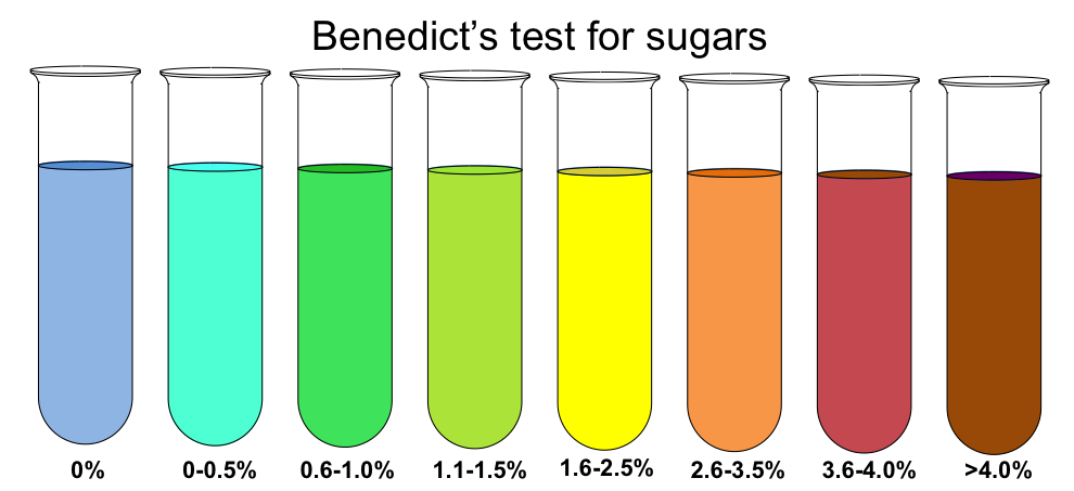
- Glucose is positive. It is the only reducing sugar on the list.
- Sucrose stays blue. Hydrolyzing it first gives a positive result: warm it with a few drops of dilute HCl in the water bath, neutralize with NaOH (the reagent needs base), then add Benedict's and heat. A positive test after hydrolysis confirms sucrose. Table sugar can carry traces of invert sugar (glucose + fructose), and long boiling can give a faint green, so compare against a blank.
- Cornstarch stays blue. Its chain has only one reducing end per thousands of glucose units.
- Ammonium chloride turns the reagent a darker, deep royal blue without heating and without a precipitate. The carbonate in the reagent deprotonates some NH₄⁺ to NH₃, and ammonia binds copper as the tetraamminecopper(II) complex. This is complex formation, not reduction:
- Calcium and magnesium salts may make the reagent cloudy (white carbonate precipitate from the reagent's carbonate). That cloudiness is white, not the colored Cu₂O.
Benedict's vs Fehling's. Fehling's solution uses the same Cu²⁺ → Cu₂O chemistry. It comes as two solutions mixed just before use: Fehling's A is CuSO₄, and Fehling's B is sodium potassium tartrate (Rochelle salt) with NaOH. Fehling's is more strongly alkaline and less stable. Benedict's is a single, milder (carbonate rather than hydroxide), long-lasting solution, which is why Supervisors supply it. Tollens' reagent (silver–ammonia complex, giving a silver mirror) also detects aldehydes.
Carbonates and a worked identification
Carbonates and hydrogen carbonates fizz with dilute acid. H⁺ converts them to carbonic acid, which breaks down into water and carbon dioxide gas. CO₂ is colorless and odorless and turns limewater milky. Every acid–carbonate reaction gives a salt + water + carbon dioxide. These are acid–base (neutralization) reactions, often also classed as double displacement followed by decomposition of H₂CO₃. The two react differently with heat. Na₂CO₃ + HCl warms the solution, but NaHCO₃ + HCl cools it slightly. With bicarbonate, little heat is released by the proton transfer. More heat is absorbed by dissolving solid NaHCO₃ and by breaking H₂CO₃ into CO₂ gas and water (HCO₃⁻(aq) + H⁺(aq) → CO₂(g) + H₂O(l), ΔH ≈ +13 kJ/mol). The reaction still goes because forming a gas raises the entropy so much. None of them is a redox reaction. Oxidation numbers follow a few rules: an element on its own is 0; a monatomic ion has its charge (Na⁺ +1, Cl⁻ −1); O is usually −2 and H usually +1; and the numbers add up to 0 in a neutral compound (to the ion's charge in a polyatomic ion). In NaHCO₃ + HCl → NaCl + H₂O + CO₂, Na stays +1, H +1, Cl −1 and O −2, and C is +4 on both sides (in HCO₃⁻: +1 + C + 3(−2) = −1, so C = +4). Nothing is oxidized or reduced.
What does not react with HCl. NaCl, KCl, LiCl and NH₄Cl share the chloride ion with HCl, so nothing happens. Calcium nitrate, magnesium sulfate, boric acid (an acid already) and the carbohydrates show no visible change at 1 M. CaSO₄ does not fizz; it may dissolve very slightly more. If a key still wants an equation, write the formal double displacement CaSO₄(s) + 2HCl(aq) → CaCl₂(aq) + H₂SO₄(aq) and add that no visible reaction occurs. No gas or precipitate forms; the acid only makes gypsum somewhat more soluble, because some sulfate picks up H⁺ to form HSO₄⁻. A sample that bubbles must contain CO₃²⁻ or HCO₃⁻.
The same acid–bicarbonate reaction explains household look-alikes that tests sometimes use as distractors (they are not on the list): baking powder (NaHCO₃ plus a dry acid) and effervescent antacid tablets such as Alka-Seltzer (NaHCO₃, citric acid and aspirin) fizz in plain water, because the solid acid and the bicarbonate react as soon as both dissolve.
Telling the three carbonates apart.
| Test | NaHCO₃ | Na₂CO₃ | CaCO₃ |
|---|---|---|---|
| Water | Dissolves moderately | Dissolves well (anhydrous warms slightly) | Insoluble, milky |
| pH | ≈ 8.3 | ≈ 11.5 | ≈ 8–9.5 (suspension) |
| Flame | Yellow | Yellow | Orange-red |
| Heating | Loses ~37% mass; water condenses | Stable | Stable at burner temperature |
| Dropwise dilute acid into solution | Bubbles at once | First drops convert CO₃²⁻ to HCO₃⁻ with little fizz; bubbling starts as more acid is added | Fizzes on the solid |
The last row explains a common question. Add one small drop of acid to a sodium carbonate solution, and the H⁺ is used up converting CO₃²⁻ to HCO₃⁻ (CO₃²⁻ + H⁺ → HCO₃⁻), so little or no CO₂ escapes at first. With bicarbonate, every H⁺ goes straight to H₂CO₃ and CO₂.
Sodium carbonate manufacture
Questions about the industrial history of the list recur.
- Leblanc process (Nicolas Leblanc, 1791): 2 NaCl + H₂SO₄ → Na₂SO₄ + 2 HCl, then Na₂SO₄ + CaCO₃ + 2 C → Na₂CO₃ + CaS + 2 CO₂. It produced polluting HCl gas and CaS waste.
- Solvay (ammonia-soda) process (Ernest Solvay, 1860s): CO₂ and NH₃ are bubbled into brine, and the less soluble NaHCO₃ precipitates: NaCl + NH₃ + CO₂ + H₂O → NaHCO₃(s) + NH₄Cl. Heating the NaHCO₃ gives Na₂CO₃. Limestone supplies the CO₂ (CaCO₃ → CaO + CO₂), and the lime regenerates the ammonia, leaving CaCl₂ as the waste product. The overall reaction is 2 NaCl + CaCO₃ → Na₂CO₃ + CaCl₂. Ammonia is essential because it keeps the solution basic enough to hold the CO₂ as HCO₃⁻.
- Hou's process (Hou Debang) skips ammonia recovery and sells the NH₄Cl as fertilizer instead of producing CaCl₂ waste.
Stoichiometry and solution preparation
Decision table
With every test now explained, the table below is the decision aid. Run at least two independent tests on every unknown and write down the test, observation and inference for each.
| Observation | Narrows the unknown to |
|---|---|
| Insoluble or only a suspension in water | Cornstarch, calcium carbonate, calcium sulfate |
| Blue-black with iodine | Cornstarch |
| Fizzes with HCl, strong yellow flame, pH ≈ 11–12 | Sodium carbonate |
| Fizzes with HCl, strong yellow flame, pH ≈ 8–8.5, loses mass on heating | Sodium hydrogen carbonate |
| Fizzes with HCl, orange-red flame, insoluble | Calcium carbonate |
| Green to brick red with Benedict's after heating | Glucose |
| Sweet, soluble, Benedict's stays blue, chars on heating | Sucrose (confirm by acid hydrolysis and retest) |
| Crimson flame | Lithium chloride (often damp or clumped) |
| Lilac flame through cobalt glass, no fizz, neutral | Potassium chloride |
| Vinegar smell with acid, no fizz, yellow flame, pH ≈ 8–9 | Sodium acetate |
| Yellow flame, no fizz, pH 7, no vinegar smell, cubic crystals | Sodium chloride |
| Ammonia smell with NaOH; deep blue with Benedict's; no flame color; pH ≈ 5 | Ammonium chloride |
| Heavy white precipitate with NaOH, no flame color | Magnesium sulfate |
| Green flame, pH ≈ 5, flaky and slippery, only moderately soluble | Boric acid |
| Very soluble, orange-red flame, no fizz, cloudy with NaOH | Calcium nitrate |
| Barely soluble, orange-red flame, no fizz | Calcium sulfate |
Worked identification from a data table
Polymers, fibers and hair (Part 3.c)
The 2027 lists are exact. The eight plastics are PETE, HDPE, LDPE, PP, PVC, PMMA, PC and solid (non-expanded) polystyrene. You never burn them, but the Supervisor may hand you burn results. The seven fibers are cotton, linen, wool, silk, nylon, polyester and spandex, and you may burn these. The five hairs are human, bat, cow, squirrel and horse, and you must know hair structure, the root and the scale classes. All three groups are polymers, so the chemistry below is one toolkit: chain structure explains density, melting, burning and solubility.
Polymer chemistry basics
A polymer is a very large molecule (macromolecule) built from many small molecules called monomers joined by covalent bonds ("poly" = many, "mer" = part). The piece that repeats along the chain is the repeat unit. For an addition polymer it has the same atoms as the monomer (ethylene CH₂=CH₂ becomes –CH₂–CH₂–); for a condensation polymer it has fewer atoms than the monomers combined, because a small molecule is lost at each link. A chain of only a few units is an oligomer. Plastics are polymers that can be molded; fibers are polymers drawn into long, thin strands; hair and wool are natural protein polymers.
Degree of polymerization (DP) is the number of repeat units in a chain. Real samples are mixtures of chain lengths, so a polymer has no single molar mass and no single sharp melting point. Tests use the number-average molar mass :
Longer chains (higher DP) tangle more and have more total intermolecular attraction per chain, so strength, toughness and melt viscosity rise with DP. That is why ultra-high-molecular-weight polyethylene is far tougher than ordinary HDPE.
Addition (chain-growth) polymerization
In addition polymerization monomers containing a C=C double bond join end to end and nothing is lost: the polymer has exactly the empirical formula of the monomer. It is a chain-growth process: a reactive chain end adds monomers one at a time, very fast, so long chains appear early while most monomer is still unreacted. The usual mechanism is free-radical polymerization:
- Initiation. An initiator such as benzoyl peroxide or AIBN splits (by heat or light) into radicals, species with an unpaired electron. A radical attacks the π bond of a monomer, pairs with one of its electrons, and leaves a new radical on the far carbon. A plain alkene cannot polymerize on its own because it has no unpaired electron to start the chain.
- Propagation. The radical chain end adds another monomer, regenerating a radical at the new end, thousands of times. Each step turns one π bond into a σ bond, so the reaction is exothermic.
- Termination. Two radical chain ends meet. In combination (coupling) they bond into one long dead chain. In disproportionation one radical takes a hydrogen atom from the other, giving one saturated chain end and one chain ending in C=C. A chain can also stop growing by chain transfer, handing its radical to another molecule, which is how branches form in LDPE.
Polyolefins (PE, PP) are also made by coordination polymerization with Ziegler–Natta catalysts (titanium chlorides such as TiCl₄ with an organoaluminum co-catalyst) or chromium-based Phillips catalysts. The monomer inserts at a metal–carbon bond, which gives linear chains (HDPE) and controls tacticity (isotactic PP). Karl Ziegler and Giulio Natta shared the 1963 Nobel Prize in Chemistry for this work.
Condensation (step-growth) polymerization
In condensation polymerization two different functional groups react, forming a link and releasing a small molecule: water (acid + alcohol → ester; acid + amine → amide), HCl (when an acid chloride or phosgene is used) or methanol (when a methyl ester is used). Each monomer needs at least two reactive groups (bifunctional) so the chain can keep growing at both ends. It is a step-growth process: any two molecules (monomers, dimers, oligomers) can join, so the average chain length stays low until almost all the groups have reacted, and very long chains form only at very high conversion. Removing the by-product (vacuum, heat, desiccant) drives the equilibrium toward polymer, which is also why PET pellets must be dried before melting: water at processing temperature hydrolyzes ester links and shortens chains.
| Addition (chain-growth) | Condensation (step-growth) | |
|---|---|---|
| Monomer feature | C=C double bond | Two reactive groups per monomer (–COOH, –OH, –NH₂, –COCl) |
| By-product | None | H₂O, HCl or CH₃OH |
| How chains grow | Monomer adds to an active chain end | Any two molecules join; long chains only near completion |
| Backbone | Usually all carbon | Contains O or N (ester, amide, carbonate links) |
| On the lists | HDPE, LDPE, PP, PVC, PS, PMMA | PETE, PC, nylon 6,6, polyester |

Chain architecture: branching, crystallinity, tacticity
A homopolymer is made from one kind of repeat unit (PP); a copolymer contains two or more (linear low-density polyethylene is ethylene copolymerized with a little 1-butene, 1-hexene or 1-octene). Chains may be linear, branched, or cross-linked into a network.
HDPE vs LDPE is the standard example. Both are polyethylene from ethylene (CH₂=CH₂) by addition, with the same formula (C₂H₄)ₙ; they are structural variants of one polymer. LDPE is made by high-pressure free-radical polymerization, where chain transfer and "backbiting" put many short and long branches on the chain. Branches keep chains from lining up, so LDPE has less crystalline order: lower density (≈0.91–0.93 g/cm³), lower melting point (≈105–115 °C), softer, more flexible and more transparent. HDPE is made at low pressure with Ziegler–Natta or Phillips catalysts and is nearly linear. Linear chains pack tightly into crystalline regions, so HDPE has higher density (≈0.94–0.97 g/cm³), higher melting point (≈130–137 °C), greater stiffness, tensile strength and chemical resistance, and is more opaque. Pick HDPE for a stiff, chemically resistant cutting board or outdoor furniture; pick LDPE for a flexible clear film or a squeeze bottle. Because LDPE is flexible, it also resists impact well (it flexes instead of cracking), which is why it is used for impact pads and drape-formed prosthetic and orthotic devices. The difference is branching, not cross-linking: neither is cross-linked.
Amorphous vs semicrystalline. In an amorphous polymer the chains are randomly coiled, like cooked spaghetti; it has a glass transition temperature () where it changes from glassy to rubbery and softens gradually instead of melting sharply, and it is usually transparent because there are no crystal boundaries to scatter light. A semicrystalline polymer has ordered crystalline regions (which melt at a melting point ) in an amorphous matrix; crystallites raise density, stiffness and solvent resistance and tend to make the material translucent or opaque. Higher crystallinity means higher density. On the list, PS, PMMA and PC are amorphous (clear), PVC is mostly amorphous, and HDPE, LDPE and PP are semicrystalline. PET can be either: quenched it is amorphous and clear (bottles), slowly cooled or stretched it crystallizes.
Tacticity describes where side groups sit along the chain. Isotactic: all on one side. Syndiotactic: alternating sides. Atactic: random. Regular (iso- or syndiotactic) chains can crystallize, raising melting point and density; atactic chains cannot. Commercial PP is mostly isotactic (semicrystalline, ≈ 160–170 °C); syndiotactic PP packs less regularly and melts lower (about 125–130 °C); atactic PP is amorphous and rubbery with no sharp melting point, so by melting point atactic < syndiotactic < isotactic; ordinary polystyrene is atactic, which is why it is amorphous, clear and brittle.

Physical versus chemical properties. A physical property is observed without changing what the substance is: density, melting point, color, hardness, solubility, refractive index. A chemical property shows up only when the substance reacts and becomes something new: flammability and how it burns, reaction with acids or bleach, decomposition on heating. Flotation tests use a physical property; burn tests use chemical properties.
Thermoplastics and thermosets
A thermoplastic is made of linear or branched chains held together only by intermolecular forces (London dispersion forces, dipole–dipole attractions, hydrogen bonds). Heat lets the chains slide past each other, so it softens and melts, and it hardens again on cooling; the cycle is physical and repeatable, so thermoplastics can be remolded and recycled (each cycle shortens some chains, so quality slowly drops). A thermoset is cured: covalent cross-links join the chains into one three-dimensional network, so it cannot melt or dissolve; strong heat chars and decomposes it instead. Thermosets are rigid, heat- and solvent-resistant and hard to recycle. Most polymers made are thermoplastics, and all eight list plastics are thermoplastics, as are nylon and polyester. Bakelite, epoxies and vulcanized rubber are thermosets. A "sliver" or heat test that gives powdery chips or charring without melting suggests a thermoset.
Additives change properties without changing the polymer. Plasticizers (for example phthalates such as DEHP) are small molecules that sit between chains, weaken chain–chain attractions and lower , turning rigid PVC into flexible PVC for tubing, blood bags and cable jackets; they also lower its density. Stabilizers (heat and UV stabilizers, for example benzophenone UV absorbers) slow degradation; UV light breaks bonds and forms radicals, causing chain scission, yellowing and brittleness, and PVC can lose HCl (dehydrochlorination) to form C=C bonds that discolor and embrittle it. Each new C=C leaves the chlorine on the next unit allylic and weakly held, so HCl comes off neighboring units one after another in a fast "zipper", sped up further by the released HCl. The result is runs of conjugated double bonds that absorb visible light (yellow, then brown, then black) and can oxidize or cross-link. Heat stabilizers work largely by scavenging HCl. Fillers (calcium carbonate, talc) cut cost and stiffen; colorants and flame retardants are also common.
Biodegradable, compostable, recyclable. Biodegradable means microorganisms can eventually convert the material to CO₂, water and biomass, with no time limit attached. Compostable is a stricter, certified claim: the item biodegrades within a set time under composting conditions and leaves no toxic residue; industrial-compost standards such as ASTM D6400 and EN 13432 require roughly 90% conversion to CO₂ within about six months at elevated composting temperatures. So every compostable plastic is biodegradable, but not every biodegradable plastic is compostable, and "industrially compostable" items (such as PLA cutlery) often do not break down in a cool home compost bin. Bio-based (made from plants) is a separate idea: bio-based polyethylene is not biodegradable, and some fossil-based polyesters are. Compostable plastics contaminate recycling streams because they melt and degrade differently from the resin being recycled. Recyclable means it can be collected and reprocessed; in practice PETE (1) and HDPE (2) are the most widely recycled.
History of plastics and fibers
| Year | Event |
|---|---|
| 1862 | Alexander Parkes shows Parkesine, a semi-synthetic plastic made from cellulose nitrate; celluloid follows around 1870 |
| 1884 | Hilaire de Chardonnet makes "Chardonnet silk" from nitrocellulose, the first manufactured (regenerated) fiber; viscose rayon follows in the 1890s |
| 1907 | Leo Baekeland makes Bakelite from phenol and formaldehyde: the first fully synthetic plastic, a thermoset |
| 1933 | ICI chemists discover polyethylene (high-pressure, branched: LDPE) |
| 1935 | Wallace Carothers's group at DuPont first makes nylon 6,6; sold from 1938–1939 (toothbrush bristles, then stockings), the first commercially successful fully synthetic fiber, developed as a silk substitute |
| 1941 | J. R. Whinfield and J. T. Dickson patent PET polyester (sold as Terylene and later DuPont's Dacron) |
| 1950s | Ziegler and Natta catalysts give linear HDPE and isotactic PP; polycarbonate developed at Bayer and GE (1953) |
| 1968 | Union Carbide starts up the first Unipol plant (Seadrift, Texas): a low-pressure, gas-phase (fluidized-bed) process for making polyethylene, first HDPE and from 1977 LLDPE |
| 1958 | Joseph Shivers at DuPont develops spandex ("Fiber K"; some sources give 1959), later sold as Lycra; key patent issued 1962. "Spandex" is an anagram of "expands" |
| 1988 | Resin identification codes introduced by the U.S. Society of the Plastics Industry; now maintained by ASTM (D7611) |
Plastics and density
Density is the most useful simple test for plastics because you are not allowed to burn them. Density = mass ÷ volume (g/cm³ = g/mL). A solid floats in a liquid less dense than itself and sinks in a liquid denser than itself; if they match it hangs suspended. A plastic floats in water if its density is below 1.00 g/cm³. Because ranges for one polymer depend on crystallinity, fillers, plasticizer and trapped air, always trust the behavior of the samples over a memorized number.

The resin identification code (RIC) is the number in a triangle molded into an item: 1 PETE (PET), 2 HDPE, 3 V (PVC), 4 LDPE, 5 PP, 6 PS, 7 Other. Code 7 covers everything else, including PC, PMMA, nylon, ABS and PLA. Since 2013 the ASTM standard uses a plain solid triangle rather than chasing arrows, to stress that the code identifies resin and is not a recycling promise. In practice, US curbside programs most widely accept 1 (PETE) and 2 (HDPE) bottles and jugs, increasingly 5 (PP); 3 (PVC), 6 (PS) and 7 (Other) are rarely recycled, and 4 (LDPE) film usually goes to store drop-off bins rather than curbside carts.
The solution densities used below are approximate values near room temperature: water 1.00 g/mL; 10% (w/w) NaCl ≈ 1.07; 25% NaCl ≈ 1.19; saturated NaCl (about 26%) ≈ 1.20; 46% (by volume) isopropyl alcohol ≈ 0.93; 70% isopropyl alcohol ≈ 0.88 (manufacturer data sheets give 0.878 g/mL at 20 °C); vegetable oil ≈ 0.91–0.93 (corn and soybean oil about 0.92). "Borderline" means the plastic's range straddles the liquid's density, so the sample may hover, float slowly or sink.
ReferenceThe eight plastics· 8 rows
Densities are typical ranges (g/cm³). W = water (1.00), 10% = 10% NaCl (1.07), Sat = saturated NaCl (1.20), IPA = 70% isopropyl alcohol (0.88), Oil = vegetable oil (≈0.92). F = floats, S = sinks. Burn results are typical reports; supervisors' charts differ, so match the descriptions you are given.
No rows match.
| Code | Plastic | Monomer(s) | Repeat unit | Type | Density | W / 10% / Sat / IPA / Oil | Typical burn result | Acetone | Key properties and uses |
|---|---|---|---|---|---|---|---|---|---|
| 1 | PETE, polyethylene terephthalate | Terephthalic acid C₆H₄(COOH)₂ (or dimethyl terephthalate) + ethylene glycol HOCH₂CH₂OH | C₁₀H₈O₄ (192.17 g/mol); ester links, benzene ring | Condensation (loses H₂O, or CH₃OH from the dimethyl ester) | 1.33–1.40 (often 1.38) | S / S / S / S / S | Yellow, slightly sooty flame; shrinks and curls from the flame, melts, bubbles, drips; sweet odor | Resistant | Clear, tough, good gas barrier; soda and water bottles, peanut-butter jars, Mylar film, polyester fiber |
| 2 | HDPE, high-density polyethylene | Ethylene CH₂=CH₂ | C₂H₄ (28.05 g/mol) | Addition (Ziegler–Natta or Phillips catalyst) | 0.94–0.97 | F / F / F / S / S | Blue flame with yellow tip, melts and drips, keeps burning; candle-wax (paraffin) odor; little smoke | Resistant | Linear, semicrystalline, opaque, stiff, chemically resistant; milk jugs, detergent and shampoo bottles, cutting boards, thin crinkly "T-shirt" grocery bags |
| 3 | PVC, polyvinyl chloride | Vinyl chloride CH₂=CHCl | C₂H₃Cl (62.50 g/mol) | Addition (free radical, often suspension) | 1.30–1.45 rigid; ≈1.16–1.35 when plasticized | S / S / S / S / S (plasticized PVC may float in Sat) | Hard to ignite, chars and softens, yellow flame with green edges, white acrid smoke (HCl), self-extinguishes | Swells or softens, especially flexible PVC | Only list plastic with a halogen; rigid pipe and siding, flexible tubing, blood bags, cable insulation |
| 4 | LDPE, low-density polyethylene | Ethylene CH₂=CH₂ | C₂H₄ (28.05 g/mol) | Addition (high-pressure free radical) | 0.91–0.93 | F / F / F / S / borderline | Like HDPE: blue flame with yellow tip, drips, keeps burning, candle-wax odor | Resistant | Branched, flexible, translucent; grocery and bread bags, cling film, squeeze bottles, six-pack rings |
| 5 | PP, polypropylene | Propylene (propene) CH₂=CHCH₃ | C₃H₆ (42.08 g/mol) | Addition (Ziegler–Natta, coordination) | 0.89–0.91 (least dense) | F / F / F / S / F | Yellow flame with blue base, drips, burns slowly, little smoke; waxy, slightly sweet or acrid odor | Resistant | Isotactic, semicrystalline, highest of the polyolefins (≈160–170 °C), "microwave safe"; bottle caps, yogurt tubs, straws, rope, N95 filter layers, banknotes |
| 6 | PS, polystyrene (non-expanded) | Styrene C₆H₅CH=CH₂ (C₈H₈) | C₈H₈ (104.15 g/mol); phenyl side group | Addition | 1.04–1.06 | S / F (just) / F / S / S | Ignites easily, bright yellow flame, dense black sooty smoke and floating soot flakes, sweet (styrene) odor, keeps burning, softens and drips | Dissolves or turns gummy | Atactic, amorphous, clear, hard, brittle; disposable cutlery, CD cases, clear cups, beads; foam (EPS) is the same polymer with ~95–98% air |
| 7 | PMMA, poly(methyl methacrylate), acrylic | Methyl methacrylate CH₂=C(CH₃)COOCH₃ | C₅H₈O₂ (100.12 g/mol); ester side group | Addition | 1.17–1.20 | S / S / F (just) / S / S | Burns steadily, blue base and yellow flame, crackles and bubbles, little smoke, fruity (ester) odor | Softens, crazes, dissolves slowly | Amorphous, glass-clear, UV-resistant; Plexiglas, Lucite, Perspex, windows, lenses, signs |
| 7 | PC, polycarbonate | Bisphenol A (BPA) + phosgene COCl₂ (or diphenyl carbonate) | C₁₆H₁₄O₃ (254.28 g/mol); two benzene rings, carbonate link | Condensation (loses HCl, or phenol from diphenyl carbonate) | 1.20–1.22 | S / S / borderline / S / S | Hard to ignite, chars and bubbles, sooty yellow flame, self-extinguishes, faint phenolic odor | Crazes and whitens (stress-cracks); not quickly dissolved | Amorphous, clear, very impact-resistant, ≈ 145–150 °C; safety glasses and helmets, headlight lenses, CDs and DVDs, water-cooler jugs |
Forms of polystyrene. The same polymer comes in several forms: solid general-purpose PS (clear, brittle: CD cases, disposable cutlery); high-impact PS (HIPS), toughened with rubber (opaque cups and trays); oriented PS (OPS), stretched into clear, stiff sheet (clamshells, bakery lids); expanded PS (EPS), beads puffed with pentane into white foam (cups, coolers, packing peanuts); and extruded PS (XPS), foam board for building insulation (the blue or pink "Styrofoam" board). Only solid, non-expanded PS is on the 2027 list.
Thermal and structural landmarks
| Plastic | Morphology | Approximate / | Why it matters |
|---|---|---|---|
| PETE | Amorphous or semicrystalline | ≈ 70–80 °C, ≈ 250–260 °C | Bottles soften in hot water; shrivels when heated |
| HDPE | Semicrystalline (high) | ≈ 130–137 °C | Survives boiling water better than LDPE |
| LDPE | Semicrystalline (lower) | ≈ 105–115 °C | Softest, most flexible |
| PP | Semicrystalline | ≈ 160–170 °C | Most heat-tolerant polyolefin |
| PVC | Mostly amorphous | ≈ 80 °C; releases HCl on strong heating | Softens around 70–80 °C before decomposing |
| PS | Amorphous (atactic) | ≈ 100 °C | Brittle, clear |
| PMMA | Amorphous | ≈ 105 °C | Clear, light-transmitting |
| PC | Amorphous | ≈ 145–150 °C | Tough at high temperature |
Three structural features answer many short questions. Benzene rings: PETE, PS and PC (PC has two per repeat unit; PS has the phenyl side group). Halogen: PVC only. Ester groups: PETE and PMMA, which is why both give sweet or fruity burning odors. PE and PP are pure hydrocarbons, nonpolar, so water, acids and acetone barely affect them; PS is nonpolar and aromatic, so acetone dissolves it by replacing the weak London forces between its chains. Their weak point is strong oxidizers: concentrated or fuming nitric acid, hot concentrated sulfuric acid and halogens such as chlorine gas attack PE and PP, especially when warm or under stress, and leave them brittle, so concentrated nitric acid is not stored in polyolefin bottles (household bleach and 3% hydrogen peroxide, by contrast, are routinely sold in HDPE). PMMA is not water-soluble either: its polar ester groups are outweighed by the hydrocarbon backbone. Water-based acrylic paint is an emulsion (latex) of tiny acrylic-polymer particles dispersed in water. It thins and cleans up with water while wet; as the water evaporates the particles touch and fuse (coalesce) into a film that water can no longer redissolve.
The density ladder
| Density (g/cm³) | Plastic | Separating liquid just above it |
|---|---|---|
| 0.89–0.91 | PP | 70% IPA (0.88) sinks it; oil (≈0.92) floats it |
| 0.91–0.93 | LDPE | 46% IPA (0.93) floats it |
| 0.94–0.97 | HDPE | Water (1.00) floats it; sinks in 46% IPA |
| — | water 1.00 | — |
| 1.04–1.06 | PS | 10% NaCl (1.07) floats it |
| 1.17–1.20 | PMMA | Saturated NaCl (1.20) floats it, just |
| 1.20–1.22 | PC | Borderline in saturated NaCl |
| 1.30–1.45 | PVC | Sinks in everything common; use the Beilstein test |
| 1.33–1.40 | PETE | Sinks in everything common; no halogen |
A density column or a set of liquids brackets the density. If a sample sinks in liquid A and floats in liquid B, then . Salt water is used because adding NaCl raises density in a controlled way; alcohol–water mixtures give densities below 1. Gradient columns built from heavier organic liquids (for example bromoform or bromobenzene mixtures, as in ASTM density-gradient methods) reach higher densities, but you will normally get salt and alcohol solutions.
Beilstein test, PVC and PETE
PVC and PETE are both well above water in density and their ranges overlap, so a density bracket rarely separates them. The Beilstein test finds the halogen. Heat a clean copper wire in a burner flame until it no longer colors the flame, touch it to the plastic so a little melts onto it, and return it to the flame. A green or blue-green flame means a halogen: chlorine from the polymer forms volatile copper chloride, and excited copper emits green light. On the 2027 list only PVC gives a positive result. The test is done by the Supervisor or reported to you, since you do not burn plastics. In burn data, PVC is the one that is hard to ignite, chars, gives acrid HCl fumes and self-extinguishes (chlorine acts as a flame retardant). Burning PVC releases hydrogen chloride and can form dioxins, and any incompletely burned plastic gives carbon monoxide and soot.
Why PETE suits carbonated drinks. PET's stiff aromatic, polar chains pack tightly, and stretch-blow molding orients and partly crystallizes the bottle wall. That makes PET strong enough to hold the pressure of dissolved CO₂, clear, and a much better barrier to CO₂ and O₂ than HDPE or PP, whose nonpolar chains let gases diffuse through, so a soda would go flat in an HDPE jug.
How much plastic is recycled. Very little overall: the US EPA estimated that only about 9% of US plastic waste was recycled in 2018, compared with about two-thirds of paper and cardboard; most plastic is landfilled or burned for energy. PET bottles and jars do much better than plastic overall: about 29% were recycled in 2018 (EPA 29.1%; the industry group NAPCOR 28.9%), and tests often round this to "about 30%".
Why PVC is rarely recycled. PVC is hard to reprocess: it releases HCl when overheated, it is compounded with many different plasticizers and stabilizers, and a small amount of PVC in a PET melt degrades at PET's processing temperature and spoils the batch. Its chlorine also makes incineration produce HCl and dioxins. Most curbside programs therefore reject code 3.
Fiber burn tests
A fiber is a thread-like strand, very long compared with its width (at least about 100 times longer than wide), that can be spun into yarn. Short natural fibers are called staple fibers; continuous strands (silk, melt-spun synthetics) are filaments. Fibers fall into natural fibers (plant cellulose: cotton, linen; animal protein: wool, silk) and manufactured fibers. Manufactured fibers are either synthetic (polymers made from petrochemicals, extruded through a spinneret: nylon, polyester, spandex) or regenerated (a natural polymer dissolved and re-formed: rayon/viscose and lyocell from wood-pulp cellulose; acetate is chemically modified cellulose). Rayon and acetate are not on the list; know only that rayon is cellulose and therefore burns like cotton, and that acetate dissolves in acetone. In the viscose process wood pulp is soaked in sodium hydroxide, reacted with carbon disulfide (CS₂) to make a soluble cellulose xanthate, and the viscous solution is extruded into an acid bath that regenerates cellulose filaments. CS₂ is volatile, flammable and toxic to the nervous system, a long-standing hazard for rayon workers. A third natural class, mineral fibers, includes asbestos (fibrous silicate minerals: heat- and fire-resistant, but the fine needle-like fibers lodge in the lungs and cause asbestosis, lung cancer and mesothelioma); glass fiber is a manufactured inorganic fiber.
Fibers are the one polymer group you may burn. Use small samples with tweezers over a heat-proof surface, observe as the fiber approaches the flame, while in the flame, and after removal, then note the odor and the residue. Burn a known reference beside the unknown.
| Family | Fibers | Approaching flame | In flame / removed | Odor | Residue |
|---|---|---|---|---|---|
| Plant cellulose | Cotton, linen | Does not shrink; ignites on contact | Burns quickly with a yellow flame; keeps burning and afterglows | Burning paper, leaves or wood (campfire) | Fine, soft gray to whitish feathery ash |
| Animal protein | Wool, silk | Curls away from flame | Burns slowly, sputters; usually self-extinguishes | Burning hair or feathers | Crushable black bead or brittle ash |
| Synthetic | Nylon, polyester, spandex | Shrinks and melts away (spandex melts but barely shrinks) | Melts and burns, often dripping | Chemical | Hard bead that will not crush (spandex: soft, sticky) |
Why drawing strengthens fibers. Melt-spun synthetic filaments are drawn (stretched several times their length) after spinning. Drawing pulls the randomly coiled chains into line along the fiber axis, so they pack into ordered, partly crystalline regions held by many intermolecular forces (hydrogen bonds between nylon's amide groups, for example). The fiber becomes stronger and stiffer along its length but less stretchy.
The most reliable distinction is protein versus cellulose. Protein fibers contain nitrogen (in every amino acid) and, in wool, a lot of sulfur (in the amino acid cysteine), so they give the burnt-hair smell, and their char layer smothers the flame, so they self-extinguish. Their high moisture content (wool holds the most water of any common fiber) also absorbs heat, which helps. Cellulose is a carbohydrate (C, H, O only) that burns cleanly like paper and leaves little mineral ash. Synthetics are thermoplastics: they melt before they burn, shrink from the flame and leave a fused bead. Watch the melt more than the smell.
ReferenceThe seven fibers· 7 rows
Moisture regain is the standard commercial value (ASTM D1909, % water held at 21 °C, 65% relative humidity). Densities are approximate (g/cm³).
No rows match.
| Fiber | Category | Source | Polymer and bond | Microscope | Burn: approach / in flame / removed | Odor | Residue | Chemical tests | Properties and uses |
|---|---|---|---|---|---|---|---|---|---|
| Cotton | Natural plant, seed fiber | Seed hairs (trichomes) of Gossypium species, separated from seeds by ginning; short fuzz left on the seed = linters | Cellulose (≈90% of the fiber): β-D-glucose joined by β-1,4-glycosidic bonds; many –OH groups | Flat, twisted ribbon with convolutions; collapsed central lumen; kidney or bean-shaped cross-section | No shrink / burns fast, yellow flame / keeps burning, afterglow | Burning paper | Soft gray feathery ash | Bleach: whitens, not dissolved; cold concentrated NaOH: swells (mercerization); concentrated H₂SO₄ (≈70%): dissolves | Regain 8.5%, density ≈1.54; absorbent, comfortable, stronger wet; most-used plant fiber; T-shirts, denim, towels, cotton balls |
| Linen | Natural plant, bast fiber | Stem (phloem, inner bark) of flax, Linum usitatissimum; freed by retting and scutching | Cellulose (less pure than cotton, with hemicellulose and some lignin) | Straight, smooth cylinders with nodes and cross-markings (like bamboo); narrow lumen; polygonal cross-section | No shrink / ignites a little more slowly than cotton, burns steadily / afterglow | Burning paper or grass | Fine gray ash | Same as cotton | Regain 12%, density ≈1.50; among the strongest plant fibers, low stretch (≈2%), stronger wet, absorbs and dries fast (cool, "hygienic"); summer clothing, tablecloths, bedding |
| Wool | Natural animal, protein (hair) | Fleece of sheep, Ovis aries (other "wools": goat = cashmere and mohair, rabbit = angora) | α-keratin (coiled α-helices) cross-linked by disulfide bonds (cysteine); peptide (amide) backbone | Overlapping imbricate scales; round to oval cross-section; natural crimp; coarse fibers may show a medulla | Curls from flame / burns slowly, sputters / self-extinguishes | Strong burnt hair (sulfur) | Crushable black bead or brittle ash | Dissolves in bleach (NaOCl); dissolves in hot 5% NaOH; fairly resistant to dilute acid | Regain ≈13.6–16% (most hygroscopic), density ≈1.31; elastic, crimped, warm, felts and shrinks; the most widely used animal fiber; sweaters, coats, carpets |
| Silk | Natural animal, protein (secreted filament) | Cocoon of the silkworm Bombyx mori (sericulture) | Fibroin (≈70–80%), glycine- and alanine-rich repeat (Gly-Ser-Gly-Ala-Gly-Ala), stacked in β-pleated sheets; coated by sericin gum (≈20–30%) | Smooth, rod-like, no scales; rounded-triangular cross-section (prism-like, gives luster); raw silk shows paired filaments in sericin | Curls from flame / burns slowly, sputters / usually self-extinguishes | Burnt hair or feathers (weaker than wool; little sulfur) | Crushable black bead | Dissolves in bleach; dissolves in concentrated HCl and H₂SO₄ | Regain 11%, density ≈1.25–1.35; one of the strongest natural fibers for its weight, lustrous, longest natural fiber (continuous filament); scarves, ties, lingerie |
| Nylon (nylon 6,6) | Synthetic polyamide | Hexamethylenediamine H₂N(CH₂)₆NH₂ + adipic acid HOOC(CH₂)₄COOH, both from petroleum | Polyamide: amide links –CO–NH–, hydrogen-bonded between chains; repeat C₁₂H₂₂N₂O₂ | Smooth, uniform rod; round or trilobal cross-section; delustrant specks (TiO₂) in dull grades | Shrinks, melts / melts, burns slowly / usually self-extinguishes; white smoke | Celery | Hard, round, cream to gray bead | Dissolves in 20% HCl, 90% formic acid and ≈70% H₂SO₄; resists bleach and acetone | Regain ≈4.5%, density ≈1.14; very strong, tough, abrasion-resistant, elastic; made to replace silk; stockings, ropes, carpets, parachutes |
| Polyester (PET) | Synthetic polyester | Terephthalic acid + ethylene glycol, same polymer as resin code 1 | Ester links –CO–O–, aromatic rings; repeat C₁₀H₈O₄ | Smooth, uniform rod; round (sometimes trilobal); delustrant specks | Shrinks, melts / melts and burns, drips / may continue slowly or self-extinguish; black sooty smoke | Sweet, aromatic chemical | Hard black or dark-brown bead | Resists bleach, acetone and most acids; hot strong alkali attacks the surface | Regain 0.4% (hydrophobic, quick-drying, wicks), density ≈1.38; wrinkle-resistant, holds shape; Dacron, Terylene; most-produced fiber; clothing, fiberfill, carpet, rope |
| Spandex (elastane, Lycra) | Synthetic segmented polyurethane | Long flexible diol (polyether or polyester "macroglycol") + diisocyanate, chain-extended with a diamine | At least 85% segmented polyurethane: flexible soft segments alternate with rigid hard segments (urethane and urea links) | Often several filaments fused together; round or dog-bone cross-section; uniform | Melts but does not shrink away / melts and burns / keeps melting or burning | Chemical, sharp (sometimes described as vinegar-like) | Soft, sticky, gummy black residue | Resists acetone; dissolves in dimethylformamide; chlorine bleach yellows and weakens it over time | Regain ≈1.3%, density ≈1.2; stretches several hundred percent (often quoted as 5–8 times its length) and snaps back; low tenacity; blended into athletic wear, swimsuits, waistbands |


Separating fibers within a family
- Cotton vs linen (both paper smell, gray ash): use the microscope. Cotton is a twisted ribbon; linen is a straight cylinder with nodes. Linen is longer, stiffer, less stretchy and slightly slower to ignite. Cotton is a seed hair; linen is a stem (bast) fiber.
- Wool vs silk (both burnt-hair smell, self-extinguish): wool has scales and crimp and a stronger sulfur smell; silk is smooth and lustrous with a triangular cross-section and a weaker smell. Wool's α-helices can stretch and recoil, so wool is more elastic (≈25–35% elongation) than silk, whose β-sheets are already extended (≈20%).
- Nylon vs polyester vs spandex (all melt to beads): nylon gives a celery smell, white smoke and a hard tan-gray bead and usually self-extinguishes; polyester gives a sweet chemical smell, black sooty smoke and a hard dark bead; spandex is the one that stretches enormously and leaves a soft, sticky, gummy residue rather than a hard bead.
- A fiber that stretches and melts but also smells of paper or hair is probably a blend (for example cotton–spandex); test separated yarns.
Fiber chemistry and treatments
Cellulose is a polymer of β-D-glucose linked β-1,4, which keeps the chains straight so they hydrogen-bond side by side into strong microfibrils (the repeat-unit formula is C₆H₁₀O₅). Its many –OH groups attract water, which is why cotton and linen absorb water, feel cool and dry slowly, and why they get stronger when wet (water plasticizes and evens the load among chains). Cellulose resists alkali but is hydrolyzed by strong acid, which breaks glycosidic bonds; humans cannot digest it because we lack cellulase.
Proteins (wool, silk) are chains of amino acids joined by peptide (amide) bonds. Hypochlorite bleach oxidizes the protein (including wool's disulfide bonds) and hot alkali hydrolyzes the peptide bonds, so protein fibers dissolve in bleach or hot NaOH, while cellulose and the synthetics do not. Never chlorine-bleach wool or silk.
Mercerization (John Mercer, 1844; tension added by Horace Lowe in 1889) treats cotton (sometimes linen) with cold, concentrated sodium hydroxide (about 20%), usually under tension, then washes and neutralizes it. NaOH swells the fiber: the flat twisted ribbon untwists into a rounder, smoother rod and the collapsed lumen nearly closes, and the cellulose crystal form converts to a more accessible form. Results: higher luster (a round, smooth surface reflects light evenly), greater strength and better dye uptake and absorbency, with less shrinkage. Wool cannot be mercerized; the alkali would destroy it.
Silk degumming removes the sericin gum by washing raw silk in hot soapy or alkaline water, leaving the soft, lustrous fibroin filaments. Fibroin's tiny glycine (side chain H) and alanine (CH₃) residues let neighboring chains pack into tight β-pleated sheets held by hydrogen bonds, which is why silk is strong but not very stretchy.
Nylon 6,6 is named for its two six-carbon monomers (hexamethylenediamine and adipic acid); it is made by condensation with loss of water (or HCl if adipoyl chloride is used), and hydrogen bonds between amide groups make it strong and somewhat water-absorbing. Nylon 6 comes from caprolactam by ring-opening. Kevlar and Nomex are aromatic polyamides (aramids), much stiffer because rigid rings and hydrogen bonds line up the chains. Polyester is the fiber form of PET. Spandex stretches because its long soft segments are coiled and can straighten out to several times their relaxed length, while hard segments cluster into "physical cross-links" that pull the chains back.
Fiber measurement terms
Denier is the mass in grams of 9,000 m of fiber; tex is grams per 1,000 m (so 1 tex = 9 denier). Tenacity is breaking force divided by linear density, usually in grams-force per denier (gf/den) or cN/tex: the fiber's strength corrected for its thickness. Elongation is how far it stretches before breaking; moisture regain is water held relative to dry mass.
Fabric construction. Fibers are spun into yarns, and yarns are woven or knitted into fabric. In a woven fabric the lengthwise yarns are the warp and the crosswise yarns that interlace with them are the weft (filling); common weaves are plain, twill (diagonal ribs, as in denim) and satin. Knits are made of interlocking loops (T-shirts, sweaters) and stretch more. Thread count, weave and yarn twist are extra class characteristics when comparing fabric pieces.
Fiber evidence: collection and analysis
Fibers are trace evidence transferred by contact, either directly (a sweater fiber onto a car seat) or indirectly (a carpet fiber to a suspect, then to a victim: secondary transfer), as Locard's exchange principle predicts. Collect visible fibers with clean forceps, lift the rest with adhesive tape lifts, or use a vacuum with a filter trap for large areas (it also picks up background debris). Package each item separately in folded paper (a druggist's fold) inside a paper envelope or bag, seal and label it with the chain of custody, and keep suspect and victim items apart to avoid cross-transfer.
In the lab, examiners first compare color, diameter, cross-section and surface features under a stereomicroscope and a compound microscope. A comparison microscope joins two microscopes by an optical bridge so the questioned and known fibers appear side by side in one field. Polarized light microscopy measures birefringence: oriented polymer chains give different refractive indices parallel and perpendicular to the fiber axis, so synthetics show bright interference colors between crossed polarizers, and the size of the effect helps classify the polymer. Microspectrophotometry compares dye spectra; FTIR spectroscopy identifies the polymer class from its bonds (ester C=O in polyester, amide bands in nylon, O–H in cellulose); pyrolysis gas chromatography identifies breakdown products. Chemical solubility tests are destructive and are used last.
Fibers are class evidence: they show that a fiber is consistent with a source type, not that it came from one garment, unless a physical fit (a torn edge matching a fabric) exists. Probative value rises when the fiber is unusual (a rare polymer, cross-section or dye; an uncommon blend), when many fibers are found, and when transfer is two-way (suspect's fibers on victim and victim's on suspect). Common fibers such as white cotton or blue denim have low probative value. A trilobal fiber (three-lobed cross-section, common in nylon carpet) sparkles because each lobe reflects light. A hollow fiber (a synthetic, usually polyester, extruded with an air-filled core) traps still air, so it insulates well for its weight; it is used as fiberfill in jackets, sleeping bags and pillows.
Hair structure
Hair is a thread of dead, keratinized cells grown from a living follicle in the skin. The study of hair is trichology. Mammals have several hair types: in humans, fine, short, lightly pigmented vellus hair ("peach fuzz") covers most of the body, while thick, long, pigmented terminal hair grows on the scalp, eyebrows, eyelashes, beard, armpits and pubic area; lanugo is the fine downy hair of a fetus. Animals have long, stiff guard hairs (the outer coat, used for species identification), soft insulating fur (wool) hairs, and tactile hairs (whiskers). Hair grows everywhere on the skin except areas such as the palms, soles and lips.
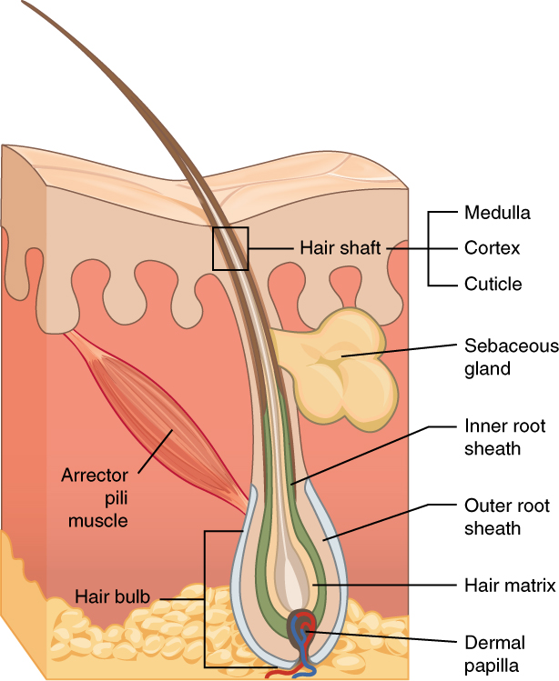
Follicle anatomy
| Structure | What it is and does |
|---|---|
| Follicle | Tube of epidermal cells reaching into the dermis that grows the hair; the only living, growing part |
| Dermal papilla | Connective-tissue knob at the base of the follicle with capillaries (and nerve endings); nourishes the matrix and signals growth |
| Hair bulb and matrix | Swollen base wrapped around the papilla; matrix keratinocytes divide rapidly and fill with keratin as they move up; melanocytes in the bulb add pigment |
| Inner and outer root sheaths | Cell layers around the forming hair; pieces stuck to a pulled growing hair form the follicular tag |
| Root | The part of the hair inside the follicle |
| Shaft | The part above the skin: cuticle, cortex, medulla |
| Sebaceous gland | Secretes oily sebum into the follicle to waterproof hair and skin; it lies near the skin surface |
| Arrector pili muscle | Smooth muscle attached to the follicle; contraction (cold, fear, via sympathetic nerves) stands the hair up and makes goose bumps |
The shaft: cuticle, cortex, medulla
From outside in, the shaft has three layers.
- Cuticle: a thin, colorless outer layer of flattened, overlapping keratin scales, with their free edges pointing toward the tip. It protects the cortex and controls how much water and chemicals get in; lifted or missing scales mean damage (bleaching, perming, weathering) or high porosity. The scale pattern is the main surface clue to species.
- Cortex: the thick middle layer and most of the hair's mass (often quoted as roughly 80–90%). It is made of spindle-shaped cells packed with keratin filaments, and gives hair its strength and elasticity. It holds the pigment granules (melanin), and also cortical fusi (small air spaces, most common near the root) and sometimes ovoid bodies (large, solid, oval pigmented bodies, more common in animal hair than human). The cortex is where color, texture and most comparison features are read.
- Medulla: the central core of loosely packed cells and air spaces. In humans it is often absent, fragmented (scattered pieces), interrupted (broken but regular) or, especially in coarse hairs such as beard hair, continuous; it may look dark in transmitted light because it holds air. Its function is unclear (possibly insulation). Many animals have a wide, continuous, patterned medulla.
Root form matters as much as the shaft (see growth phases below). The tip shows whether a hair was cut (square, angled), split or naturally tapered.
Keratin, melanin and hair chemistry
Keratin is the fibrous structural protein of hair, wool, nails and the outer skin; hair is made of α-keratin, whose chains coil into α-helices that twist together into intermediate filaments (α-keratin is found in mammals; the harder β-keratin, built of β-sheets, occurs in bird feathers, beaks and claws and in reptile scales). Keratin is unusually rich in the sulfur-containing amino acid cysteine; pairs of cysteines on neighboring chains form covalent disulfide (–S–S–) bonds, which give hair its strength, insolubility and set shape. Hair is held together by disulfide bonds (covalent), hydrogen bonds and salt (ionic) bridges. The elements in hair are C, H, N, O and S; burning releases sulfur compounds, hence the foul burnt-hair smell, and nitrogen compounds.
- Curl comes mainly from the shape of the follicle (round follicle → round, straight hair; oval or flattened follicle → oval or flat, curly hair) and the uneven distribution of disulfide cross-links. Water breaks hydrogen bonds temporarily (a wet set lasts until the next wash).
- A permanent wave uses a reducing agent (ammonium thioglycolate) to break disulfide bonds, the hair is reshaped on rods, and an oxidizer (hydrogen peroxide) re-forms disulfide bonds in new positions.
- Melanin, made from the amino acid tyrosine by the enzyme tyrosinase in melanocytes of the hair bulb, is passed as granules into the cortex cells. Eumelanin gives brown to black; pheomelanin gives red to yellow. Dark hair is rich in eumelanin; blond hair has little melanin; red hair has a high proportion of pheomelanin, typically from loss-of-function variants of the MC1R receptor gene (inherited recessively). Gray or white hair grows when bulb melanocytes stop making pigment; air in the shaft makes it look white. With age, hair also grows more slowly, strands become finer, and many follicles stop making new hairs. Hormones (testosterone-related male- and female-pattern baldness) and genes drive much of this thinning.
- Bleaching (hydrogen peroxide, often with ammonia) oxidizes melanin into colorless products; eumelanin breaks down first, so dark hair passes through red-orange and yellow stages, and the oxidizer also damages the cuticle and cortex. Dyes coat or penetrate the cortex; untreated new growth near the root shows the natural color.
Hair species identification
Forensic examiners identify species from guard hairs using three features together: cuticle scale pattern, medulla form and width, and pigment distribution, plus diameter, length, color banding and root shape.
Scale patterns are best seen on a scale cast or by SEM. To make a cast, brush a thin layer of clear nail polish on a slide, lay the hair in it, let it nearly dry (a few minutes), then peel the hair off; the impression left in the polish shows the scales under the microscope. The medulla is seen by mounting the whole hair in water or mounting medium in transmitted light. The three patterns:
- Imbricate: flattened scales overlapping like roof shingles, with smooth to wavy (crenate) margins and several scales around the shaft circumference. Found on human hair and on most mammal guard hairs, including cow, horse and squirrel (and dog, cat and wool).
- Coronal: each scale ring wraps all the way around the shaft like a crown or a stack of paper cups or funnels. Typical of fine hairs of bats and of some small rodents.
- Spinous (petal): triangular scales that project from the shaft like petals; seen near the base of some furbearer hairs such as mink and seal. No list species is defined by it.
Medulla forms: absent, fragmental, interrupted (discontinuous) and continuous; in animals the medulla is often wide and patterned (ladder-like uniserial or multiserial in many rodents and rabbits, lattice in deer, amorphous in humans).
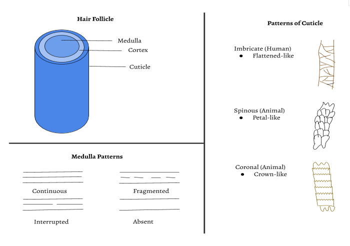
The medullary index (MI) compares the width of the medulla to the width of the whole hair:
ReferenceHairs on the 2027 list· 5 rows
Descriptions are for typical dorsal guard hairs (body coat) unless noted; individuals and body regions vary, so compare with the reference slides provided. Diameters are approximate midshaft values.
No rows match.
| Hair | Diameter | Scale pattern | Medulla and MI | Pigment | Distinguishing features |
|---|---|---|---|---|---|
| Human (scalp) | ≈50–100 µm (fine to coarse) | Imbricate, flattened, smooth to slightly wavy margins | Absent, fragmental, interrupted or continuous; thin, amorphous; MI usually < 1/3 | Evenly distributed, denser toward the cuticle (outer cortex); same color along the shaft, no banding | Uniform diameter, often cut or treated tip; club or bulb root; may show dye or bleach |
| Bat | Very fine (≈7–20 µm, most about 7–13 µm (one survey of Texas bats)) | Coronal: stacked funnel or cup-shaped scales, each ring encircling the shaft; margins may be toothed (dentate) | No medulla (MI = 0) | Light, varies by species | The only list hair with coronal scales; extremely fine; no medulla |
| Cow | Coarse | Imbricate, crenate (regular wavy) margins | Continuous, wide medulla filling about 2/3 of the shaft (MI ≈ 0.6–0.7) | Often little; may be streaky or granular | Coarse, thick continuous medulla with thin cortex; animal-type index |
| Squirrel (eastern gray, Sciurus carolinensis) | ≈75–80 µm | Imbricate, crenate, wavy and broken rows | Continuous, wide (more than half the shaft), flattened and nodose (ladder-like), with cortical intrusions | Often in color bands along the shaft (agouti banding of light and dark zones) | Wide continuous medulla plus banded pigment |
| Horse | Coarse (body ≈100–120 µm; mane and tail much coarser) | Imbricate, flattened, irregular wavy rows | Narrow; fragmental or interrupted to continuous; often about 1/3 of the shaft or less in body hairs | Usually even, may be dark | Large diameter and stiffness; MI can fall in the "human" range, so do not rely on MI alone |
Human versus animal hair
| Feature | Human | Typical animal |
|---|---|---|
| Medulla | Absent, fragmental or thin; amorphous | Present, continuous, wide, often patterned (ladder, lattice) |
| MI | Usually < 1/3 | Usually ≥ 1/2 |
| Pigment | Evenly spread, denser toward the cuticle | Denser toward the medulla; often in bands |
| Color along shaft | Uniform | Often banded (agouti) or changing abruptly |
| Scales | Imbricate | Imbricate, coronal or spinous, often more prominent |
| Shape | Fairly uniform diameter along the shaft | Often a widened "shield" near the tip; diameter varies |
| Root | Club or bulb | Often elongated, spade- or brush-shaped |

Hair growth, body area and evidence
Growth phases and the root
Each follicle cycles independently, so a scalp sheds hairs continuously (about 50–100 head hairs a day).
| Phase | What happens | Duration (scalp) | Share of scalp hairs | Root appearance |
|---|---|---|---|---|
| Anagen (growth) | Matrix cells divide; hair lengthens about 1 cm per month | 2–7 years (often ~1,000 days or more) | ≈80–90% | Pulled: stretched, pigmented root, often with a follicular tag (root-sheath tissue) |
| Catagen (transition) | Growth stops, follicle shrinks and detaches from the papilla's blood supply; the club forms | ≈2–3 weeks | ≈1–2% | Elongated, tapering root partway to a club |
| Telogen (resting) | Follicle dormant; hair held only by a keratinized club root; a new anagen hair starts below | ≈3 months (≈100 days; 2–4 months) | ≈10–15% (FBI: 10–18%) | Club-shaped, unpigmented bulb, no tissue: naturally shed |
| Exogen (shedding) | The club hair is released, often pushed out by the new hair | — | — | Same as telogen |
Most hairs found at scenes are shed telogen hairs. A hair with a stretched root and a follicular tag was in anagen and was pulled out forcibly, which suggests a struggle. Hair from a decomposing body may show a dark postmortem root band near the root. Hair does not keep growing after death; the skin dries and retracts, so hair and nails look longer.
Dating with hair growth
A typical scalp has roughly 100,000 hairs (often quoted as 100,000–150,000), and a person normally sheds on the order of 50–100 scalp hairs a day, which is why shed telogen hairs are so common at scenes. Scalp hair grows about 1 cm per month (≈0.3–0.4 mm per day, ≈12–15 cm or about 5–6 in per year). New growth starts at the root, so the oldest part of a hair is the tip. This lets examiners estimate how long ago hair was dyed or bleached and, by cutting a hair into segments, roughly when a drug or poison (for example arsenic) was taken in, because substances in the blood become trapped in the growing keratin. In a drug test, washed hair segments are digested and screened by immunoassay (for example ELISA). Any positive is confirmed and quantified by GC-MS or LC-MS/MS. Trace elements such as arsenic can be measured by neutron activation analysis (NAA). The hair is irradiated with neutrons in a reactor, which turns some stable atoms into radioactive isotopes, and the energies of the gamma rays they emit identify each element; the intensities give the amounts. NAA of Napoleon's preserved hair found high arsenic.
Body area and ancestry
Hairs from different body areas look different, and examiners compare head and pubic hairs because they vary most between people. Head hairs are the longest, with a fairly uniform diameter and often a cut or treated tip. Pubic hairs are coarse and wiry, with marked diameter changes (buckling) and a continuous to discontinuous medulla. Facial (beard and mustache) hairs are coarse, can be triangular in cross-section, often have a wide (sometimes doubled) medulla and razor-cut tips. Limb hairs are short, arc-shaped, with abraded or tapered tips and trace to discontinuous medullae. A known sample should contain many hairs (the FBI advises at least 25 full-length hairs from different areas; many analysts collect about 50) because one person's hairs vary.
Forensic literature (FBI Laboratory, 2000) also describes broad ancestry-associated traits in head hair. Hairs from people of European ancestry tend to be straight or wavy, round to oval in cross-section, with fine to medium, evenly distributed pigment granules. Hairs from people of East Asian ancestry tend to be straight, coarse and circular in cross-section with the largest diameter, a thick cuticle, a continuous wide medulla, and larger pigment granules in patchy groups. Hairs from people of African ancestry tend to be curly or tightly coiled, flattened in cross-section with apparent diameter variation, and have large pigment granules grouped in clumps. These are population tendencies, not rules: mixed ancestry and individual variation are common, infant hairs are hard to classify, and microscopy cannot reliably give age or sex.
Hair as evidence
Microscopic hair comparison is class evidence: it can show that a hair is consistent with a person's hair, never that it came from that person. Hair is also trace evidence, transferred by Locard's exchange principle, directly or by secondary transfer. Hair analysis can indicate species, body area, likely ancestry group, whether the hair was cut, crushed, burned, dyed or bleached, and whether it was shed or pulled; it cannot identify an individual. In 2015 the FBI acknowledged that its examiners' testimony had overstated microscopic hair associations in the large majority of reviewed trial transcripts, so modern practice confirms microscopic associations with DNA.
DNA from hair. A pulled anagen hair with a follicular tag carries nucleated cells, so it can give nuclear DNA (STR profiling) that can individualize. A shed telogen hair or a rootless shaft has little or no nuclear DNA because the cells are fully keratinized, but it contains many copies of mitochondrial DNA, which can be sequenced. mtDNA is inherited only from the mother and is shared by maternal relatives, so it narrows the source but does not individualize. Hair is collected with forceps, packaged in paper folds, and kept separate by source.
Chromatography and spectroscopy (Part 3.d)
The word chromatography comes from the Greek chroma (color) and graphein (to write), because the first separations were of colored plant pigments. The Russian botanist Mikhail Tsvet invented the method in the early 1900s by washing leaf extracts through a column of powdered chalk (calcium carbonate) and watching the pigments separate into colored bands. Paper chromatography was developed in 1944 by Consden, Gordon and Martin; Martin and Synge shared the 1952 Nobel Prize in Chemistry for partition chromatography.
In paper chromatography, the paper is the stationary phase: water held in the cellulose fibers. The solvent creeping up the paper is the mobile phase. Each compound partitions between the two phases. Compounds that bind the paper more strongly (usually more polar) move less. Draw the origin line in pencil, because pen ink would itself run, and start with the line above the solvent level. A lane that splits into several spots is a mixture. The developed strip or plate with its pattern of separated spots is the chromatogram. For column, gas and liquid methods, the chromatogram is the detector's plot of peaks against time.

How the separation works
Three ideas explain every chromatography question.
- Capillary action moves the solvent. The solvent is pulled up between the paper fibers (or the silica grains) because it adheres to the surface more strongly than its own molecules hold together. The solvent, not the paper, carries the sample.
- Partition and adsorption hold the sample back. In paper chromatography a compound partitions (dissolves back and forth) between the water bound in the paper and the moving solvent. In partition chromatography in general, both phases are liquids that do not mix (immiscible liquids): one is held stationary on a solid support (water on cellulose or silica), and the other flows past it. In thin-layer chromatography (TLC) it mainly adsorbs (sticks) to the surface of the silica or alumina and is knocked loose by the solvent. Adsorption means sticking to a surface only. Absorption means being taken up into the whole bulk of a material, the way a sponge, or the paper's fibers, soaks up solvent. Either way, the more time a compound spends in the stationary phase, the slower it moves.
- "Like dissolves like." Polar compounds are attracted to polar phases; nonpolar compounds to nonpolar ones. A pigment that is very soluble in the mobile phase and weakly attracted to the stationary phase travels far (high ). One that is insoluble in the solvent stays on the origin ( = 0).
Normal phase versus reverse phase. Paper and ordinary silica TLC are normal-phase systems: polar stationary phase, less polar solvent, so the most polar compounds travel the shortest distance. Reverse-phase chromatography flips this: the stationary phase is made nonpolar (for example, silica coated with long C₁₈ hydrocarbon chains) and the mobile phase is polar (water mixed with methanol or acetonitrile), so polar compounds now move fastest. Reverse phase is the usual mode for HPLC.
Choosing the solvent. A more polar solvent competes better for the polar stationary phase and pushes everything farther, raising all the values; too polar a solvent sends every spot to the front, and too nonpolar a solvent leaves them all near the origin. In a hexane–ethyl acetate mixture, raising the share of ethyl acetate (the more polar component) makes polar compounds travel farther. Water-soluble (washable) marker inks separate well in water. Permanent-marker dyes are nonpolar and barely move in water, so use a less polar solvent such as isopropyl (rubbing) alcohol, which dissolves them. A good mobile phase moves every component off the origin without carrying any to the front. Adjust its polarity until the spots you care about sit mid-plate, at Rf roughly 0.3–0.7 (about 0.5 is ideal).
Advantages and limits of paper chromatography. It is cheap, fast, needs tiny samples and almost no equipment, and lets you compare an unknown ink directly with suspects' pens. But it separates less sharply and more slowly than TLC, works best for colored or easily stained compounds, gives only values (not an identity on its own), depends on solvent, paper and temperature, and is hard to use for nonpolar or insoluble dyes. TLC is faster and sharper; GC and HPLC are more sensitive and quantitative. TLC also works on a wider variety of chemicals. Plates come with different coatings (silica, alumina, reverse-phase) and withstand almost any organic solvent, so TLC can separate nonpolar and colorless compounds, such as drugs, and then reveal them with UV light or a stain.
Running a chromatogram
- Draw the origin line in pencil about 1–2 cm above the bottom. Graphite does not dissolve and run; pen ink would separate along with your sample.
- Spot the sample small and concentrated: touch, let it dry, touch again. For a pen or marker, draw a small dot or short line on the origin. For a juice, food color or plant pigment, apply drops with a capillary tube, toothpick or cotton swab, letting each dry before the next so the spot stays small but concentrated. A big wet spot spreads into a smear.
- Put the solvent in the chamber so its level is below the origin line. If the spots sit in the solvent they dissolve into the pool instead of traveling up.
- Cover the chamber. A lid (a watch glass, plastic wrap, a Petri dish) lets solvent vapor saturate the air, so the solvent does not evaporate off the paper or plate as it rises. An uncovered chamber gives a slow, uneven front and irreproducible values.
- Remove the strip before the front reaches the top and mark the solvent front at once, before it evaporates. First let the solvent run most of the way, to within about 1 cm of the top. A long run spreads the spots farther apart, and because every distance is larger, the same ±1 mm measuring error makes a smaller error in Rf. A short run gives crowded spots and unreliable Rf comparisons.
- Make colorless spots visible: under UV light on plates with a fluorescent indicator, with iodine vapor, or with a chemical stain.
- Run known reference samples (and, if possible, a co-spot of unknown plus reference) on the same strip in the same solvent.
| Problem seen | Likely cause |
|---|---|
| Spots streak into long smears | Too much sample loaded, or the sample is not fully soluble |
| No spots, or faint lane | Spots were below the solvent line and washed into the pool, or sample too dilute |
| Crooked or uneven front | Chamber not covered and saturated, strip touching the wall, or strip not vertical |
| All spots crowded at the front | Solvent too polar (normal phase) |
| All spots stuck near the origin | Solvent not polar enough, or dye insoluble in it |
A good chromatogram has small, round, well-separated spots (or, from an instrument, sharp, narrow, fully resolved peaks on a flat, stable baseline), a straight solvent front, and reference samples run beside it under the same conditions.
Rf values
The retention factor compares how far a spot travels with how far the solvent travels:

What an value tells you:
- High (near 1): strong affinity for the mobile phase, weak for the stationary phase. In normal phase, the compound is relatively nonpolar.
- Low (near 0): strong affinity for the stationary phase. In normal phase, the compound is relatively polar (or insoluble in the solvent).
- Three spots in one lane mean the ink is a mixture of at least three dyes. One spot does not prove a pure substance; two dyes can share an in one solvent, so a second solvent is the better test.
- depends on the solvent, the stationary phase, temperature, and how saturated the chamber is. It is valid to compare values only when these are the same, which is why references are run on the same strip.
- is called the retention factor (some texts say retardation factor). Column methods describe the same balance with the retention (capacity) factor , related by .
Record the solvent with each chromatogram, since a different solvent changes every Rf value.
Thin-layer chromatography (TLC) uses the same Rf ratio on a plate coated with silica. Silica is polar and the solvent is usually less polar, so the more polar compounds stick and travel shorter distances, which gives them lower Rf values.
Amino acids are a common version of this question. They differ only in their side chains. Amino acids with nonpolar side chains (alanine, valine, leucine, isoleucine, phenylalanine) travel farthest on silica. Those with acidic side chains (aspartic and glutamic acid) or basic ones (lysine, arginine, histidine) are charged and very polar, so they stay near the origin.
Other chromatography methods
Tests often ask you to name or compare methods beyond paper and TLC. All of them use the same stationary-versus-mobile idea.
| Method | Stationary phase | Mobile phase | Separates by | Forensic use |
|---|---|---|---|---|
| Paper | Water held in cellulose paper | Liquid solvent (most often water, or water mixed with an alcohol such as ethanol or isopropanol) | Partition (polarity, solubility) | Inks, dyes, food colors |
| Thin-layer (TLC) | Silica gel or alumina on a glass, plastic or aluminum plate | Liquid solvent | Mainly adsorption (polarity) | Inks, dyes, drugs; fast screening |
| Column | Silica or alumina packed in a vertical tube | Liquid solvent dripping down by gravity | Adsorption (polarity) | Purifying a component for further testing |
| Gas (GC) | Thin liquid or polymer film coating a long, narrow, heated column | Inert carrier gas: helium, nitrogen or hydrogen | Volatility (boiling point) and interaction with the film | Fire accelerants, alcohol, drugs, volatile residues |
| High-performance liquid (HPLC) | Very fine particles in a steel column, often reverse phase | Liquid pumped at high pressure | Polarity (and other interactions) | Drugs, explosives, dyes that would decompose in GC |
| Size-exclusion (gel permeation) | Porous beads | Liquid | Molecular size: large molecules come out first because they cannot enter the pores and take the short path | Polymers and proteins (molar-mass distribution) |
| Ion-exchange | Charged resin | Buffer | Charge | Ions, proteins |
In column, gas and liquid chromatography the components come out of the end of the column one at a time. Elution is the process of the mobile phase carrying components through and out of the stationary phase; the mobile phase is the eluent and the liquid or gas coming out with the separated components is the eluate, and a stronger solvent "elutes" a compound faster. The time from injection to a component's peak is its retention time. Under fixed conditions (same column, temperature program and gas flow), a compound always has the same retention time, so it is compared against standards the way is. In GC, compounds with lower boiling points (more volatile) generally spend more time in the gas and come out first. A run works like this. A microliter of sample, usually dissolved in a volatile solvent, is injected into a heated port, where it vaporizes. The carrier gas sweeps the vapor through a long coiled column inside an oven, and each component reaches the detector at its own retention time and is recorded as a peak. GC is both qualitative and quantitative: the retention time, compared with standards, indicates what a component is, and the peak's area is proportional to how much of it is present.
Mass spectra
A mass spectrometer ionizes molecules, sorts the resulting ions by their mass-to-charge ratio (m/z), and records how many ions reach the detector at each m/z. The plot shows m/z on the horizontal axis and relative abundance on the vertical axis. Three features matter for this event:
- The molecular ion peak (M⁺) is the ion of the intact molecule. For a singly charged ion, its m/z equals the molecular mass calculated with the most common isotope of each element (close to the molar mass). It is usually the highest-m/z peak with a meaningful intensity, apart from the isotope peaks just above it (a small M+1 from carbon-13, and an M+2 about one-third as tall as M for one chlorine or as tall as M for one bromine).
- The base peak is the tallest peak and is assigned 100% relative abundance. It is the most abundant ion (often the most stable fragment), and it may or may not be the molecular ion.
- The fragmentation pattern is the set of smaller peaks. Matching it to a reference spectrum identifies the compound.
How a mass spectrometer works
The instrument has three main parts: an ion source, a mass analyzer and a detector, all kept under high vacuum so the ions do not collide with air molecules. In the classic electron-ionization instrument the sample must be in the gas phase, so liquids and solids are vaporized first. The classic magnetic-sector instrument works in four stages:
- Ionization. A beam of high-energy electrons knocks an electron out of each molecule, making a positive ion (the molecular ion, M⁺). Excess energy makes many molecular ions break into smaller fragments. Only the charged pieces are detected; neutral fragments are lost.
- Acceleration. An electric field speeds all the positive ions up into a narrow beam.
- Deflection. A magnetic field bends the beam. Ions with a smaller mass-to-charge ratio bend more; heavier ions (or ions with less charge) bend less. Changing the field strength steers each m/z onto the detector in turn. The physics: the magnetic force on a moving charge is F = qvB, and by Newton's second law (F = ma) it bends the ion into a circle of radius r = mv/(qB). All ions are accelerated through the same voltage V, so r = √(2mV/q)/B, which grows with m/z: light ions curve more tightly than heavy ones.
- Detection. The detector counts the ions arriving at each m/z, and the computer plots the spectrum, scaling the largest peak to 100%.

Modern instruments often use other analyzers (quadrupole, time-of-flight) and other ion sources (electrospray ionization, ESI, for large biomolecules), but the spectrum is read the same way. J. J. Thomson built the first instrument of this kind (around 1912) and used it to show that neon has two isotopes; his student F. W. Aston built the mass spectrograph and won the 1922 Nobel Prize in Chemistry for discovering isotopes of many elements with it.
The 31 peak in that example is CH₂OH⁺, formed when the C–C bond breaks; it is typical of primary alcohols. A stable molecule such as an aromatic ring often survives ionization, so its molecular ion can also be the base peak, as in the spectrum below.

Reading peaks: losses, fragments and isotopes
Work from the molecular ion downward. The gap between M⁺ and a fragment is the mass of the neutral piece that fell off.
| Loss from M⁺ | Neutral piece lost | Suggests |
|---|---|---|
| 1 | H | Many compounds; aldehydes often show a strong M−1 |
| 15 | CH₃ | A methyl group |
| 17 | OH | Alcohol or carboxylic acid |
| 18 | H₂O | Alcohol (dehydration) |
| 27 | HCN | Nitrogen in a ring (pyridine and similar) |
| 28 | CO or C₂H₄ | Carbonyl compound, or an ethylene unit |
| 29 | CHO or C₂H₅ | Aldehyde, or an ethyl group |
| 31 | OCH₃ | Methyl ester or methyl ether |
| 45 | COOH or OC₂H₅ | Carboxylic acid, or ethyl ester |
| Fragment ion m/z | Common identity |
|---|---|
| 15 | CH₃⁺ |
| 29 | C₂H₅⁺ or CHO⁺ |
| 31 | CH₂OH⁺ (primary alcohols) |
| 43 | C₃H₇⁺ (propyl) or CH₃CO⁺ (acetyl, from ketones and acetates) |
| 45 | COOH⁺ or C₂H₅O⁺ |
| 77 | C₆H₅⁺ (phenyl, a benzene ring) |
| 91 | C₇H₇⁺ (tropylium, from a benzyl group such as in toluene) |
| 105 | C₆H₅CO⁺ (benzoyl) |
Isotope peaks come from molecules that contain a heavier isotope:
- M+1. Carbon-13 makes up about 1.1% of carbon, so each carbon in the molecule adds about 1.1% to the M+1 peak (relative to M). An M+1 peak about 11% as tall as M⁺ suggests roughly ten carbons. It is not a heavier impurity. The peak in which every atom is its most common isotope is the monoisotopic peak. In most small organic molecules it is the tallest peak of the molecular-ion cluster. Above roughly 2,000 Da (large peptides and proteins), a molecule has so many atoms that the heavier isotope peaks outgrow it, and in very large proteins it is too small to detect.
- Chlorine. ³⁵Cl and ³⁷Cl occur in about a 3 : 1 ratio (roughly 76% and 24%), so a compound with one chlorine shows M and M+2 peaks in a 3 : 1 height ratio.
- Bromine. ⁷⁹Br and ⁸¹Br occur in nearly equal amounts, so one bromine gives M and M+2 of about equal height (1 : 1).
- Nitrogen rule. A molecule made of C, H, O, N, S and halogens has an odd nominal molar mass only if it contains an odd number of nitrogen atoms. An odd M⁺ (79 for pyridine) signals nitrogen.
Strengths. Mass spectrometry is extremely sensitive: a usable spectrum needs only a tiny sample (micrograms or far less; modern instruments detect nanogram to picogram amounts), and the fragmentation pattern is specific enough to act as a chemical "fingerprint", which is why GC-MS is the confirmatory test in drug and fire-debris labs.
Limits of mass spectrometry. It destroys the sample, needs the sample converted into gas-phase ions, and cannot easily tell apart stereoisomers (mirror-image or cis/trans forms), because they have the same formula and very similar fragmentation. Structural isomers often can be told apart, because they break at different bonds.
Useful hints: a loss of 15 from M⁺ suggests a methyl group, a loss of 18 suggests water, and a small peak at M+1 is usually the carbon-13 isotope, not a heavier molecule.
Other spectroscopy you may be asked about
Spectroscopy measures how matter absorbs or emits light (or, in mass spectrometry, how ions move). Tests sometimes contrast these with mass spectra:
| Method | What it measures | What it tells you | Forensic examples |
|---|---|---|---|
| Infrared (IR, usually FTIR) | Absorption of infrared light by vibrating bonds | Functional groups: C=O near 1700 cm⁻¹, broad O–H about 3200–3550 cm⁻¹, C–H just below 3000 cm⁻¹ | Identifying fibers, plastics, paints and drugs non-destructively |
| UV–visible | Absorption of UV and visible light | Colored and conjugated compounds; concentration | Dyes, inks |
| Atomic emission (flame test, ICP) | Light emitted by excited atoms | Which elements are present | Metal ions in powders, glass, soil |
| Nuclear magnetic resonance (NMR) | Response of nuclei (¹H, ¹³C) in a magnetic field | Carbon–hydrogen framework | Confirming a drug's structure |
| Mass spectrometry | Mass-to-charge ratio of ions | Molar mass and fragments | GC-MS of accelerants and drugs |
Physical evidence (Part 3.e)
Part 3.e lists nine physical-evidence topics, and the Supervisor picks some of them: 1–2 at Regionals, 1–3 at State and 3–5 at Nationals. Together they are worth about 15% of the score, but their results also feed the crime-scene analysis (Part 3.f). This section covers fingerprints, DNA, blood spatter and blood typing; glass, entomology, seeds and pollen, tracks and soil, and bullets follow. Questions ask you to recognize a pattern, run a short calculation, explain the chemistry or biology behind a test, or judge what an observation proves.
Fingerprints
Skin and ridge formation
Fingerprint ridges are anchored at the boundary between the epidermis and the dermis (the dermal papillae and primary ridges), which is why surface damage heals with the same pattern and only damage that reaches the dermis scars. The ridges themselves are made by the epidermis: primary ridges are folds of its deepest (basal) layer growing down into the dermis, and the dermal papillae fill in between them. So when a test asks which skin layer forms fingerprints, the expected answer is usually the epidermis (its basal layer, the stratum basale), with the dermal papillae at the junction holding the pattern in place. The ridges you see are epidermis, but they form where the stratum basale meets the dermal papillae of the dermis. So when a key asks which layer fingerprints come from, it usually wants the dermis (dermal papillae).
Skin has three layers. The outer epidermis is a keratinized stratified squamous epithelium with no blood vessels of its own. Beneath it the dermis is a connective-tissue layer with blood vessels, nerves, sweat-gland coils and, in hairy skin, hair follicles and oil glands. The dermis has two parts: a thin upper papillary layer and a thicker, tougher reticular layer. Below the dermis lies the hypodermis (subcutaneous fat), which is not strictly part of the skin.
The epidermis is renewed from the bottom up. Its deepest layer, the stratum basale (basal or generating layer), is one row of dividing cells. New keratinocytes are pushed upward through the stratum spinosum and stratum granulosum, filling with the tough protein keratin as they die. Thick skin of the palms and soles has a clear stratum lucidum, and all skin ends in the stratum corneum, flat dead keratin-filled cells that flake off. Keratinocytes make up about 90–95% of epidermal cells; melanocytes (pigment) and Merkel cells (touch) sit in the basal layer.

Friction ridges (also called papillary or epidermal ridges) are the raised lines on the fingers, palms, toes and soles. This hairless ridged skin is glabrous skin; it has many sweat pores but no hair follicles or oil glands. Ridges improve grip and touch sensitivity. The study of ridge patterns is dermatoglyphics; using them for identification is dactyloscopy (also called dactylography), and a fingerprint itself is sometimes called a dactylogram.
How ridges form. In the embryo, swellings called volar pads rise on the fingertips (about 7–8 weeks of gestation) and then shrink relative to the growing finger ("regress") from about 11 weeks; by about 16 weeks they have merged into the finger's contour. At about 10.5 weeks, cells of the basal layer begin to proliferate and fold down into the dermis as primary ridges. The pattern follows the stresses on the skin surface at that moment:
| Volar pad when ridges start | Resulting pattern |
|---|---|
| High, round, symmetric | Whorl (ridges circle the pad's apex); high ridge counts |
| Asymmetric ("leaning") | Loop, opening toward the low side |
| Low or absent | Arch; low ridge counts |
Primary ridges keep forming until about 16 weeks, when the minutiae become set. Secondary ridges appear between them at about 15–17 weeks and keep maturing until about 24 weeks, and dermal papillae, the bumps of dermis that project up into the epidermis, form from about 23–24 weeks onward. So the pattern is fixed during the second trimester; sources give the overall window as roughly 10–24 weeks, with the arrangement settled by about 16–17 weeks. A 2023 study (Glover and colleagues, in Cell) showed how the pattern is laid down. Ridges start at a few sites on each fingertip (often three: the center of the pad, the tip near the nail, and the crease at the base of the fingertip) and spread outward as waves of new ridges. As the waves grow toward each other they collide and merge, and where and when they meet decides whether a loop, whorl or arch forms. Genes influence the general pattern type and ridge counts (total finger ridge count is strongly inherited), but the fine detail comes from random local differences in growth, pad shape, timing and the fetus's position and movement ("developmental noise"). That is why identical twins, who share their DNA, still have different fingerprints, and why your left and right hands differ.
Permanence. Ridges are anchored by the primary ridges and dermal papillae and are reproduced continuously by the basal layer, so they keep the same arrangement from before birth until decomposition. They grow larger with the hand but do not change pattern. Cuts, blisters and burns that stay in the epidermis heal without a trace. Damage that destroys the basal layer and reaches the dermis (a deep cut or a third-degree burn) heals with a permanent scar, which then becomes one more identifying feature. Adermatoglyphia is a very rare inherited condition (linked to the gene SMARCAD1) in which a person is born with no fingerprints (nicknamed "immigration delay disease" because border fingerprint scanners fail). Fingerprints can also be missing or faint in a few rare ectodermal syndromes, such as Naegeli–Franceschetti–Jadassohn syndrome and dermatopathia pigmentosa reticularis, and can be worn down temporarily by abrasive work or some chemotherapy drugs.

Glands and latent-print residue
Three kinds of glands matter. Their secretions are what a latent print is made of, and each development method targets one part.
| Gland | Where | Secretion | Role in prints |
|---|---|---|---|
| Eccrine sweat glands | All over the body; densest on palms, soles and forehead | Watery sweat for cooling: about 99.0–99.5% water, plus chloride, sodium and potassium salts, amino acids, urea, lactic acid and ammonia | The only glands in friction ridge skin; their pores open along the ridge tops |
| Sebaceous (oil) glands | With hair follicles; face, scalp, chest; none on palms or soles | Sebum: fatty acids, glycerides, wax esters, squalene, cholesterol | Picked up when you touch your face or hair, so most latent prints also carry oils |
| Apocrine glands | Armpits and groin, opening into hair follicles | Thicker, milky secretion with proteins and lipids; odor once bacteria act on it | Rarely contribute to fingerprints |
A fresh latent print is therefore mostly water, with small amounts of inorganic material (chloride and other salts) and organic material (amino acids, urea, fatty acids and other lipids). Water evaporates within hours, so old prints are richer in lipids than in water-soluble components, and prints that were wet lose their salts and amino acids. These facts decide which method still works.
| Residue component | Water-soluble? | Target of |
|---|---|---|
| Water | yes | Cyanoacrylate (initiates polymerization), powders (stick to moist residue) |
| Chloride (Cl⁻) | yes | Silver nitrate |
| Amino acids (amine groups) | yes | Ninhydrin, DFO, 1,2-indanedione |
| Fatty acids and other lipids | no | Iodine, physical developer, small particle reagent, Sudan black, powders |
| Proteins and heme in blood prints | — | Amido black, leucocrystal violet |
Three kinds of prints. Patent prints are visible because the finger was coated in something colored (blood, ink, paint, grease, dirt); photograph them first. Latent prints are invisible deposits of the residue above and must be developed. Plastic prints are three-dimensional impressions in soft material (wax, putty, wet paint, soap, chocolate); photograph them with oblique light and, if needed, cast them. All three are questioned prints found at the scene. An exemplar (known print) is taken deliberately from a known person for comparison, for example an inked or live-scan ten-print card or elimination prints.
The eight pattern types
The rules name eight patterns in three families. Two landmarks decide them. A delta is the point where ridges from three directions meet, or where two ridges that ran side by side split apart around the pattern; it looks like a small triangle or a Greek Δ. The core is the approximate center of the pattern (in a loop, on or near the innermost recurving ridge). A recurve is a ridge that turns back on itself.
| Family | Type | Deltas | How to recognize it |
|---|---|---|---|
| Arch | Plain arch | 0 | Ridges enter one side, rise gently in the middle and leave the other side, like a wave |
| Arch | Tented arch | 0 (sometimes a delta-like point beneath the upthrust, but no recurving ridge) | A sharp spike, steep upthrust or angle in the center, like a tent pole |
| Loop | Ulnar loop | 1 | One or more ridges enter, recurve and leave on the same side, opening toward the little finger (ulna side) |
| Loop | Radial loop | 1 | Same shape, but opening toward the thumb (radius side) |
| Whorl | Plain whorl | 2 | At least one ridge makes a full circuit (circle, oval or spiral); a line between the two deltas crosses the central circular ridges |
| Whorl | Central pocket loop whorl | 2 | Looks like a loop with a small whorl ("pocket") in its center; the line between the deltas does not cross the inner circuit |
| Whorl | Double loop whorl | 2 | Two separate loops with separate shoulders, often curling around each other in an S or yin-yang shape |
| Whorl | Accidental whorl | 2 or more | A combination of two different patterns (other than a plain arch), or anything that fits no other class |

Frequencies. Loops are the most common family, at roughly 60–65% of all fingers, whorls are next at about 30–35%, and arches are the rarest at about 5% (tented and plain arches together). Exact figures vary by population and by how examiners classify borderline prints; Galton's own sample of 5,000 digits had about 6.5% arches. Within loops, ulnar loops are far more common than radial loops; radial loops occur mostly on the index fingers. The plain arch and the radial loop are therefore often named as the rarest single types, and the ulnar loop as the most common.
Radial or ulnar from a print. The names refer to the bones of the forearm: the radius is on the thumb side, the ulna on the little-finger side. To decide from a print you must know which hand it came from. On a standard ten-print card (rolled prints, viewed face up), the thumb side of a right-hand print is on the left and the little-finger side is on the right. So on the right hand a loop that opens (its ridges flow out) to the right is ulnar, and one that opens to the left is radial. On the left hand it is reversed: opening to the left is ulnar. A lone print of unknown hand can only be described by its slant; FBI records code loops as LS or RS (left or right slant).
Ridge count and tracing. In a loop, the ridge count is the number of ridges that cross an imaginary straight line from the delta to the core, not counting the delta and core themselves. Ridge counts subdivide loops in classification and help compare prints. Whorls are subdivided by ridge tracing: follow the ridge below the left delta toward the right delta and note whether it passes inside, meets, or passes outside it.
Minutiae and identification
Minutiae (Galton details, Galton characteristics, ridge characteristics) are the places where individual ridges end, split or do something unusual. Their type, position and direction within the pattern are what make a print individual.
| Term | What it looks like |
|---|---|
| Ridge ending | A ridge that stops abruptly |
| Bifurcation (fork) | One ridge splits into two |
| Island (short ridge, independent ridge) | A short ridge not connected to the ridges around it |
| Dot | An island so short it is about as long as it is wide |
| Enclosure (lake, eye) | A ridge splits and rejoins, enclosing an oval space |
| Spur (hook) | A short branch off a longer ridge (a bifurcation with one very short branch) |
| Bridge (crossover) | A short ridge joining two parallel ridges |
| Trifurcation | One ridge splits into three |
| Double bifurcation | A ridge forks and one branch forks again soon after, giving three branches in two steps |
| Delta | Pattern landmark: the triangular meeting point of three ridge systems |
| Core | Pattern landmark: the approximate center of a loop or whorl |
| Loop (as a ridge feature) | A ridge that recurves, entering and leaving on the same side |

Three levels of detail. Examiners describe level 1 detail (pattern type and overall ridge flow: class evidence), level 2 detail (minutiae and their positions: individualizing) and level 3 detail (pore positions, ridge edge shapes, ridge widths, seen only in very clear prints).
Class versus individual. A pattern type is shared by millions of people, so "both prints are ulnar loops" only fails to exclude. A match rests on enough minutiae in the same type, position and relationship, with no unexplained difference. A difference in pattern type excludes at once.
Other body prints and features. Fingerprints are not the only marks that identify people. Teeth can be matched to dental records and bite marks (forensic odontology), and ear prints are left when someone presses an ear to a door or window. Lip prints (cheiloscopy), palm and sole prints, and tongue prints, which are still being studied as a biometric, are also used.
How many points? There is no fixed minimum number of matching minutiae in the United States. In 1973 the International Association for Identification (IAI) concluded that no scientific basis exists for requiring a set number, and US examiners decide on the quality and quantity of all the detail together. Some countries have used numerical standards (the UK used 16 points until 2001; some others have used 12). Older textbooks and some keys quote "8–16" or "12 points"; treat these as historical or foreign standards.
ACE-V is the standard examination method: Analysis (is the latent print usable; what detail and distortion does it show?), Comparison (side by side with the known print), Evaluation (conclude identification, exclusion or inconclusive) and Verification (a second qualified examiner repeats the examination independently).
Databases. An AFIS (Automated Fingerprint Identification System) digitizes prints, maps their minutiae, and searches a database for the closest candidates. A human examiner always makes the final comparison. The FBI's national system was IAFIS (Integrated Automated Fingerprint Identification System), launched in 1999 and holding fingerprints and criminal histories. The FBI's NGI (Next Generation Identification) system began replacing it in 2011 with improved fingerprint matching and fully replaced it in 2014, adding palm prints, face, iris and other biometrics. NCIC (National Crime Information Center) is the FBI's broader crime-information database.
Henry classification
Before computers, ten-print cards were filed with the Henry system, developed in British India in the 1890s by Edward Henry with Azizul Haque and Hem Chandra Bose, and adopted by Scotland Yard in 1901. Its first step, the primary classification, depends only on where the whorls are (any of the four whorl types counts; arches and loops count as zero).
Fingerprint development methods
Development makes latent residue visible. Powder dusting is the oldest and most commonly used method, because it is quick, cheap and works at the scene on smooth surfaces. Physical methods stick to the residue (powders); chemical methods react with one component of it. The key question is the surface: porous surfaces (paper, cardboard, raw wood) absorb the residue, so you need a reagent that reacts with what soaked in; nonporous surfaces (glass, metal, glossy plastic, painted surfaces) leave the residue sitting on top, where it is fragile and best fixed by cyanoacrylate or lifted with powder; semi-porous surfaces (glossy magazine paper, waxed cardboard, some leathers and plastics) can go either way.
ReferenceDevelopment methods and their chemistry· 16 rows
The first four are named in the rules; the others appear often on tests.
No rows match.
| Method | Best surface | Reacts with or sticks to | Chemistry | Result |
|---|---|---|---|---|
| Powder dusting (regular black, white, gray; metallic flake) | Nonporous, smooth, fresh prints | Moisture and oils | Physical adhesion; no reaction | Print in the powder's color; lifted with tape |
| Magnetic powder | Nonporous and some semi-porous; textured plastic, leather | Moisture and oils | Iron particles carried by a magnetic wand, so no bristles touch the print | Less brush damage; not on ferromagnetic metal |
| Fluorescent powder | Multicolored or patterned backgrounds | Moisture and oils | Dye-coated powder viewed under an alternate light source (ALS) with goggles or filter | Glowing print on a busy background |
| Iodine fuming | Porous (paper, cardboard), also some nonporous | Lipids (fats, oils); also water | Solid iodine sublimes to violet vapor, which dissolves in or is absorbed by the oily residue (a physical process, not a covalent reaction) | Yellow-brown print that fades within minutes to hours as iodine sublimes away; photograph at once, or fix it (starch → blue-black; α-naphthoflavone/benzoflavone); nondestructive, so it can come first |
| Ninhydrin | Porous (paper, cardboard, raw wood) | Amino acids (primary amine, –NH₂, groups) | Two ninhydrin molecules and one amino acid form a purple dye, Ruhemann's purple, plus CO₂ and an aldehyde | Purple print; slow (hours to days) unless sped up with heat and humidity (steam iron, humidity oven) |
| DFO (1,8-diazafluoren-9-one) | Porous | Amino acids | Ninhydrin analog; the product is highly fluorescent | Faint pink print that glows under blue-green light viewed through an orange filter; more sensitive than ninhydrin; use before ninhydrin |
| 1,2-indanedione | Porous | Amino acids | Ninhydrin analog; zinc chloride often added | Pale pink, strongly fluorescent print |
| Silver nitrate (AgNO₃) | Porous (paper, raw wood) | Chloride ions from sweat | ; light reduces AgCl to metallic silver | Print turns gray, red-brown or black on exposure to light (UV speeds it); background darkens over time, so photograph it; destructive; largely replaced by physical developer |
| Physical developer (PD) | Porous, especially paper that has been wet | Water-insoluble lipids | Silver ions are reduced to silver metal that deposits on the fatty residue | Dark gray-black print; used last in the paper sequence because it is destructive |
| Cyanoacrylate (superglue) fuming | Nonporous (glass, plastic, metal, guns, bags) | Water and basic or anionic residue components (amines, amino acids, lactate) | Monomer vapor (ethyl or methyl cyanoacrylate) undergoes anionic polymerization started by these nucleophiles, building white poly(cyanoacrylate) along the ridges; done in a chamber with heat (warming plate) and about 80% humidity to speed it | Hard, stable white print; can be stained (rhodamine 6G, basic yellow 40, gentian violet) or powdered for contrast |
| Small particle reagent (SPR) | Wet nonporous surfaces (dew-covered cars, items from water) | Lipids | Suspension of fine molybdenum disulfide (MoS₂) particles that cling to fatty residue | Dark gray print; lifted like powder |
| Vacuum metal deposition (VMD) | Smooth nonporous, especially plastic bags | Residue film | Thin layers of gold then zinc are evaporated in a vacuum chamber; the metals deposit differently on ridges and background | Very sensitive; expensive equipment |
| Amido black | Prints in blood | Proteins | Protein stain (dye binds blood proteins) | Blue-black ridges |
| Leucocrystal violet (LCV) | Prints in blood | Heme | Heme catalyzes H₂O₂ oxidation of colorless LCV to crystal violet | Violet-purple ridges |
| Gentian violet (crystal violet) | Sticky side of adhesive tape | Fatty residue | Dye stains the sebaceous components | Purple print |
| Sudan black | Greasy or contaminated surfaces | Lipids | Fat-soluble dye | Blue-black print |
ReferenceChoosing a method by surface· 8 rows
No rows match.
| Surface | Examples | First choices (in order) |
|---|---|---|
| Porous | Paper, envelopes, cardboard, raw wood | Visual and ALS → (iodine) → DFO or 1,2-indanedione → ninhydrin → physical developer; silver nitrate as an older alternative |
| Nonporous | Glass, glossy plastic, metal, painted walls, DVD cases, guns | Visual and ALS → cyanoacrylate → dye stain → powder; powder alone for a quick field job |
| Semi-porous | Glossy magazines, waxed paper, some leathers | Cyanoacrylate, powders, or the porous sequence; test both |
| Wet | Car windows with dew, items recovered from water | Small particle reagent (nonporous); physical developer (paper) |
| Blood | Bloody prints on any surface | Photograph → amido black or leucocrystal violet |
| Adhesive tape | Sticky side of duct or packing tape | Gentian violet, sticky-side powder |
| Textured or dark nonporous | Leather, textured plastic | Magnetic or fluorescent powder; cyanoacrylate with a fluorescent stain |
| Skin of a body | — | Cyanoacrylate fuming, then powder (difficult) |
Sequencing. Always go from the least to the most destructive: visual inspection and photography, then optical methods (oblique light, alternate light source, lasers), then nondestructive chemistry (iodine), then reagents that change the item. On paper, DFO or indanedione must come before ninhydrin, and physical developer comes last because its washing steps remove the water-soluble residue the others need. On nonporous items, cyanoacrylate comes before powder, because the polymer locks the fragile residue in place. Biological swabbing for DNA is planned in, since some reagents and heating reduce DNA yield.
Dusting and lifting. Choose a powder that contrasts with the surface (black on light, white or gray on dark, fluorescent on multicolored). Twirl a soft fiberglass or feather brush lightly over the area in the direction of the ridges, building up the print gradually; too much powder fills the furrows, too much pressure smears the ridges. Photograph the developed print with a scale, then lift it with clear lifting tape (or a hinge lifter or gel lifter for curved surfaces), smooth out air bubbles, and press the tape onto a backing card of contrasting color. Label the card with the case number, item, location, date, time and initials, and seal it, because the lift is now the evidence. A plastic print is photographed with oblique lighting and may be cast; a print developed by cyanoacrylate is stable enough to photograph at leisure; an iodine print must be photographed or fixed immediately.
Fingerprint history
| Year | Person or event | Contribution |
|---|---|---|
| 1686 | Marcello Malpighi | Described ridges and loops under the microscope (no identification use) |
| 1823 | Jan Evangelista Purkinje | Described and classified nine ridge patterns |
| 1858 onward | William Herschel (India) | Required handprints and fingerprints on contracts; showed his own prints did not change over decades |
| 1880 | Henry Faulds | Letter in Nature proposing fingerprints left at crime scenes to identify offenders |
| 1892 | Francis Galton | Finger Prints: statistical evidence of uniqueness and permanence; three pattern families (arch, loop, whorl); minutiae ("Galton details"); often called the father of fingerprinting |
| 1891–1892 | Juan Vucetich (Argentina) | Early classification system; the 1892 Francisca Rojas case, the first murder solved by a fingerprint (a bloody thumbprint); afterward Argentina became the first country to abolish anthropometry and file criminal records by fingerprints alone, making fingerprinting its official method of criminal identification |
| 1897–1901 | Edward Henry (with Haque and Bose) | Henry classification system; adopted by Scotland Yard in 1901 |
| 1902 | Harry Jackson (England) | First conviction in England based on fingerprints (burglary) |
| 1903 | Will West case (Leavenworth, Kansas) | Two prisoners with almost identical Bertillon body measurements but different fingerprints; helped fingerprints replace bertillonage in the US |
| 1905 | Stratton brothers (England) | First murder convictions in England based on fingerprints |
| 1911 | Thomas Jennings (Illinois) | First US murder conviction based on fingerprints, upheld on appeal |
| 1924 | FBI Identification Division | National fingerprint repository |
| 1954 | Odén and von Hofsten | Proposed ninhydrin for developing latent prints (ninhydrin's purple product was described by Siegfried Ruhemann in 1910) |
| Late 1970s | Japan's National Police Agency | Cyanoacrylate fuming discovered; adopted by US labs in the early 1980s |
| 1999 | FBI | IAFIS goes live; replaced by NGI (2011–2014) |
DNA and PCR
DNA structure
DNA (deoxyribonucleic acid) is a polymer whose monomer is the nucleotide. Each nucleotide has three parts:
- a five-carbon sugar, deoxyribose, whose carbons are numbered 1′ to 5′;
- a phosphate group attached to the 5′ carbon;
- a nitrogenous base attached to the 1′ carbon.
Nucleotides link by phosphodiester bonds between the phosphate of one and the 3′ carbon of the next, forming a sugar-phosphate backbone with the bases sticking out. Each strand therefore has a 5′ end (free phosphate) and a 3′ end (free –OH). Two strands wind into a right-handed double helix, with the bases paired in the middle by hydrogen bonds. The strands run in opposite directions (antiparallel): one 5′→3′, the other 3′→5′. At neutral pH each phosphate carries a negative charge, so DNA is a polyanion; this is why it moves toward the positive electrode in electrophoresis.
| Base | Class | Rings | Pairs with | Hydrogen bonds |
|---|---|---|---|---|
| Adenine (A) | Purine | 2 (fused 6- and 5-membered) | Thymine (T) in DNA; uracil (U) in RNA | 2 |
| Guanine (G) | Purine | 2 | Cytosine (C) | 3 |
| Cytosine (C) | Pyrimidine | 1 (6-membered) | Guanine | 3 |
| Thymine (T) | Pyrimidine | 1 | Adenine | 2 |
| Uracil (U, RNA only) | Pyrimidine | 1 | Adenine | 2 |
Every pair joins a purine to a pyrimidine, which keeps the helix the same width all along. Because G–C pairs have three hydrogen bonds, G/C-rich DNA is harder to separate (denature) than A/T-rich DNA. (Mnemonic: "Pure As Gold" for purines A and G; "CUT the py" for pyrimidines C, U, T.) A nucleoside is only a sugar plus a base (for example adenosine); adding a phosphate makes it a nucleotide.


Chargaff's rules (Erwin Chargaff, about 1950): in double-stranded DNA the amount of A equals T and G equals C, so purines = pyrimidines (A + G = 50%). The ratio (A + T) : (G + C) varies between species. The rule follows directly from base pairing.
DNA versus RNA.
| Feature | DNA | RNA |
|---|---|---|
| Sugar | Deoxyribose (H at the 2′ carbon) | Ribose (–OH at the 2′ carbon) |
| Bases | A, T, G, C | A, U, G, C |
| Strands | Usually double-stranded helix | Usually single-stranded |
| Stability | More stable: no 2′-OH to attack the backbone, and T (methylated U) helps detect mutation | Less stable: the 2′-OH makes it easy to hydrolyze, especially in base |
| Role | Long-term genetic storage | Messenger (mRNA), ribosomal (rRNA), transfer (tRNA) and others |
| Where (eukaryotes) | Nucleus and mitochondria | Made in the nucleus, works mainly in the cytoplasm |
Why thymine in DNA but uracil in RNA. Cytosine slowly loses its amino group (deamination) and becomes uracil. Uracil pairs with A, so after replication a C–G pair would become T–A. Because DNA normally contains no U, the repair enzyme uracil-DNA glycosylase removes every U it finds, and thymine's extra methyl group marks T as a correct base. RNA is short-lived and is not the permanent copy of the genes, so the cheaper base, uracil (made without the methylation step), is good enough there.
The central dogma: DNA is copied by replication, transcribed into RNA (transcription), and RNA is translated into protein (translation). Three bases make a codon. A gene is a stretch of DNA that codes for a product; most of the human genome (about 3.1 billion base pairs per set of chromosomes) does not code for protein, and forensic markers come from these non-coding regions, which vary more between people and reveal nothing about health or appearance.
History. Friedrich Miescher isolated "nuclein" (DNA) in 1869. Hershey and Chase (1952) showed DNA, not protein, carries the genes of a virus. In 1953 James Watson and Francis Crick proposed the double helix, using X-ray diffraction data from Rosalind Franklin (her "Photo 51") and Maurice Wilkins; Watson, Crick and Wilkins shared the 1962 Nobel Prize (Franklin had died in 1958). Meselson and Stahl (1958) proved replication is semiconservative.
Mutations are changes in the base sequence. A substitution (point mutation) swaps one base and may be silent (same amino acid), missense (different amino acid) or nonsense (premature stop codon). An insertion or deletion of bases not in multiples of three causes a frameshift. The extra or missing repeat units that create new STR alleles arise mainly from replication slippage, which is why STR loci are so variable between people.
DNA replication
Before a cell divides it copies its DNA by semiconservative replication: each new double helix has one original strand and one new strand. The steps at a replication fork:
- Helicase unwinds the helix and breaks the hydrogen bonds between bases, opening the fork. Single-strand binding proteins keep the strands apart, and topoisomerase (gyrase in bacteria) relieves the twisting strain ahead of the fork.
- Primase lays down a short RNA primer, because DNA polymerase cannot start a strand from nothing; it can only add to an existing 3′ –OH.
- DNA polymerase reads the template 3′→5′ and builds the new strand 5′→3′, adding nucleotides that pair with the template.
- The leading strand is made continuously toward the fork. The lagging strand runs the other way, so it is made in short pieces, Okazaki fragments, each with its own primer.
- RNA primers are removed and replaced with DNA (by DNA polymerase I in bacteria, which removes the primer with its 5′→3′ exonuclease activity while filling the gap with DNA), and DNA ligase seals the gaps in the backbone. In E. coli, DNA polymerase III, the main copying enzyme, has 5′→3′ polymerase and 3′→5′ exonuclease (proofreading) activity. DNA polymerase I has both of those plus a 5′→3′ exonuclease, which lets it remove the RNA primer ahead of it while filling the gap with DNA.
- Proofreading: DNA polymerase checks each new base and removes mismatches with its 3′→5′ exonuclease activity, which keeps the error rate very low.

How PCR works
The rules point to the Nobel Prize educational game on PCR, so expect "how DNA is copied" to mean both cell replication and the polymerase chain reaction (PCR). PCR makes millions to billions of copies of one chosen region of DNA, so a trace sample (a nanogram or less) can be typed. It does in a tube what replication does in a cell, but heat replaces helicase and synthetic DNA primers replace primase.
Reagents.
| Ingredient | Role |
|---|---|
| Template DNA | The sample containing the target region |
| Two primers (forward and reverse) | Short single-stranded DNA pieces (often 18–25 bases) complementary to the two ends of the target; they define which region is copied and give polymerase a 3′ end to extend |
| dNTPs (dATP, dTTP, dGTP, dCTP) | The four nucleotide building blocks |
| Taq polymerase | Heat-stable DNA polymerase from the hot-spring bacterium Thermus aquaticus |
| Buffer with Mg²⁺ | Keeps the pH right; magnesium ions are a cofactor the polymerase needs |
| Water | Brings the reaction to volume |
A thermocycler changes the temperature automatically. Each cycle has three steps:
| Step | Typical temperature | What happens |
|---|---|---|
| 1. Denaturation | 94–98 °C (often 95 °C) | Hydrogen bonds break and the two strands separate |
| 2. Annealing | about 50–65 °C (set a few degrees below the primers' melting temperature) | Primers bind (anneal) to their complementary sites on each single strand |
| 3. Extension (elongation) | about 72 °C | Taq adds dNTPs to the 3′ end of each primer, building new strands 5′→3′ |
A run is usually 25–35 cycles of a few minutes each. The extension step must last long enough for the polymerase to copy the whole target, so its length depends on the product length and on the enzyme's speed. For Taq the rule is about 1 minute per 1,000 bases (under a minute for short products), with a final extension of about 5 minutes at the end of the run. After the first few cycles, nearly all products are exactly the length between the two primers.

Why Taq? Every cycle heats the mixture to about 95 °C. A normal polymerase (from E. coli or human cells) would denature at that temperature and stop working after the first cycle, so the DNA would not be amplified. In the original 1983–1985 method, fresh E. coli enzyme had to be added by hand after every heating step. Thermus aquaticus, found in Yellowstone hot springs by Thomas Brock in the 1960s, has a polymerase that survives repeated heating and works best near 72–80 °C, so it is added once. Its drawback is that it lacks 3′→5′ proofreading, so it makes more errors than proofreading enzymes such as Pfu (from Pyrococcus furiosus) or Vent (from Thermococcus litoralis).
History. Kary Mullis conceived PCR in 1983 at Cetus Corporation and shared the 1993 Nobel Prize in Chemistry for it (with Michael Smith, who was honored for a different method). Taq polymerase was adopted for PCR in 1988. Fast, cheap amplification made large-scale DNA sequencing practical and helped make projects such as the Human Genome Project (1990–2003) possible.
Primers and melting temperature. The annealing temperature of a primer depends on its length and its G/C content, because G-C pairs have three hydrogen bonds and A-T pairs have two. The rough rule Tm ≈ 2 × (A + T) + 4 × (G + C), in °C (the Wallace rule), is accurate only for short primers (under about 14 bases); for longer primers it overestimates Tm but still ranks primers correctly, which is how tests use it; for longer primers, use the number of G/C bases together with the length only to rank primers against each other. Adding bases never lowers the melting temperature, so G/C fraction alone can mislead when lengths differ.
| PCR problem | Likely cause |
|---|---|
| No product | Polymerase not heat-stable, a missing reagent (often Mg²⁺ or primers), annealing temperature too high, inhibitors in the sample (heme, soil humic acids, indigo dye from denim) |
| Extra unexpected bands | Annealing temperature too low, so primers bind to partly matching sites |
| Product from a blank (negative control) | Contamination with outside DNA |
| A locus missing in a profile (dropout) | Too little template DNA, or degraded DNA (long fragments lost first) |
Gel electrophoresis
Gel electrophoresis separates DNA fragments by size.
- Pour the gel. Agarose (a polysaccharide from seaweed, typically 0.7–2%) is melted in buffer and set with a comb to form wells. Higher agarose percentages have smaller pores and separate small fragments better. Polyacrylamide gels have much finer pores and can separate fragments differing by a single base.
- Add buffer. The gel sits in a buffer such as TAE or TBE, which carries the current and holds the pH so the DNA stays negatively charged.
- Load. DNA is mixed with loading dye, which contains glycerol (so the sample sinks into the well) and a tracking dye (such as bromophenol blue) to show how far the run has gone. A DNA ladder (marker) of fragments of known sizes goes in one lane.
- Run. The wells sit at the negative electrode (cathode, black). When voltage is applied, negatively charged DNA moves toward the positive electrode (anode, red): "run to red". All DNA has about the same charge per unit length, so the gel's mesh sorts it by size: smaller fragments move faster and farther; larger fragments stay near the wells.
- Stain and view. DNA is invisible until stained: ethidium bromide fluoresces orange under UV (it intercalates between bases and is a mutagen), safer dyes such as SYBR stains are used under blue light, and methylene blue is used in classrooms.
Other factors that change how far DNA runs. Higher voltage and longer run time move all bands farther (too high a voltage overheats and smears the gel). A higher agarose percentage makes smaller pores, which slows large fragments more and gives better separation of small ones. Shape matters for uncut circular plasmid DNA: compact supercoiled DNA runs fastest, linear DNA in the middle, and floppy nicked (open) circular DNA slowest, even at the same length.
Fragment size is estimated by comparing a band's distance to the ladder bands. Distance traveled decreases roughly linearly as the logarithm of size increases (a plot of log bp against distance is close to a straight line with a negative slope), so interpolate between the two nearest ladder bands rather than using a straight ruler scale.
| Problem on the gel | Cause |
|---|---|
| Smears instead of sharp bands | Degraded DNA, too much DNA loaded, voltage too high (overheating), contamination |
| Bands "smiling" (curved) | Uneven heating; voltage too high |
| DNA ran off the bottom | Ran too long, or electrodes reversed (then it runs out the top of the wells) |
| No bands | No DNA, failed PCR, forgot to stain |

Other blotting methods sometimes appear: a Southern blot transfers DNA from a gel to a membrane and detects a sequence with a labeled probe (used in RFLP typing); a Northern blot detects RNA; a Western blot detects proteins.
RFLP, VNTR and STR typing
Restriction enzymes (restriction endonucleases) are bacterial enzymes that cut DNA at specific recognition sites, usually 4–8 bases long and palindromic: the site reads the same 5′→3′ on both strands. EcoRI (from E. coli) cuts G^AATTC, leaving single-stranded sticky ends; others such as SmaI (CCC^GGG) leave blunt ends. Bacteria use them as a defense, cutting up the DNA of invading viruses (bacteriophages) while protecting their own DNA by adding methyl groups at the same sites. DNA ligase can join cut ends. The cut is the hydrolysis of one phosphodiester bond in the sugar-phosphate backbone of each strand; ligase re-forms that bond.
| Method | What varies | How it is read | Pros and cons |
|---|---|---|---|
| RFLP (restriction fragment length polymorphism), the original DNA fingerprinting (1984–1990s) | Fragment lengths after cutting, because people differ in restriction sites and in the number of repeats between them | Restriction digest → gel electrophoresis → Southern blot → probe → X-ray film showing bands | Highly discriminating but needs much more DNA, intact (not degraded), and weeks of work |
| VNTR (variable number tandem repeat; minisatellites) | Number of copies of a repeat unit roughly 10–100 bases long | First analyzed by RFLP, later by PCR | Long alleles; poor results on degraded DNA |
| STR (short tandem repeat; microsatellites), the current standard | Number of copies of a short repeat unit, usually 4 bases (2–6) long, e.g. (GATA)ₙ | PCR with fluorescent primers → capillary electrophoresis → electropherogram | Needs tiny amounts of DNA, works on partly degraded samples, many loci at once (multiplex), fast and automated |
An STR allele is named by its number of repeats: a person with 8 and 11 repeats of (AGAT) at a locus is "8,11" there. Each person has two alleles at each locus, one from each parent; a heterozygote has two different numbers, a homozygote has one number twice. STR locus names encode their location: D8S1179 is DNA on chromosome 8, a Single-copy sequence, number 1179. Others have gene-based names (TH01, TPOX, vWA, FGA, CSF1PO).
CODIS (Combined DNA Index System) is the FBI's software and national database network linking local, state and national DNA indexes (the national level is NDIS). It holds profiles from crime scenes (forensic index), convicted offenders, arrestees and missing persons. Its standard was 13 core STR loci from 1997 until January 1, 2017, when it expanded to 20 core loci. Kits also amplify amelogenin, a sex marker: the gene has slightly different lengths on the X and Y chromosomes, so a female shows one peak (X) and a male two (X and Y).
Capillary electrophoresis replaces the slab gel with a thin polymer-filled glass capillary. PCR products carry fluorescent dyes; as fragments pass a window near the end of the capillary, a laser excites the dyes and a detector records the color and the time, which is converted to fragment length.
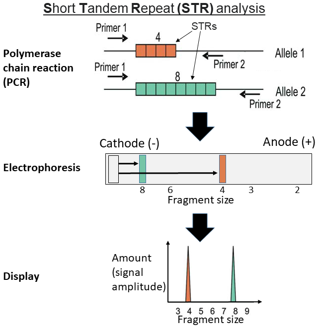
Reading electropherograms and gels
An electropherogram is the printout of capillary electrophoresis. The x-axis is fragment size in base pairs (small on the left), the y-axis is signal strength in relative fluorescence units (RFU), and each peak is an allele. Loci are shown in labeled boxes above the trace, grouped by dye color, and each peak is tagged with its allele number. Read the profile locus by locus:
- One peak at a locus: homozygous (the same allele from both parents); the peak is usually about twice as tall as each peak of a heterozygote.
- Two peaks: heterozygous.
- More than two peaks at several loci: a mixture of DNA from two or more people.
- A small peak one repeat shorter than a real allele is usually a stutter artifact of PCR, not an allele.
- A missing allele or locus (dropout) usually means too little or degraded DNA.

Matching. A suspect is included (consistent with the evidence) only if the alleles agree at every locus shown; one clear mismatch at any locus excludes that suspect (assuming a clean single-source sample). On a gel, compare band positions lane by lane: a match has every band at the same distance from the wells. Identical twins have the same STR profile.
Paternity and kinship. At every locus a child has one allele from the mother and one from the father. Find the allele the child must have received from the mother; the other one (the obligate paternal allele) must be present in the true father.
Mitochondrial and Y-chromosome DNA
| Feature | Nuclear STRs | Mitochondrial DNA (mtDNA) | Y-STRs |
|---|---|---|---|
| Inherited from | Both parents (half each) | Mother only, to all her children | Father to son only |
| Copies per cell | 2 (one set from each parent) | Hundreds to thousands | 1 |
| Discrimination | Individual | Shared by all maternal relatives; class-like | Shared by all paternal male relatives |
| Best use | Most evidence | Hair shafts without roots, old bones and teeth, degraded remains, maternal relatives as references | Sexual-assault mixtures (detects the male DNA among much female DNA), male lineage, missing persons |
mtDNA is a small circular molecule (about 16,569 base pairs) in the mitochondria. Because each cell holds many copies, it often survives when nuclear DNA does not. Analysts usually sequence its two hypervariable regions in the control region rather than counting repeats. Mitochondrial DNA also identifies species. Sequencing a gene such as cytochrome b, or testing it with species-specific PCR, shows whether DNA is human or animal and which animal it came from, because the gene differs between species but varies little within one.
DNA sources, collection and contamination
DNA is in every cell that has a nucleus, plus mitochondria.
| Source | DNA available | Notes |
|---|---|---|
| Blood | Nuclear DNA from white blood cells | Mature red blood cells have no nucleus, so they carry no nuclear DNA. Liquid blood is kept with EDTA (purple-top tube), which also protects DNA |
| Saliva, cheek swabs | Nuclear, from shed epithelial (cheek) cells | Reference samples are taken with a buccal swab; cigarette butts, cups, envelope flaps, bite marks |
| Semen | Nuclear, from sperm (and other cells) | Differential extraction separates sperm from victim cells |
| Hair with a root (follicle, follicular tag) | Nuclear | Pulled hairs often carry roots; shed (telogen) hairs often do not |
| Hair shaft only | Mitochondrial only | See the hair section for hair anatomy |
| Skin cells (touch DNA) | Nuclear, small amounts | Handled objects, steering wheels, clothing collars |
| Bone, teeth | Nuclear and mitochondrial | Best source for old or burned remains; tooth pulp is protected |
| Urine, feces, sweat | Small, variable | Few cells |
Extracting DNA. Before PCR the DNA must be freed and cleaned: (1) lyse cells and nuclear membranes with detergent (and heat); (2) remove proteins, including histones and DNA-cutting nucleases, with proteinase K or by salt or phenol–chloroform extraction; (3) precipitate the DNA with cold ethanol or isopropanol, or bind it to silica (forensic labs use Chelex or silica-column kits); (4) quantify it so the right amount goes into PCR. In the classroom strawberry extraction, dish soap breaks the membranes, salt neutralizes the phosphate charges so the strands clump, and cold alcohol brings the DNA out of solution as white threads.
Degradation, contamination and custody. Heat, moisture, sunlight (UV) and bacteria break DNA into fragments, which is why wet biological evidence is air-dried and stored in paper, not plastic (plastic traps moisture and promotes mold and bacteria), and kept cool. Collectors wear gloves, masks and fresh tools for each item, package items separately, and collect elimination samples from investigators and other people with legitimate access. Labs run negative controls (blanks) to catch contamination. A documented chain of custody records every hand the evidence passed through.
History. Alec Jeffreys discovered DNA fingerprinting (today usually called DNA profiling or DNA typing) (RFLP of repeated sequences) at the University of Leicester in 1984. It was first used in an immigration case in 1985, then in the Narborough murders: in 1986 it exonerated Richard Buckland, the first suspect, and after a mass screening of local men, Colin Pitchfork was convicted in 1988. Robert Melias (England) and Tommy Lee Andrews (Florida) were convicted with DNA evidence in 1987, and commercial DNA testing began that year. PCR-based STR typing replaced RFLP in the 1990s; the UK Forensic Science Service began STR casework in 1994 with a four-locus "quadruplex", and the UK National DNA Database, the world's first, opened in 1995 using STRs; CODIS began as a pilot in 1990 and the national index in 1998.
Blood spatter
Bloodstain pattern analysis (BPA) reads the physics of how blood left the body and reached a surface. Blood is a fairly viscous liquid held together by surface tension (cohesion), which pulls a falling drop into a sphere, not a teardrop. A drop does not break up in free fall unless something strikes it. A typical free-falling drop has a volume often quoted near 0.05 mL, although the volume really depends on what it drips from. Blood is also class evidence (it can be typed) and DNA evidence, so photograph patterns before sampling.
Bloodstain terminology
Modern texts group patterns as passive, transfer, spatter (projected) and altered stains, and the SWGSTAIN terminology (now carried on by OSAC and the ASB standards) prefers mechanism-based terms such as "impact pattern" (impact spatter) over the older low/medium/high velocity labels. Tests use both vocabularies.
ReferenceBloodstain pattern terms· 21 rows
No rows match.
| Term | Group | What it is | What it tells you |
|---|---|---|---|
| Passive drop (drip stain) | Passive | A drop falling under gravity alone | Round on a flat smooth surface; a source that was still bleeding |
| Drip trail | Passive | A line of drops from a moving source | Path of a bleeding person or a dripping weapon; elongation shows direction of movement |
| Drip pattern (blood into blood) | Passive | Drops falling into a pool or wet stain, with small satellite stains around | A source that stayed in one place and kept dripping |
| Pool | Passive | Blood collecting on a level surface | A victim remained there; volume and drying hint at time |
| Flow | Passive | Blood moving under gravity along a surface | Direction shows the slope or the body's position when it flowed; changes in direction show the body was moved |
| Saturation | Passive | Blood soaked into absorbent material | Contact with a bleeding source |
| Transfer (contact) stain | Transfer | Made when a bloody object touches a clean surface | Can preserve the object's shape: handprint, shoe print, hair swipe, weapon outline |
| Swipe | Transfer | Blood carried onto a clean surface by a moving bloody object | Feathered edge toward the end of the motion |
| Wipe | Transfer | An object moving through an existing wet stain, altering it | Shows someone moved through the scene after the stain formed |
| Cast-off | Spatter | Drops flung from a moving bloody object (bat, hammer, fist) in arcs or lines | Minimum number of blows = number of cast-off trails + 1 (the first blow only opens the wound); also direction of swing, which hand, ceiling height |
| Arterial spurt / gush | Spatter (projected) | Blood pumped from a breached artery under pressure | Large stains in arcs or zigzags rising and falling with the heartbeat; the victim was alive |
| Expirated (expired) | Spatter | Blood blown from the nose, mouth or airways | Small stains, often with air bubbles (vacuoles, "bubble rings") and diluted by saliva; may look like high-velocity mist |
| Impact spatter | Spatter | Blood broken into drops by a force striking the blood source | Size of drops relates to the force; distribution points back to the source |
| Forward spatter | Spatter | Spatter traveling in the same direction as the force (for a gunshot, out of the exit wound) | Typically finer and more abundant than back spatter |
| Back spatter (blowback) | Spatter | Spatter traveling back toward the force (from the entrance wound) | Can land on the shooter, the gun, or inside the barrel (blowback) |
| Satellite stain (secondary spatter) | Spatter | Small stains around a parent stain, from droplets that break off on impact | Common on rough surfaces and from drops falling from greater heights |
| Void | Altered | A blank area in a pattern where an object or person blocked the blood | Shows something or someone was present and later moved |
| Skeletonization | Altered | The center of a partly dried stain is wiped or flakes away, leaving its dried outer rim | The stain had started drying before it was disturbed (edges dry first) |
| Clot / serum separation | Altered | Blood clots within minutes; the clot later retracts and squeezes out yellowish serum | A clotted stain with a serum ring shows time passed between bleeding and the stain's disturbance |
| Diluted | Altered | Blood mixed with water or other fluid | Cleanup attempt, rain |
| Insect stains | Altered | Spots made by flies feeding on blood | Can mimic spatter; no consistent direction |
Velocity and drop size
The classic categories describe the speed of the force or object that struck the blood source, not the speed of the drops. More energy breaks the blood into smaller drops.
| Category (old name) | Force speed (commonly cited) | Typical stain diameter | Typical sources |
|---|---|---|---|
| Low velocity | up to about 1.5 m/s (5 ft/s) | about 4 mm and larger (some sources say 3 mm) | Gravity drips, drip trails, pools, transfer |
| Medium velocity | about 1.5–7.5 m/s (5–25 ft/s) | about 1–4 mm | Blunt force (beatings with a bat or fist), stabbing, often cast-off |
| High velocity | about 30 m/s (100 ft/s) and above | under 1 mm; a fine mist | Gunshots, explosions, high-speed machinery; expirated blood can also be fine |
Cutoffs differ slightly between textbooks; use any the Supervisor gives. A single small stain does not prove a gunshot: you need a pattern of many tiny stains, the context and other evidence.
What a drop does on impact. A drop hitting a surface goes through four stages: contact and collapse (the bottom flattens), displacement (blood is pushed out into a rim), dispersion (the rim may break into spines and satellite droplets) and retraction (surface tension pulls the edges back). How far it gets through those stages depends mainly on the surface.
Surface texture. On a hard, smooth, nonporous surface (glass, tile, polished metal) a drop leaves a round stain with a smooth edge and few satellites. On a rough or porous surface (concrete, wood, carpet, paper towel) the same drop ruptures, leaving spines (pointed rays), scalloped edges and satellite stains. Compare stain sizes only on the same kind of surface.
Drop height. A drop accelerates as it falls until air resistance balances gravity. For a typical drop, this terminal velocity is about 7.5 m/s (25 ft/s). The stain gets larger as the fall height increases, but the growth slows and levels off; most of the change happens in the first meter or two, and above the height at which the drop nears terminal velocity the diameter no longer changes. Height estimates from diameter work only if you test the same blood volume on the same surface, which is why a single stain's height cannot be read from its size. Sources disagree on the exact plateau height.

Angle of impact and direction
The shape of a single drop reflects the angle at which it hit. A drop striking a surface at 90 degrees makes a nearly circular stain. As the angle drops, the stain elongates into an ellipse, and the narrow end is the tail. The tail points in the direction the drop was traveling, so the origin of the spatter lies on the opposite side from the tail. A stain that is almost circular means an angle near 90°; a long, thin stain means a shallow (glancing) angle, below about 30°.
Measure W across the widest part and L along the long axis of the elliptical body only: fit an ellipse to the main stain and leave the tail and any spines out of the length. Since W can never exceed L, W/L is always 1 or less; if a question lists numbers the other way round, the smaller one is the width.
Direction. The tail, spines and satellite stains are on the side the drop was moving toward. The directionality angle is the angle of the long axis on the surface (for example, measured from vertical on a wall); it tells you which way to draw the line back toward the source.
Area of convergence and area of origin
To locate the area of origin, draw a line along the long axis of each elongated stain, extending it backward from the tail toward the origin. In two dimensions, the lines from several stains should converge on a common region. That region on the surface is the area of convergence (2D): it locates the source in the plane of the surface but not its height. The area of origin (3D) adds the height above that point. Analysts speak of an "area" rather than a "point" because measurements and blood flight are imprecise. For the height, measure the distance d from a stain to the point of convergence and use the stain's angle:
d is the distance along the surface from the stain to the area of convergence; θ is the stain's angle of impact; h is the height of the source above the surface. This tangent method assumes straight-line flight, so it overestimates height slightly (real drops curve downward under gravity), and the error grows for distant stains.
Stringing is the physical version: at the scene, strings are run from each stain back along its directionality line, rising at its angle of impact, and the region where they meet is the area of origin. Software and the tangent method do the same thing numerically.

What spatter can reconstruct. Positions of victim and attacker (standing, sitting, lying), number of blows (cast-off trails; the first blow usually makes no spatter because blood is not yet exposed), sequence of events (a stain on top of another is later), movement after injury (drip trails, flows changing direction), whether the scene was cleaned or the body moved (wipes, voids, diluted stains), and whether a victim was alive (arterial patterns).
Blood typing
Blood composition
An adult has roughly 5 liters of blood. Spun in a centrifuge, it separates into:
| Component | Share of blood | Contents and function |
|---|---|---|
| Plasma | about 55% | Straw-yellow (amber) liquid, about 92% water (sources give 90–92%), with proteins (albumin, globulins including antibodies, fibrinogen), salts, glucose, hormones, wastes. Serum is plasma without the clotting proteins (the fluid left after blood clots) |
| Red blood cells (erythrocytes) | about 45% of blood (the hematocrit, roughly 37–52% depending on sex) | Carry O₂ and some CO₂ using hemoglobin. Human RBCs are biconcave discs about 7–8 µm across with no nucleus and no mitochondria when mature, leaving more room for hemoglobin; they live about 120 days |
| White blood cells (leukocytes) | under 1% (with platelets, the thin "buffy coat") | Immune defense (neutrophils, lymphocytes, monocytes, eosinophils, basophils). Nucleated: the source of nuclear DNA in blood |
| Platelets (thrombocytes) | under 1% | Cell fragments without nuclei that start clotting |
By number, the order is not the same as by volume. A microliter of blood holds about 4–6 million red cells, 150,000–400,000 platelets and 4,500–11,000 white cells, so red cells > platelets > white cells.
Clotting needs platelets and a cascade of plasma clotting factors that ends with soluble fibrinogen being turned into a fibrin mesh. Hemophilia is an inherited, X-linked (so mostly male) disorder in which one clotting factor is missing or low (factor VIII in hemophilia A, factor IX in hemophilia B), so blood clots slowly and bleeding lasts longer.
All blood cells are made from hematopoietic stem cells in the red bone marrow (the process is hematopoiesis). Among white cells, neutrophils are the most numerous (about 50–70%), then lymphocytes (about 20–40%), monocytes (about 2–8%), eosinophils (about 1–4%) and basophils (under 1%); the mnemonic "Never Let Monkeys Eat Bananas" gives the order. Neutrophils, eosinophils and basophils are granulocytes (granular leukocytes, with many visible cytoplasmic granules). Lymphocytes and monocytes are agranulocytes. By size, monocytes are the largest white cells (about 12–20 µm) and small lymphocytes the smallest (about 7–10 µm, close to a red cell). Hemoglobin is a protein of four globin chains, each holding a heme group with an iron ion (Fe²⁺) at its center that binds O₂; iron deficiency causes anemia. Heme is also what presumptive blood tests detect. The study of blood is hematology; the study of serum and other body fluids, including antigen-antibody tests, is serology.
Anticoagulants. Liquid blood clots within minutes outside the body. Samples for typing or DNA are collected into tubes with an anticoagulant: EDTA (purple or lavender top; it binds the calcium ions clotting needs and is preferred for DNA) or sodium citrate (light blue top; also binds calcium). Heparin (green top) works differently and can inhibit PCR.
ABO and Rh antigens
ABO and Rh are the two most important of many blood group systems: the International Society of Blood Transfusion recognizes more than 40 systems (for example Kell, Duffy, Kidd and MNS), but routine typing and this event use only ABO and Rh. Anti-A and anti-B antibodies are not present at birth: a newborn makes its own only from about 3–6 months of age, apparently in response to A- and B-like sugars on gut bacteria and food, so newborns are usually forward-typed only (any anti-A or anti-B found is the mother's IgG that crossed the placenta).
Red cells carry surface antigens, sugar chains or proteins that the immune system can recognize. Antibodies are Y-shaped proteins in the plasma that bind a specific antigen. When antibodies meet their antigen on red cells, each antibody can link two cells, so the cells clump together: agglutination. In a transfusion, agglutination and the destruction of red cells (hemolysis) can be fatal.
The ABO system was discovered by Karl Landsteiner in 1901 (Nobel Prize in Physiology or Medicine, 1930). The A and B antigens are the same base sugar chain (the H antigen) with one extra sugar added: N-acetylgalactosamine for A, galactose for B; type O cells carry the unmodified H antigen. People naturally make antibodies against the A and B antigens they lack, early in life, without any transfusion.
| Blood type | Antigens on red cells | Antibodies in plasma | Can receive red cells from | Can give red cells to |
|---|---|---|---|---|
| A | A | Anti-B | A, O | A, AB |
| B | B | Anti-A | B, O | B, AB |
| AB | A and B | None | A, B, AB, O (universal recipient) | AB |
| O | Neither | Anti-A and anti-B | O only | A, B, AB, O (universal donor) |

The Rh system. Rh stands for rhesus: Landsteiner and Alexander Wiener described the factor in 1940 after antibodies raised against rhesus macaque red cells also reacted with most human red cells. Clinically "Rh-positive" means the red cells carry the D antigen; Rh-negative people lack it. Unlike anti-A and anti-B, anti-D antibodies are not present naturally: an Rh− person makes them only after exposure to Rh+ blood (a transfusion or pregnancy).
Hemolytic disease of the newborn (HDN; erythroblastosis fetalis). An Rh− mother carrying an Rh+ baby can be exposed to fetal blood at delivery and become sensitized. The first Rh+ baby is usually unaffected; in a later Rh+ pregnancy, her anti-D (IgG) crosses the placenta and destroys fetal red cells, causing anemia and jaundice, an enlarged liver and spleen and, in severe cases, swelling of the whole body (hydrops fetalis). An injection of anti-D immune globulin (RhoGAM) during and after pregnancy prevents sensitization. ABO incompatibility between mother and baby also occurs but is usually milder.
Transfusion compatibility. For red cells, the donor's antigens must not meet the recipient's antibodies. Rh− recipients should get Rh− blood; Rh+ recipients can get either.
ReferenceRed-cell transfusion compatibility· 8 rows
✔ = compatible donor (row) for the recipient (column).
No rows match.
| Donor ↓ / Recipient → | O− | O+ | A− | A+ | B− | B+ | AB− | AB+ |
|---|---|---|---|---|---|---|---|---|
| O− | ✔ | ✔ | ✔ | ✔ | ✔ | ✔ | ✔ | ✔ |
| O+ | — | ✔ | — | ✔ | — | ✔ | — | ✔ |
| A− | — | — | ✔ | ✔ | — | — | ✔ | ✔ |
| A+ | — | — | — | ✔ | — | — | — | ✔ |
| B− | — | — | — | — | ✔ | ✔ | ✔ | ✔ |
| B+ | — | — | — | — | — | ✔ | — | ✔ |
| AB− | — | — | — | — | — | — | ✔ | ✔ |
| AB+ | — | — | — | — | — | — | — | ✔ |
So O− donates to everyone and receives only O−; AB+ receives from everyone and donates only to AB+. For plasma transfusions the rule reverses (AB plasma has no anti-A or anti-B, so AB is the universal plasma donor). People with the very rare Bombay phenotype (hh) make no H antigen, so they have anti-A, anti-B and anti-H and can receive only Bombay blood, although they type as O on a standard card.
US frequencies (Stanford Blood Center figures; other US sources differ by a point or two):
| Type | O+ | A+ | B+ | O− | A− | AB+ | B− | AB− |
|---|---|---|---|---|---|---|---|---|
| Share of US population | 37.4% | 35.7% | 8.5% | 6.6% | 6.3% | 3.4% | 1.5% | 0.6% |
O+ is the most common type and AB− the rarest of the eight; about 85% of people are Rh+. Frequencies differ between populations (type B is more common in parts of Asia, for example). "Rh-null" blood, which lacks all Rh antigens, is extremely rare.
Typing with antisera
Forward (cell) typing mixes drops of the sample with antisera: anti-A, anti-B and anti-D (anti-Rh). Agglutination (visible clumping, a grainy or curdled look) means the cells carry that antigen; a smooth, uniform mixture means they do not. A control well (the sample with no antibody, or with the reagent's diluent) must stay smooth; if the control clumps, the test is invalid. Typing cards and slides label the wells anti-A (often blue reagent), anti-B (often yellow), anti-D and control.
| Anti-A | Anti-B | Anti-D | Type |
|---|---|---|---|
| + | − | + | A+ |
| + | − | − | A− |
| − | + | + | B+ |
| − | + | − | B− |
| + | + | + | AB+ |
| + | + | − | AB− |
| − | − | + | O+ |
| − | − | − | O− |
Reverse (back) typing checks the result from the other side: the sample's serum or plasma is mixed with known A cells and B cells. Type O plasma clumps both (it has anti-A and anti-B); type A plasma clumps B cells; type B plasma clumps A cells; type AB plasma clumps neither. Forward and reverse results must agree.

Blood type inheritance
The ABO gene has three common alleles: Iᴬ and Iᴮ are codominant (both are expressed, giving AB), and i (O) is recessive to both. The RhD gene has a dominant D (Rh+) and a recessive d (Rh−).
| Phenotype | Possible genotypes |
|---|---|
| A | IᴬIᴬ, Iᴬi |
| B | IᴮIᴮ, Iᴮi |
| AB | IᴬIᴮ |
| O | ii |
| Rh+ | DD, Dd |
| Rh− | dd |
The ABO antigens are also present in other body fluids (saliva, semen) in about 80% of people, called secretors; this let early forensic serologists type stains other than blood.
Presumptive and confirmatory blood tests
Before typing, an examiner must show a stain is blood, and then that it is human. Presumptive tests are fast, sensitive screening tests: a negative result means blood is very likely absent, but a positive result only means "possibly blood". Confirmatory tests prove blood is present.
Most presumptive tests rely on the peroxidase-like activity of heme: the iron in hemoglobin catalyzes the oxidation of a colorless reagent by hydrogen peroxide (H₂O₂), producing a color or light. Anything else that catalyzes the same reaction gives a false positive: plant peroxidases (horseradish, potato, other vegetables), and chemical oxidants or metal salts (copper and nickel salts, bleach) that oxidize the reagent even without blood.
Kastle-Meyer (KM) test. The reagent is phenolphthalin, the colorless reduced form of the indicator phenolphthalein, kept in base (with zinc to keep it reduced). Procedure: rub the stain with a moistened swab; add a drop of ethanol (exposes the heme and improves sensitivity); add KM reagent and wait: if it turns pink before peroxide is added, an oxidant is present and the test is invalid; then add hydrogen peroxide. An immediate bright pink means heme catalyzed the oxidation of phenolphthalin to phenolphthalein, which is pink in base:
The reagent is oxidized, not reduced. KM is presumptive, does not distinguish human from animal blood, and does not destroy the sample's DNA.
ReferenceBlood tests· 11 rows
No rows match.
| Test | Type | Reagent and principle | Positive result | Notes |
|---|---|---|---|---|
| Kastle-Meyer | Presumptive (color) | Phenolphthalin + H₂O₂, oxidized by heme | Pink | Two-step method screens out oxidants |
| Leucomalachite green (LMG) | Presumptive (color) | Colorless leucomalachite green + H₂O₂ in acid | Blue-green | Also used to enhance bloody prints |
| Tetramethylbenzidine (TMB); Hemastix strips | Presumptive (color) | TMB + peroxide (Hemastix are TMB-impregnated strips made for urine tests) | Green to blue-green | Quick field screening |
| o-Tolidine, benzidine | Presumptive (color) | Older benzidine-type reagents | Blue | Benzidine dropped as a carcinogen |
| Luminol | Presumptive (light) | Luminol (C₈H₇N₃O₂) + oxidant in base; iron in heme catalyzes the oxidation, and the excited product emits light (chemiluminescence) | Blue glow in a dark room | Detects very dilute, old or cleaned-up blood; sprayed over large areas; bleach, copper and horseradish give false positives. BlueStar is a brighter luminol formulation |
| Fluorescein | Presumptive (fluorescence) | Reduced fluorescein oxidized by heme + H₂O₂ | Fluoresces under an alternate light source | Alternative to luminol |
| H₂O₂ alone | Crude screen | Catalase in blood breaks down H₂O₂ to water and O₂ | Foaming | Very unspecific (many cells have catalase) |
| Teichmann (hemin crystal) test | Confirmatory (microcrystal) | Dried stain heated with glacial acetic acid and a halide salt (NaCl) | Brown rhomboid hemin (hematin chloride) crystals | Ludwig Teichmann, 1853 |
| Takayama (hemochromogen) test | Confirmatory (microcrystal) | Pyridine and glucose in base, gently heated | Pink-red feathery or needle pyridine hemochromogen crystals | Takayama, 1912 |
| Precipitin test | Species of origin | Antiserum against human proteins (raised in an animal) mixed with the stain extract | A line or ring of precipitate where antibody meets human protein | Paul Uhlenhuth, 1901; antisera for other species identify animal blood |
| Immunochromatographic strips (e.g. for human hemoglobin) | Confirms human blood | Antibodies to human hemoglobin on a test strip | A line, like a pregnancy test | Modern routine confirmation |
Order of work. Document and photograph the stain, use presumptive tests on a small portion (luminol is sprayed only after photographs of visible stains), confirm blood, confirm human origin, then type it (ABO or, today, DNA). Luminol and some color tests can dilute or affect later tests, so a sample is set aside for DNA first when possible.
Blood cells by animal class
The rules ask you to sort a slide or picture as human, other mammal, bird, or reptile/amphibian. Two features do almost all the work: whether the red cells have a nucleus, and their size compared with a scale bar or with human cells. Check a stained smear under low power, then high power; the nucleus stains dark purple.
| Group | Shape | Nucleus | Approximate length | Notes |
|---|---|---|---|---|
| Human | Round, biconcave disc (pale center) | No | about 7–8 µm | Compare other cells against this size |
| Other mammals | Round, biconcave | No | about 3–9 µm (e.g. dog ≈ 7 µm, goat ≈ 3–4 µm) | Human versus other mammal is mainly size; camels, llamas and alpacas have oval red cells but still no nucleus |
| Birds | Oval (elliptical) | Yes, oval and central | about 11–16 µm | Thrombocytes are also nucleated |
| Reptiles | Oval | Yes | about 12–23 µm | Often larger than bird cells, though many lizards overlap the bird range |
| Amphibians | Oval | Yes | about 20–25 µm in frogs; some salamanders up to about 70 µm | Largest red cells of all vertebrates |

Why mammals lose the nucleus. Mammalian red cells eject their nucleus (and lose mitochondria) as they mature in the bone marrow. This leaves more room for hemoglobin, keeps the cells small and flexible enough to squeeze through capillaries, and stops them using the oxygen they carry; the cost is that they cannot repair themselves and live only about 120 days. Birds, reptiles and amphibians keep the nucleus. Fish red cells are also oval and nucleated, though fish are not one of the rules' categories.
Reptiles and amphibians are ectotherms ("cold-blooded"). Their metabolism makes little body heat, so they control body temperature with external heat: they bask in the sun or move to warmer or cooler places. Birds and mammals are endotherms that make their own heat.
Glass and refractive index
What glass is. Glass is a hard, brittle, amorphous solid: its atoms are disordered like a liquid's, so it breaks with smooth, curved conchoidal surfaces instead of along cleavage planes. Most glass is about 70% or more silica (sand) melted with fluxes that lower the melting point. In soda-lime glass, soda (Na₂O) is the flux, but silica melted with soda alone gives water-soluble "water glass" (sodium silicate). Lime (CaO) is the stabilizer: it makes the glass insoluble, harder and chemically durable. The table lists the types tests ask about.
| Type | Composition and treatment | Refractive index (approx.) | Density (g/cm³, approx.) | Breaks into | Typical uses |
|---|---|---|---|---|---|
| Soda-lime | SiO₂ with Na₂O (from soda ash, Na₂CO₃) and CaO (from limestone) | 1.51–1.52 | 2.5 | Sharp shards with radial and concentric cracks | Windows, bottles, jars (most common glass) |
| Borosilicate | SiO₂ with boron oxide (B₂O₃); low thermal expansion | ≈ 1.47 | ≈ 2.2–2.3 | Shards | Lab glassware, Pyrex-type ovenware, heat-resistant cookware |
| Lead ("crystal", "flint") | SiO₂ with lead(II) oxide (PbO) | ≈ 1.54–1.7 (rises with lead content) | ≈ 3.0 and up | Shards | Decorative crystal, optical glass |
| Fused silica (quartz glass) | Nearly pure SiO₂ | ≈ 1.46 | ≈ 2.2 | Shards | High-temperature and UV optics, lab ware |
| Tempered (toughened) | Soda-lime glass heated and quickly cooled so the surface is in compression | as soda-lime | as soda-lime | Small, blunt cube-like pieces ("dicing"); the whole pane fails at once | Car side and rear windows, shower doors, glass tables |
| Laminated | Two sheets of glass bonded to a plastic interlayer (usually polyvinyl butyral, PVB) | as its glass | — | Cracks in a spider-web pattern but the pieces stay stuck to the plastic | Windshields; with polycarbonate layers, "bullet-resistant" glass |
The refractive index (n) of a material is the ratio of the speed of light in a vacuum to the speed of light in that material. Ordinary soda-lime glass has an index near 1.5. When light crosses a boundary between materials with different indices, it bends according to Snell's law:
Light entering a denser (higher-index) medium slows down and bends toward the normal; light leaving it into a lower-index medium bends away from the normal. The normal is an imaginary line perpendicular to the surface at the point where the ray hits.
Critical angle. When light goes from a higher-index material into a lower-index one, there is an angle of incidence beyond which no light escapes and all of it reflects back (total internal reflection). Setting the refracted angle to 90° gives . For glass (1.52) into air (1.00), θc = arcsin(1/1.52) ≈ 41°.
Brewster's angle. At one angle of incidence, given by (about 57° for air into glass of 1.52), the reflected and refracted rays are exactly 90° apart. The reflected light is then completely linearly polarized, vibrating perpendicular to the plane of incidence (parallel to the surface). The refracted light that Snell's law describes is only partially polarized.
Measuring the index of a fragment. Forensic labs use the immersion method: the fragment is placed in a liquid whose refractive index is known (and, in the lab, changed by heating a calibrated silicone oil on a hot stage, as in automated GRIM instruments) and viewed under a microscope. Common liquids for a classroom version: water and methanol (both about 1.33), ethanol (about 1.36), glycerol and many vegetable oils (about 1.47), and clove oil (about 1.54). A clear borosilicate rod seems to vanish in glycerol or vegetable oil because the indices match, so no light bends at its surface.
The Becke line test uses this bending to estimate an unknown glass fragment's index. A fragment is immersed in a liquid of known index, and the microscope is focused on the edge. A bright halo, the Becke line, appears at the boundary. When you raise the objective (increase the distance between the objective and the slide), the Becke line moves into the material with the higher refractive index. When the fragment's index equals the liquid's, the edge disappears. Many glasses share an index, so this narrows sources without individualizing.
Density is the other quick physical comparison. Fragments are dropped into a liquid mixture adjusted until a known glass just suspends; a questioned fragment that sinks is denser, one that floats is less dense. Borosilicate (about 2.2–2.3 g/cm³) is noticeably lighter than soda-lime (about 2.5), and lead glass is much heavier. Elemental analysis (for example by mass spectrometry or X-ray fluorescence) can discriminate further.

Ordering the breaks. Rules ask which holes or fractures came first. Two ideas settle most questions. First, a crack stops when it runs into an existing crack, so a crack that terminates at another crack is the later of the two; the earlier crack is the one that runs through uninterrupted. Second, for a single impact, radial cracks (spokes from the impact point) form first and concentric cracks (rings) form afterward. Radial cracks start on the face of the glass opposite the impact, where the glass is stretched, and concentric cracks start on the impact face. A bullet hole is also a clue: the hole is usually wider, with a cone-shaped chip, on the side the bullet exited. With several holes, line them up by asking whose radial cracks stop at whose.
Why the cracks form where they do. A push on a pane bows it outward. The far (reverse) side is stretched (tension), and glass fails in tension, so radial cracks start on the reverse side and run outward from the impact. As the push continues, the glass around the impact is bent the other way, putting the impact side in tension along rings, so concentric cracks start on the impact side. Looking at the broken edge of a radial crack, you see curved stress lines (rib marks, also called conchoidal lines or Wallner lines; the fine feathery marks running across them are hackle marks). The 3R rule: on a Radial crack, the stress lines are at Right angles to the Reverse side (the side away from the force). On a concentric crack the rule flips: the lines meet the impact side at right angles. This tells you which side of a window the blow came from even after the pieces are removed. Some answer keys loosely call the concentric cracks "conchoidal fractures". The two crack types are radial and concentric; "conchoidal" describes the curved fracture surfaces and the stress lines on their edges.
Projectiles. A high-speed bullet punches a fairly clean, round hole with a cone-shaped crater that is wider on the exit side, and leaves few cracks. A slower object (a rock or a fist) spreads its force, makes more radial and concentric cracking and may shatter the pane. Tempered glass does not keep a crack pattern: it falls apart into small cubes. Laminated glass keeps its spider-web pattern because the plastic layer holds the pieces.
Glass type is found from the refractive index. Typical values are about 1.47 for borosilicate (laboratory) glass and roughly 1.51 to 1.52 for window and bottle glass, with lead crystal higher. Match your fragment's index to the table you are given, and remember that two glasses with similar indices cannot be told apart by this test alone.
Time of death: entomology and postmortem changes
Entomology questions here estimate the time since death from the insects found on a body. The method rests on two facts: insects colonize a body in a predictable succession, and their development speed depends on temperature. Blowflies (Calliphoridae) are typically the first insects to find a body. They lay eggs in moist openings and wounds, and the eggs hatch into larvae that feed and grow. The larvae pass through several stages before they pupate and emerge as adults. The life-cycle order is egg, larva, pupa, adult.
The time estimated is the postmortem interval (PMI), the time since death. Insect evidence really measures the time since the insects arrived, which is a minimum PMI: the body may have been dead (for example, indoors, wrapped, buried or in cold weather) for some time before flies could reach it.
The blow fly life cycle
Flies undergo complete metamorphosis. A larva grows by molting; each stage between molts is an instar. Blow flies have three larval instars. They are told apart by size and by the slits in the posterior spiracles (breathing openings at the rear): one slit per spiracle in the first instar, two in the second and three in the third. The third instar is the longest larval stage and the main feeding stage: during its feeding phase the larva eats and grows fastest and reaches its maximum length and weight. The third instar feeds, then stops feeding and wanders away from the body to find a place to pupate (the post-feeding or prepupal stage). The pupa forms inside the hardened, darkening skin of the last larva, called the puparium; empty puparia mean adults have already emerged.
Development times vary by species and especially by temperature, so any table is approximate. As a rough guide for a common blow fly in warm weather (about 21–27 °C, 70–80 °F):
| Stage | What you see | Approximate time since eggs were laid |
|---|---|---|
| Eggs | Clusters of tiny white eggs in the eyes, nose, mouth, wounds | Hatch in roughly 8 hours to 1 day |
| 1st instar | Tiny larvae, a few mm | About day 1–2 |
| 2nd instar | Larger larvae, feeding in masses | About day 2–3 |
| 3rd instar (feeding) | Large maggots, about 1–2 cm, in a heat-producing maggot mass | About day 3–5 |
| Post-feeding (prepupa) | Larvae leave the body and wander into soil or under objects | About day 5–8 |
| Pupa | Brown, barrel-shaped puparium, darkening with age; the pupal stage itself usually lasts about a week or more in warm weather, much longer when cool | About day 7–12 or longer |
| Adult emerges | Empty puparia left behind | Roughly 2–3 weeks |
Beetles and other insects arrive later, once the body changes. In rough order, blowflies and flesh flies come while the body is fresh and bloated, other flies and early beetles during active decay, and dermestid beetles and mites when the remains are dry. Flies find a body by smell. Bacteria breaking down the tissue release sulfur compounds (dimethyl disulfide and dimethyl trisulfide) and amines, and the odor of this microbial decay draws blow flies and flesh flies. They then lay eggs or larvae in wounds and natural openings. Because development depends on temperature, the stage of the oldest larvae, combined with the temperature history of the scene, is what estimates the interval. Cold slows development and warm speeds it up, so an estimate must account for temperature. A standard way to use temperature is accumulated degree-hours (ADH): the development a species needs is a fixed total of degree-hours above a base temperature. For illustration only (the numbers are invented), suppose a species needs 1,200 degree-hours above 10 °C to reach pupation and the scene averaged 25 °C. Each hour contributes 15 degree-hours, so the time is 1,200 ÷ 15 = 80 hours. The estimate is a range, and the strongest answers say which evidence sets the minimum and which sets the maximum.
Insects can also carry evidence. A blood-fed mosquito (or other blood-feeding insect) found at a scene holds human blood that can be DNA-typed for a few days after feeding, linking a person to the place; maggots that fed on a body can be tested for drugs or toxins in the tissue they ate.
Insect succession and decomposition
| Decomposition stage | Body condition | Typical insects |
|---|---|---|
| Fresh | From death until bloating; no odor obvious to people | Blow flies (Calliphoridae) and flesh flies (Sarcophagidae) arrive and lay eggs or larvae |
| Bloat | Gut bacteria produce gas; body swells; odor begins | Flies peak; predatory beetles start to arrive |
| Active decay | Skin breaks, gases escape, maggot masses feed heavily, strong odor, body mass falls fast | Large maggot masses; rove beetles (Staphylinidae), clown beetles (Histeridae) and carrion beetles (Silphidae) prey on maggots |
| Advanced decay | Most soft tissue gone; maggots leave to pupate | Fewer flies; beetles increase; cheese skippers (Piophilidae) on fermenting remains |
| Dry or skeletal | Bones, cartilage, dried skin and hair; little soft tissue is left, so insect activity drops sharply | Hide and skin beetles (Dermestidae, Trogidae), clothes moths (Tineidae, which eat hair), mites (Acari) |
The order of arrival tested most often: Calliphoridae → Sarcophagidae → Coleoptera (beetles) → Lepidoptera (moths) and Acari (mites). Coffin flies (scuttle flies, family Phoridae) are tiny flies found in the active and advanced decay stages. They can squeeze through small gaps and burrow into soil, so they are often the only flies on bodies that are sealed indoors or buried, even inside coffins. Insects found on a body can also show that it was moved (species from a different habitat) or that an area was a wound (eggs laid away from natural openings early on). Decomposition itself starts at death. Cells run out of oxygen and their own enzymes begin to digest them (autolysis) long before bloating, so even the first flies arrive on a body that is already decomposing.
The rules frame this topic around the insects, so the insect evidence should carry your estimate. Body changes such as livor mortis (settled blood), algor mortis (cooling) and rigor mortis (stiffening) only help in the first day or so, and only if the scenario gives them; use them as a cross-check, not as the main method. After about 72 hours these body changes are of little use, so insect evidence is often described as the most accurate (and sometimes the only) way to estimate the postmortem interval beyond three days. Insects can also be used earlier; 72 hours is when they become the best method, not when they start to work.
Postmortem changes
| Change | What happens | Commonly cited timing (varies with temperature, body size and activity before death) |
|---|---|---|
| Pallor mortis | Paleness as circulation stops | Within about 15 minutes to 2 hours |
| Algor mortis | The body cools toward the surrounding temperature | Roughly 1.5 °F (about 0.8 °C) per hour at first, under average conditions |
| Livor mortis (lividity, hypostasis) | Blood settles by gravity into the lowest parts, giving purple-red discoloration; pressure points stay pale | Visible after about 30 minutes to 2 hours; becomes fixed (no longer blanches when pressed) after about 8–12 hours |
| Rigor mortis | Muscles stiffen because ATP runs out and actin–myosin cross-bridges stay locked | Begins about 2–6 hours after death, in small muscles (eyelids, jaw) first; full at about 12 hours; passes off over about 24–48 hours (36 hours is a common figure) |
| Putrefaction | Bacterial breakdown: green discoloration, bloating, marbling, odor | Begins within days; much faster in heat |
Fixed livor in a pattern that does not match the body's position shows the body was moved after the lividity set. A rough cooling estimate is the Glaister equation: hours since death ≈ (98.7 °F − rectal temperature in °F) ÷ 1.5. For example, a rectal temperature of 89.7 °F gives (98.7 − 89.7) ÷ 1.5 = 6 hours. It assumes a normal starting temperature and average conditions, so treat it as a rough cross-check.
History. The first recorded use of insects in a death investigation is from 13th-century China: the official Song Ci (Sung Tz'u) described a case in which flies gathered on only one of the villagers' sickles, drawn by invisible traces of blood, and its owner confessed. In 1894 the French veterinarian Jean Pierre Mégnin described the successive waves of insects that colonize a corpse in La Faune des Cadavres.
Seeds and pollen
You will see pictures, not live samples: seeds or pollen from the scene beside pictures from each suspect, or from different regions. Compare features in a fixed order. For seeds: overall shape, size against the scale bar, surface texture, and attachments such as wings, hooks or hairs (which also hint at how the seed travels and so where it could have been picked up). For pollen: the number and type of apertures (furrows called colpi, round pores, or both), the outline in polar view, and the wall ornamentation (smooth, spiny, netted). Pollen is produced in huge numbers and clings to clothing and hair, so a match links a suspect to a place or season rather than to the exact spot. State it that way: "the pollen on suspect B's jacket matches the flowering shrub at the scene, consistent with B having been near it."
Palynology is the study of pollen and spores; forensic palynology applies it to legal cases. It is especially well developed in New Zealand, where it has been used routinely in casework.
Pollen grain structure. A pollen grain carries the male gametophyte of a seed plant. Its wall has two layers: the tough outer exine, made of sporopollenin, one of the most decay-resistant biological polymers known (so pollen survives for years and even fossilizes), and the thin inner intine, made of cellulose and pectin. Inside are the living cells (a tube or vegetative cell and a generative cell). So the three main parts of a grain are the exine, the intine and the cytoplasm they enclose. Pollen identification uses:
- Apertures: thin spots where the pollen tube emerges. Long furrows are colpi (for example, three colpi, "tricolpate", typical of eudicots); round holes are pores; a single furrow (monosulcate) is typical of monocots and many gymnosperms.
- Shape and size: spherical, oval, triangular in polar view; most grains are about 10–100 µm.
- Ornamentation of the exine: smooth (psilate), spiny (echinate, for example sunflower family), netted (reticulate), warty (verrucate).
- Air sacs: pines and many other conifers have two balloon-like sacs that help wind carry the grain.
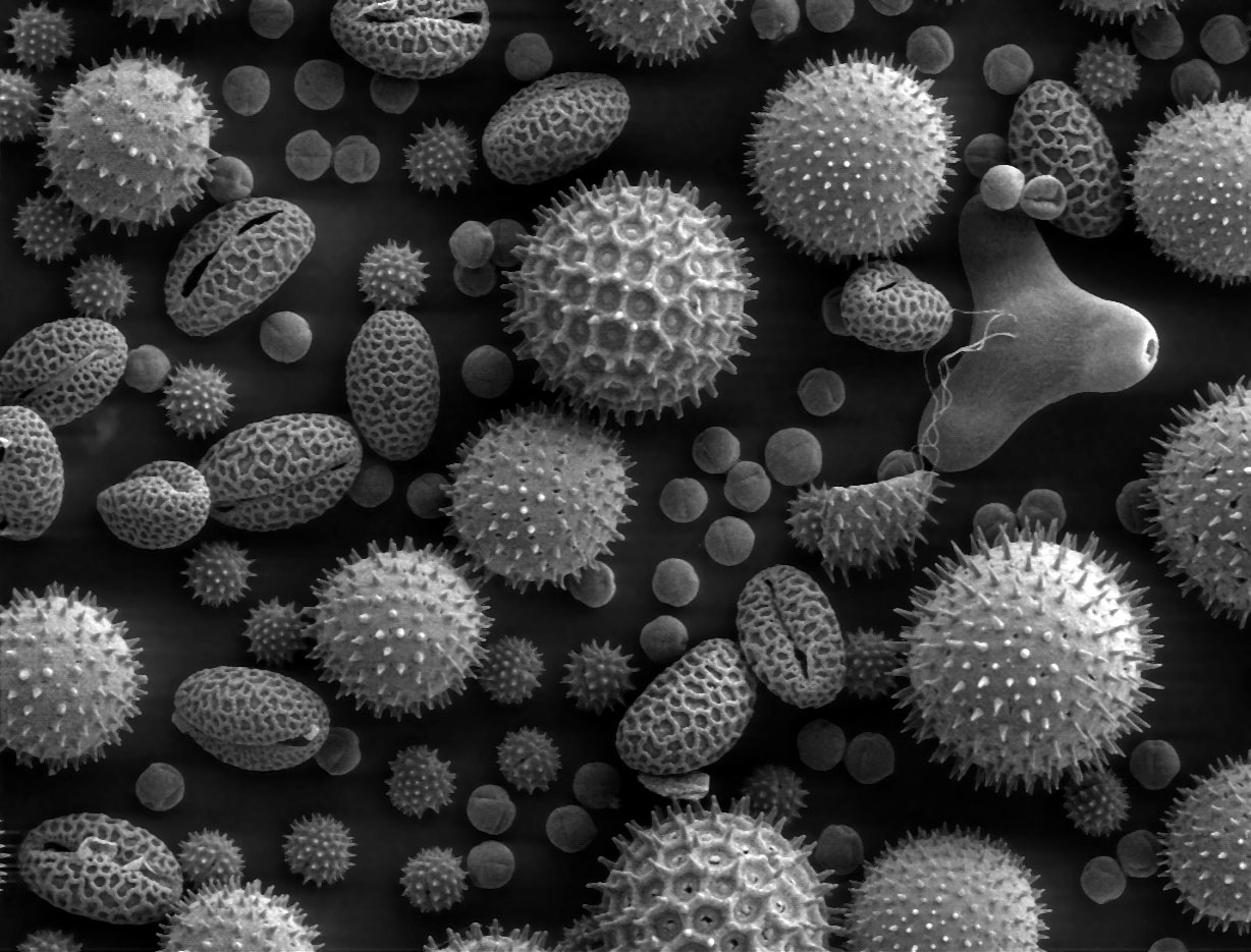
Wind versus insect pollen. Wind-pollinated (anemophilous) plants such as grasses, pines and oaks release enormous numbers of small, light, smooth grains that drift far, so finding them proves little about a specific place. Insect-pollinated (entomophilous) plants make fewer, larger, sticky or spiny grains that travel only on animals. A suspect carrying a lot of insect-pollinated pollen was probably in direct contact with that plant. Pollen also reveals season (what was flowering) and region (plants that grow only in certain areas or countries), which is how questions about "pollen from different country regions" are answered.
Strengths and limits. Pollen is microscopic (needs a microscope), abundant, durable, and easily transferred to clothes, hair, shoes and vehicles. But it requires expert identification and a reference collection, it is time-consuming, wind pollen is widespread, and it is class evidence.
Seeds. A seed has three main parts: the seed coat (testa, protective outer layer), the embryo (young plant: radicle, shoot and one or two cotyledons) and stored food (the endosperm, or food stored in the cotyledons). Angiosperms (flowering plants) enclose seeds in a fruit; gymnosperms (conifers) bear "naked" seeds on cones. Both are seed plants (spermatophytes). Seeds compare by size, shape, color, surface pattern and dispersal structures: wings and plumes for wind (maple, dandelion), hooks and barbs that catch on clothing and fur (burdock, beggar-ticks), fleshy fruits eaten by animals, and buoyant coats for water. Hooked or sticky seeds on a suspect's clothing or a car's tires can tie them to a field. Seeds in stomach contents can show a victim's last meal. Grass seeds such as fescue identify grassy areas.
Tracks and soil
Footprints and tire tracks are matched in two stages. First the class features: outsole or tread design, size, and for tires the tread width. These exclude suspects quickly; a different tread pattern is a clean elimination. Then individual features: cuts, nicks, stones lodged in the tread and uneven wear, which can tie a print to one shoe or tire. Compare the scene impression with the suspect's item at the same scale, and remember a print may be partial.
Impressions: types, recovery and reading
- Two-dimensional (patent or latent) impressions are prints on a hard surface: a muddy or bloody shoe print on a floor, or a dust print on paper. Photograph them, then lift them with an electrostatic dust lifter (a charged film that attracts the dust), gel lifters or adhesive lifters; latent shoe prints can be enhanced with powders or chemicals like fingerprints.
- Three-dimensional (plastic) impressions are depressions in a soft material: soil, sand, snow, mud. Photograph them, then cast them. Dental stone (a hard gypsum product) is the standard casting material today; it replaced plaster of Paris because it is stronger and shows more detail. Snow prints are first sprayed with a wax (snow print wax) so the warm casting mix does not melt them. Standards for examining footwear and tire impressions were written by SWGTREAD, the Scientific Working Group for Shoeprint and Tire Tread Evidence. Its work now continues through NIST's Organization of Scientific Area Committees (OSAC).
- Photography rules: an overall photo showing location, then examination-quality close-ups with a scale in the frame and the camera perpendicular (90°) to the impression, using low-angle (oblique) light to bring out depth.
Reading the trail. A series of footprints (a trackway) shows direction, stride and gait. Separate sets of prints show how many people were present, and prints that lead in from or out to a door or window mark the points of entry and exit. Running gives a longer stride and deeper toe impressions with lighter heels; carrying a heavy load deepens the whole print and shortens the stride; a limp shows uneven stride. Wear patterns show how the wearer walks. Wearing shoes of the wrong size does not change the outsole size in the print, though it can change stride, gait and the pressure pattern. For vehicles, the track width (distance between left and right tires) and the wheelbase (front to rear axle) narrow the vehicle class; tread pattern identifies the tire brand and model; cuts and wear individualize.
Soil composition and texture
Soil is a mixture of mineral particles (sand, silt and clay from weathered rock), organic matter (humus from decayed plants and animals), water, air and living organisms. A textbook "ideal" loam is roughly 45% minerals, 5% organic matter, 25% water and 25% air by volume.
Soil texture is set by the proportions of three particle sizes. The USDA limits are:
| Particle | Diameter (USDA) | Feel when rubbed wet | Properties |
|---|---|---|---|
| Sand | 0.05–2.0 mm | Gritty | Large pores: drains fast, holds little water and few nutrients, well aerated |
| Silt | 0.002–0.05 mm | Smooth, floury | Intermediate |
| Clay | less than 0.002 mm | Sticky and plastic, forms a ribbon | Tiny particles and pores: holds water and nutrients, drains slowly, poorly aerated |
Porosity versus permeability. Porosity is the fraction of a soil that is open space; permeability is how easily water flows through it. Clay has high total porosity (many tiny pores) but very low permeability, because the pores are so small that water clings to the particles and barely moves; sand has less total pore space but large, connected pores, so it drains fast.
Geologists use the slightly different Wentworth scale for sediment: sand 1/16–2 mm (0.0625–2 mm), silt 1/256–1/16 mm (about 0.0039–0.0625 mm) and clay below 1/256 mm. The scales disagree near the sand–silt boundary: a particle 0.053 mm across is very fine sand on the USDA scale but silt on the Wentworth scale.

The soil texture triangle names the twelve USDA texture classes. Milton Whitney, chief of the USDA Bureau of Soils, published the first texture chart in 1911 (Bureau of Soils Bulletin 78), a right-triangle plot of clay against silt. The first equilateral triangle followed in 1927, and the twelve-class version used today dates from 1951. When a test gives percentages without the triangle, these boundary rules reproduce it:
ReferenceUSDA texture classes from percentages· 12 rows
Percentages are of the mineral fraction (sand + silt + clay = 100). Check clay first.
No rows match.
| Class | Rule (USDA) |
|---|---|
| Clay | clay ≥ 40%, sand ≤ 45%, silt < 40% |
| Silty clay | clay ≥ 40%, silt ≥ 40% |
| Sandy clay | clay ≥ 35%, sand > 45% |
| Clay loam | clay 27–40%, sand 20–45% |
| Silty clay loam | clay 27–40%, sand ≤ 20% |
| Sandy clay loam | clay 20–35%, silt < 28%, sand > 45% |
| Loam | clay 7–27%, silt 28–50%, sand ≤ 52% |
| Silt loam | silt ≥ 50% with clay 12–27%, or silt 50–80% with clay < 12% |
| Silt | silt ≥ 80%, clay < 12% |
| Sandy loam | clay 7–20%, sand > 52%, silt + 2 × clay ≥ 30; or clay < 7%, silt < 50%, silt + 2 × clay ≥ 30 |
| Loamy sand | silt + 1.5 × clay ≥ 15 and silt + 2 × clay < 30 |
| Sand | silt + 1.5 × clay < 15 |
Common soil types described in words (tests use these informal names): sandy soil is gritty, light, drains fast and is low in nutrients; silty soil is smooth and fertile but compacts; clay soil is sticky when wet, hard when dry, drains poorly and holds nutrients; loam is a balanced mix of sand, silt and clay with humus, the best for most plants; peaty soil is dark, spongy, very high in organic matter, holds water and is acidic; chalky soil is stony and free-draining over chalk or limestone and is alkaline.
Soil horizons. A vertical cut through soil shows layers (horizons): O (organic litter and humus), A (topsoil, darkened by humus), E (leached, pale zone, not always present), B (subsoil, where clay, iron oxides and other material washed down from above accumulate), C (partly weathered parent material) and R (bedrock). Soil on a shovel or deep footprint can mix horizons.

Color is usually the first comparison, recorded with a Munsell soil color chart (hue, value and chroma) and always compared at the same moisture, because a soil darkens when wet. Dark brown to black means high organic matter; red means iron oxides such as hematite (iron oxidized in well-drained soil); yellow-brown means hydrated iron oxides such as goethite; gray or blue-gray mottles mean waterlogged, oxygen-poor (reducing) conditions; white crusts mean carbonates or salts. A soil that is light gray to white throughout has little organic matter. Pale minerals dominate it: quartz sand (as in a leached E horizon) or carbonates.
pH. Most soils fall between about 4 and 9, and most plants grow best in slightly acidic to neutral soil (about pH 6–7.5). Peat and soils under heavy rainfall and conifer forests are acidic (bog peat is often pH 3–4), because rain leaches away basic cations and decaying organic matter releases acids. Chalky and limestone soils and desert soils are alkaline (basic), because carbonates and salts are not leached away where rainfall is low. Texture does not fix pH by itself, but it changes how fast pH drifts: sandy soils have little clay or humus to hold basic cations, so in rainy climates they are easily leached and often acidic, while clay- and humus-rich soils buffer pH and change it slowly.
Comparing soil as evidence. Compare color (dry and moist), texture and particle-size distribution (sieving), mineral content (with a polarizing microscope) or by X-ray diffraction (XRD), which identifies the crystalline minerals (quartz, feldspars, clays) from their diffraction patterns, density (a density-gradient tube separates particles into bands), pH, organic content, and inclusions such as pollen, seeds, plant fragments and diatoms. Soil is class evidence. Moisture content and organic material change as a sample dries or ages, so compare stable properties (minerals, particle sizes) when samples were collected at different times. In 1904 the German forensic chemist Georg Popp used coal and mineral grains (including hornblende) on a handkerchief left at a murder scene, and soil layers on the suspect's trousers, to link him to the scene, one of the first uses of soil as evidence; in a 1908 case he read the soil layers on a suspect's shoes.
Soil questions give you a composition (color, texture or grain size, mineral content, organic matter, sometimes pH) for the scene and for samples taken from each suspect. Compare row by row. A suspect whose soil differs in a stable property, such as mineral makeup, can be excluded; a match is class evidence that places the suspect in a similar setting. Soil from a shoe can mix layers from several places, so a partial match deserves a cautious sentence, not an exclusion.
Bullet striations
A rifled barrel has spiral lands (raised strips) and grooves cut along it, which spin the bullet and scratch it. First compare class characteristics: caliber, the number of lands and grooves, their widths, and the twist direction (left or right). Any difference excludes that gun. Then compare the fine striations, the tiny scratches from imperfections in the barrel, by lining up the scene bullet beside a bullet test-fired from the suspect gun, as in a comparison microscope image. Matching striation patterns across several lands support the same barrel. On cartridge casings, the firing-pin impression, breech-face marks, and extractor and ejector marks play the same role. Say in the write-up which features were class and which were individual.

How a gun fires. Pulling the trigger releases the hammer or striker, which drives the firing pin into the primer. The primer's shock-sensitive compound explodes and ignites the propellant (modern smokeless powder, mainly nitrocellulose). The hot gas pushes the bullet down the barrel, converting chemical energy into kinetic energy. In a semi-automatic, the extractor then pulls the empty case from the chamber and the ejector flips it out of the gun. Each of these parts can leave marks.
Caliber and gauge. Caliber is the bore diameter of a rifled barrel (or the bullet's diameter), given in hundredths or thousandths of an inch (a .45 caliber is 0.45 inch) or in millimeters (9 mm). Shotguns are described by gauge: the number of lead balls of the bore's diameter that make one pound, so a 12-gauge bore is wider than a 20-gauge. Most shotgun barrels are smooth (no rifling), so shot pellets carry no rifling striations.

Why rifling, and why the marks are unique. The spin makes the bullet gyroscopically stable, so it flies point-first, straighter and farther. The bullet is slightly wider than the bore between the lands, so the lands cut land impressions into it and the grooves leave raised groove impressions. Tool marks left when the barrel was made, and later wear, corrosion and damage, are random at the microscopic level, so they engrave a pattern of fine striations that repeats from shot to shot in the same barrel but differs between barrels, even consecutive barrels from the same tool.
Comparing bullets. A comparison microscope joins two microscopes with an optical bridge so the questioned bullet and a test-fired bullet (fired into a water tank or cotton box) appear side by side in one split view. The examiner rotates them to line up matching striations. The American Calvin Goddard, using the comparison microscope built by his colleague Philip Gravelle, pioneered this method in the 1920s and used it in the 1929 St. Valentine's Day Massacre investigation. Images of bullets and cases are searched in the ATF's NIBIN (National Integrated Ballistic Information Network) using the IBIS imaging system; the NIBIN name dates from 1997, and under a 1999 FBI–ATF agreement the FBI's DRUGFIRE and the ATF's IBIS programs were unified on the IBIS platform (DRUGFIRE was phased out; ATF took full control in 2003).
Gunshot residue (GSR). Firing sprays a cloud of particles from the primer and burned powder onto the shooter's hands, face and clothes, and onto a close target. Primer residue contains lead, barium and antimony, which fuse into characteristic particles identified by scanning electron microscopy with energy-dispersive X-ray analysis (SEM-EDS), the standard confirmatory method. Older tests looked for nitrates and nitrites from the powder: the paraffin (dermal nitrate) test with diphenylamine, now abandoned because fertilizers, tobacco and many other things also contain nitrates, and the Griess test for nitrites, still used on clothing to estimate shooting distance. The sodium rhodizonate test detects lead. Burned and unburned powder grains driven into skin at close range cause stippling (powder tattooing); the size of the pattern estimates muzzle-to-target distance. GSR washes off and transfers easily, so a positive only shows the person fired, handled or was near a fired gun.
Ballistics is divided into internal (inside the gun, from firing pin to muzzle), external (the bullet's flight) and terminal (what happens when it hits).
Crime-scene analysis (Part 3.f)
The written analysis is the largest part of the score, so structure it. A strong write-up has five parts:
- A short summary of the scene: where the body or object was, what was found and what question you are answering.
- An evidence table with one row per evidence item: what it is, what the test showed, whether it is class or individual, and which suspects it affects.
- Suspect-by-suspect reasoning that says why each suspect is kept or excluded. The rules require you to explain why the chosen suspect was selected and why the others were not, so exclusions count as much as inclusions.
- The conclusion, stated with its strength: which suspect the evidence most supports, and what evidence would change the answer.
- Limitations and alternatives: what the evidence cannot rule out, and any alternative explanation the Supervisor's questions raise.
Locard's exchange principle (contact transfers material) is the logic behind it: hair, fibers, soil and blood link a person to a place only as strongly as the evidence is specific.
| Evidence | Class or individual | Reason |
|---|---|---|
| Fingerprint pattern type | Class | Many people share a pattern type |
| Fingerprint minutiae arrangement | Individual when enough detail matches | Arrangements of minutiae are unique enough to individualize, when there is enough of them |
| DNA profile across the loci shown | Individual (except identical twins) | Matching many loci is very specific |
| ABO blood type | Class | Shared by many people |
| Polymer type (for example PETE) | Class | Mass-produced items share a resin |
| Fiber family (cotton, wool, nylon) | Class | Common to many garments |
| Glass refractive index | Class, narrowing the source | Many glasses share a similar index |
| Bullet striations | Individual when the striations match | Scratches from one barrel repeat across bullets |
Never state that a class match identifies a person; say it is "consistent with" the suspect. Build an evidence-by-suspect matrix first, then write one paragraph per suspect from it:
| Evidence | Suspect A | Suspect B | Suspect C |
|---|---|---|---|
| Powder at scene: sodium carbonate | Carbonate in pocket: consistent | No carbonate: neutral | Carbonate in car: consistent |
| Fiber on victim: wool | Wool coat: consistent | Cotton only: neutral | Wool scarf: consistent |
| Shoe print: tread pattern 2 | Pattern 2: consistent | Pattern 1: excluded | Pattern 2, matching cut: individual match |
| Blood drop type B | Type B: consistent | Type O: excluded | Type B: consistent |
Here B is eliminated twice by class evidence, A is consistent but never individually matched, and C is tied to the scene by an individual feature, so the written analysis names C and explains, item by item, why A and B were not chosen.
Kinds of evidence
| Term | Meaning | Example |
|---|---|---|
| Physical evidence | Any object or material that can link a person, place or object to a crime | Glass, soil, fibers, prints, tool marks |
| Biological evidence | Physical evidence from a living thing | Blood, saliva, hair, pollen, insects |
| Trace evidence | Small amounts of material transferred by contact, often microscopic | Fibers, hairs, pollen, glass fragments, soil, GSR |
| Transient evidence | Temporary evidence that is easily changed or lost and must be documented at once | Odors, temperature, impressions in snow, the position of a body, a blood-spatter pattern before it is disturbed |
| Pattern evidence | Evidence read from its shape or arrangement | Spatter, glass fracture patterns, tire and shoe prints |
| Class evidence | Shared by a group; narrows the pool | Blood type, fiber type, tread design, glass index |
| Individual evidence | Can be traced to one source with high certainty | Fingerprint minutiae, a full DNA profile, striations, a fracture match of two broken pieces |
| Direct evidence | Proves a fact without inference | An eyewitness who saw the act, a confession, a video recording of the act |
| Circumstantial (indirect) evidence | Implies a fact through inference | Fingerprints, DNA, fibers, a suspect's motive |
| Known (exemplar, reference) sample | Collected from a known source to compare | A suspect's blood sample, fibers from the suspect's coat |
| Questioned sample | Of unknown origin, from the scene | The fiber found on the victim |
| Control (blank) sample | Material near the evidence that tests the background | Unstained carpet next to a bloodstain |
Probative value is how strongly a piece of evidence helps prove a relevant fact. Common materials (white cotton, blue denim) have low probative value; unusual ones (a rare fiber blend, an unusual pollen) have high value.
Processing the scene
- Secure and protect the scene: first officer, establish a perimeter, keep a log of everyone who enters, preserve transient evidence.
- Survey and document before anything is moved: notes, photographs (overall, mid-range, and close-up with a scale), video and sketches. A rough sketch is drawn on site with measurements taken from fixed points (by triangulation or a baseline); a finished, scaled sketch is drawn later.
- Search using a systematic pattern suited to the scene: line or strip (searchers walk in parallel lines; large outdoor areas), grid (a double strip search at right angles; thorough), spiral (inward or outward from a center; one searcher, open areas), zone or quadrant (divided into sections, for rooms and vehicles) and wheel or ray (outward from a center like spokes, rarely used).
- Collect and package each item separately to prevent cross-contamination. Dry biological evidence goes in paper bags or envelopes (plastic traps moisture and causes mold and bacterial growth that destroy DNA). Fire debris goes in airtight metal cans so volatile accelerants do not evaporate. At a fire scene, accelerant residues are first located with trained accelerant-detection dogs or portable hydrocarbon detectors ("sniffers"); the lab then analyzes the vapors (headspace) above the sealed debris by GC-MS. Trace evidence goes in a folded paper packet (druggist's fold or bindle) inside an envelope. Liquid blood samples get an anticoagulant and are refrigerated.
- Chain of custody: a written record of every person who collected, handled, transferred or analyzed each item, with dates and times, and sealed, initialed packaging. A break in the chain can make evidence inadmissible.
- Submit known, questioned and control samples to the lab.
Locard's exchange principle, from the French criminologist Edmond Locard (who set up an early police laboratory in Lyon in 1910), states that every contact leaves a trace: the criminal takes something from the scene and leaves something behind.
The crime lab and the courtroom
The word forensic comes from the Latin forensis, "of the forum": the public place where Roman legal cases were argued. Forensic science is science applied to legal questions.
A full-service crime laboratory is divided into units: physical science (chemistry, physics and geology: drugs, glass, soil, paint, fibers), biology (blood, body fluids, DNA, hair), firearms (guns, bullets, cartridge cases, shooting reconstruction, tool marks), latent prints (fingerprints), questioned documents, toxicology and photography, plus evidence collection.
Reasoning. Deductive reasoning goes from general rules to a specific conclusion that must follow ("all insects are cold-blooded; flies are insects; so flies are cold-blooded"). Inductive reasoning goes from specific observations to a probable general conclusion. A crime-scene write-up uses both: observations are combined into the most probable explanation, and each suspect is tested against it.
Admissibility. In US courts, scientific evidence was long judged by the Frye standard (1923: the method must be generally accepted in its scientific field). Federal courts and many states now use the Daubert standard (1993), under which the judge weighs whether the method has been tested, peer reviewed, has a known error rate and standards, and is generally accepted.
Forensic history and vocabulary
Tests regularly ask "who" and "what is the study of" questions. These are the names worth knowing.
| Person | Contribution |
|---|---|
| Song Ci (China, 1247) | The Washing Away of Wrongs, an early handbook of death investigation; the first recorded use of insects (flies on a sickle) |
| Mathieu Orfila (1814) | Treatise on poisons; called the father of toxicology |
| Jan Evangelista Purkinje (1823) | Described and classified fingerprint patterns |
| William Herschel (1858–1870s, India) | First to use fingerprints for identification in practice: handprints and fingerprints on contracts and records; noticed that prints persist over time |
| Henry Faulds (1880) | Proposed using fingerprints left at crime scenes to identify criminals |
| Alphonse Bertillon (1879–1880s) | Anthropometry ("bertillonage"): identifying criminals by body measurements; also standardized crime-scene and mug-shot photography. Often called the father of criminal identification |
| Francis Galton (1892) | Finger Prints: first statistical study showing prints are unique and permanent; three basic pattern types (arch, loop, whorl); minutiae ("Galton details"). Often called the father of fingerprinting |
| Juan Vucetich (Argentina, 1891–1892) | Fingerprint classification system; the 1892 Francisca Rojas case was the first murder solved by fingerprints |
| Edward Henry (1897–1901) | Henry classification system, adopted by Scotland Yard and most English-speaking countries |
| Will West case (1903) | Two prisoners at Leavenworth with nearly identical Bertillon measurements but different fingerprints; helped fingerprints replace bertillonage in the US |
| Hans Gross (1893) | Criminal Investigation; coined "criminalistics" and urged scientific methods in police work |
| Karl Landsteiner (1901) | Discovered the ABO blood groups (Nobel Prize 1930); with Wiener, the Rh factor (1940) |
| Leone Lattes (1915) | Method to determine ABO type from dried bloodstains |
| Georg Popp (1904) | Early use of soil and mineral evidence in a murder case |
| Edmond Locard (1910) | Police laboratory in Lyon; exchange principle |
| August Vollmer (1920s) | Police chief who set up the first US police crime laboratory (Los Angeles, 1923) |
| Calvin Goddard (1920s) | Comparison microscope for firearms identification (instrument built by Philip Gravelle, 1925) |
| Mikhail Tsvet (1900s) | Invented chromatography |
| J. J. Thomson and F. W. Aston (1910s) | First mass spectrometer; mass spectrograph and isotopes |
| James Watson, Francis Crick, Rosalind Franklin (1953) | Structure of DNA (Franklin's X-ray images were key) |
| Alec Jeffreys (1984) | DNA fingerprinting; cleared Richard Buckland (1986) and convicted Colin Pitchfork (1988), the first murder conviction based on DNA |
| Kary Mullis (1983) | Invented PCR (Nobel Prize in Chemistry 1993) |
| Term | Field |
|---|---|
| Criminalistics | Examination of physical evidence in a crime lab |
| Dactyloscopy | Fingerprint identification |
| Serology | Blood and other body fluids (antigens and antibodies) |
| Hematology | Study of blood |
| Toxicology | Poisons and drugs in the body |
| Forensic entomology | Insects in legal investigations |
| Forensic palynology | Pollen and spores as evidence |
| Forensic botany | Plants as evidence (seeds, leaves, wood, pollen) |
| Trichology | Hair and scalp |
| Ballistics | Projectiles and firearms |
| Forensic pathology | Cause and manner of death (autopsy) |
| Forensic anthropology | Skeletal remains |
| Forensic odontology | Teeth and bite marks |
| Abbreviation | Stands for |
|---|---|
| PMI | Postmortem interval |
| ADD / ADH | Accumulated degree days / hours |
| AFIS / IAFIS / NGI | Automated Fingerprint Identification System / the FBI's Integrated AFIS / Next Generation Identification (IAFIS's successor) |
| ACE-V | Analysis, Comparison, Evaluation, Verification |
| CODIS | Combined DNA Index System |
| STR / PCR / RFLP | Short tandem repeat / polymerase chain reaction / restriction fragment length polymorphism |
| NIBIN / IBIS | National Integrated Ballistic Information Network / Integrated Ballistic Identification System |
| GSR | Gunshot residue |
| GC-MS / TLC / HPLC | Gas chromatography–mass spectrometry / thin-layer chromatography / high-performance liquid chromatography |
| FTIR / SEM | Fourier-transform infrared spectroscopy / scanning electron microscope |
| RI / MI / Rf | Refractive index / medullary index / retention factor |
| RIC | Resin identification code (the recycling number on plastics) |
Common pitfalls
Sucrose gives a negative Benedict's test
Sucrose is non-reducing and turns positive only after acid hydrolysis. Do not rule out a sugar on one negative result.
Sodium hides potassium
A yellow flame can mask lilac. Check through cobalt glass.
A faint yellow flame is usually contamination
Trace sodium is on almost everything. A sodium salt gives an intense, lasting yellow; confirm with pH, HCl and solubility.
Use the water result, not the name
PETE, PVC, PMMA and polycarbonate sink; HDPE, LDPE and PP float; non-expanded polystyrene sinks only just. "High-density" describes packing, not buoyancy.
Boric acid is monoprotic
It is a weak Lewis acid that accepts OH⁻; it does not give up three protons. One mole of NaOH neutralizes one mole of H₃BO₃.
Measure Rf from the origin line
Both distances run from the origin line, spot center to solvent front. Measuring from the paper edge changes every value.
Medullary index uses diameters
MI = medulla diameter ÷ hair diameter. Double a radius before dividing, and convert both widths to the same unit.
Blood tails point the way the drop traveled
Draw the axis backward from the tail to find the origin. Drawing it forward reverses your answer.
Angles to the surface are not angles to the normal
Snell's law uses angles from the normal. If a question gives an angle to the glass surface, subtract it from 90° first.
Pick the development method from the surface
Ninhydrin for porous paper; cyanoacrylate and powder for nonporous surfaces; iodine fades, so photograph it quickly.
Insects give a minimum time since death
The oldest stage present dates the first egg-laying, not the death itself. Say "at least" and account for temperature.
Class evidence is not an identification
A pattern type, blood type, polymer, fiber family or glass index narrows the pool. Only an individual match, such as a multi-locus DNA profile or enough matching minutiae, supports naming one person. Fingerprints also do not change with age.
How tournaments test this
Scio.ly holds 10,372 archived Forensics tournament questions (10,304 from Division C). The practice bank holds 10,342 questions, all Division C: about 2,900 exported from archive tests, about 2,700 from a 2025 import of tests the archive also holds, and about 4,700 from older tournament tests the archive does not have.
| Format | Archive count | Archive share | Bank count | Bank share |
|---|---|---|---|---|
| Free response (FRQ) | 8,540 | 82.3% | 8,464 | 81.8% |
| Multiple choice | 1,292 | 12.5% | 1,521 | 14.7% |
| Multiple select | 246 | 2.4% | 140 | 1.4% |
| Numeric | 145 | 1.4% | 0 | 0% |
| True/false | 143 | 1.4% | 217 | 2.1% |
| Matching | 6 | 0.1% | 0 | 0% |
About five in six questions are free response, so practice one-sentence mechanisms and a short justification for every conclusion.
The topic tags on the October 2026 bank give a picture of where the points are:
| Topic | Share of questions | Section |
|---|---|---|
| Qualitative analysis (the solids and their tests) | 20% | Qualitative analysis of salts, Flame tests, Benedict's and iodine tests |
| Plastics | 12% | Plastics and density |
| Fingerprints | 11% | Fingerprints |
| Chromatography and spectra | 11% | Rf values, Mass spectra |
| Hair | 9% | Hair structure |
| DNA | 9% | DNA and PCR |
| Fibers | 8% | Fiber burn tests |
| Blood typing | 6% | Blood typing |
| Glass | 3% | Glass and refractive index |
| Entomology and decomposition | 3% | Time of death |
| Soil, seeds and pollen | 3% | Tracks and soil, Seeds and pollen |
| Blood spatter | 2% | Blood spatter |
| Impressions and ballistics | 1% | Bullet striations |
| Crime-scene analysis | 1% | Crime-scene analysis |
The written questions lean heavily on recall: formulas and common names of the solids, uses, monomers and resin codes, the three hair layers and growth phases, PCR steps, fingerprint methods and their chemistry, and "who discovered" history. Calculations are a small share, but they recur: Rf, the angle of impact, the height of origin, the medullary index, refractive index from Snell's law, molar mass and isotope patterns from a mass spectrum, ADH/ADD, Punnett squares for blood types, and solution and hydrate stoichiometry. Practice each until it takes under a minute.
Frequent phrases include "crime scene" (1,191 questions), "chemical formula" (793), "flame test" (447) and "burn test" (241), so write a balanced equation for any reaction you cite.
The most frequent correct answers are PETE (51 questions), synthetic (38), cornstarch (36), wool (36), sodium carbonate (34), animal (33), silk (33), cotton (32) and calcium carbonate (30).
Condensed facts
A one-page summary of the numbers and tables this guide teaches. Copy what you need onto your own sheet and add any Supervisor-specific charts.
| Reaction | Net ionic equation |
|---|---|
| Bicarbonate + acid | HCO₃⁻ + H⁺ → H₂O + CO₂ |
| Carbonate + acid | CO₃²⁻ + 2H⁺ → H₂O + CO₂ |
| CaCO₃ + acid | CaCO₃(s) + 2H⁺ → Ca²⁺ + H₂O + CO₂ |
| Acetate + acid | C₂H₃O₂⁻ + H⁺ → HC₂H₃O₂ |
| Mg²⁺ / Ca²⁺ + NaOH | M²⁺ + 2OH⁻ → M(OH)₂(s) |
| NH₄⁺ + NaOH | NH₄⁺ + OH⁻ → NH₃(g) + H₂O |
| Heating baking soda | 2NaHCO₃ → Na₂CO₃ + H₂O + CO₂ |
| Gypsum → plaster | CaSO₄·2H₂O → CaSO₄·½H₂O + 3/2 H₂O |
| Plastic | Code | Monomer | Type | Density (g/cm³) | Burn clue |
|---|---|---|---|---|---|
| PETE | 1 | Terephthalic acid + ethylene glycol | Condensation | 1.33–1.40 | Sooty yellow, drips, sweet |
| HDPE | 2 | Ethylene | Addition | 0.94–0.97 | Blue/yellow, drips, candle wax |
| PVC | 3 | Vinyl chloride | Addition | 1.30–1.45 | Green edge, HCl smoke, self-extinguishes |
| LDPE | 4 | Ethylene (branched) | Addition | 0.91–0.93 | Like HDPE |
| PP | 5 | Propylene | Addition | 0.89–0.91 | Drips, waxy |
| PS | 6 | Styrene | Addition | 1.04–1.06 | Dense black soot; dissolves in acetone |
| PMMA | 7 | Methyl methacrylate | Addition | 1.17–1.20 | Fruity, crackles, little smoke |
| PC | 7 | Bisphenol A + phosgene | Condensation | 1.20–1.22 | Chars, self-extinguishes |
Liquids: water 1.00; 10% NaCl ≈ 1.07; saturated NaCl ≈ 1.20; 70% isopropyl alcohol ≈ 0.88; vegetable oil ≈ 0.92.
| Fiber | Family | Burn and smell | Microscope |
|---|---|---|---|
| Cotton | Plant (seed), cellulose | Burns fast, afterglow, burning paper, soft gray ash | Twisted flat ribbon |
| Linen | Plant (bast, flax) | Like cotton | Nodes like bamboo |
| Wool | Animal, keratin | Curls away, self-extinguishes, burnt hair, crushable bead | Scales |
| Silk | Animal, fibroin + sericin | Like wool, weaker smell | Smooth, triangular cross-section |
| Nylon 6,6 | Synthetic polyamide | Melts, celery smell, hard bead | Smooth rod |
| Polyester (PET) | Synthetic | Melts, black smoke, sweet smell, hard bead | Smooth rod |
| Spandex | Synthetic polyurethane (≥ 85%) | Melts, soft sticky residue | Fused filaments |
| Formula | Use |
|---|---|
| Chromatography, 0–1, same solvent only | |
| Human usually < 1/3; many animals ≥ 1/2 | |
| ; | Glass; angles from the normal |
| Angle of impact of a bloodstain | |
| Height of origin | |
| Insect development, minimum PMI | |
| Hours since death ≈ (98.7 − rectal °F) ÷ 1.5 | Algor mortis, rough |
| ; pH = −log[H⁺] | Solutions |
Test yourself
These questions come from archived tournament tests and the question bank, with keys re-checked against this guide and limited to topics in the 2027 outline.
- 1
If the color for the Benedict's test is green, what is the percent of reducing sugar in the sample solution?
2 questions from this set are no longer available.
Practice more Forensics questions →
- 1
A polymer floats in water and feels waxy. Identify it.
From: Dick Smith Memorial Invitational - PA, 2025-26, Division C
Practice more Forensics questions →
- 1
What is the purpose of ninhydrin with common fingerprint development?
2 questions from this set are no longer available.
Practice more Forensics questions →
- 1
When a piece of glass is submerged in a liquid of a different refractive index, what is the blurry halo around its edge called?
From: University of Georgia Invitational, 2023-24, Division C
Practice more Forensics questions →
Related guides
No forensics subtopic guides exist yet. The guides below cover the chemistry, materials and biology behind this event:
- Chemistry Lab for reactions, indicators and lab technique.
- Disease Detectives for another case-based event that rewards clear reasoning.
- Designer Genes for DNA structure, PCR and electrophoresis.
- Botany for seed structure and dispersal.
References
- Science Olympiad (soinc.org): the 2027 rules manual, Forensics C, which sets the level table, substance and polymer lists, physical-evidence topics and score weights; the event page links the chemistry lab equipment list.
- SciOly Wiki, Forensics: https://scioly.org/wiki/index.php/Forensics (linked for navigation only; no text is reproduced here).
- Nobel Prize educational game on PCR, linked from the rules: http://educationalgames.nobelprize.org/educational/chemistry/pcr/
- OpenStax, Chemistry 2e (free online): https://openstax.org/details/books/chemistry-2e. Use for reactions, acids, indicators and solubility.
- OpenStax, Biology 2e (free online): https://openstax.org/details/books/biology-2e. Use for DNA structure, replication, repair and PCR.
- Saferstein, R., Criminalistics: An Introduction to Forensic Science (textbook). Standard reference for fibers, glass, paint and drugs.
- James, S. H. and Nordby, J. J. (eds.), Forensic Science: An Introduction to Scientific and Investigative Techniques (textbook). Covers fingerprint development and crime-scene methods.
- Bevel, T. and Gardner, R., Bloodstain Pattern Analysis with an Introduction to Crime Scene Reconstruction (textbook). Source for stain geometry, angle of impact and area of origin.
- Caddy, B. (ed.), Forensic Examination of Glass and Paint (textbook). Source for refractive index measurement and fracture analysis.
- Byrd, J. H. and Castner, J. L. (eds.), Forensic Entomology: The Utility of Arthropods in Legal Investigations (textbook). Source for insect succession and development.