Protein Modeling
2027 season
On this page(20 sections)
Protein Modeling is a two-part Division C event built around one real protein. Before the tournament, you and your partner study a crystal structure on the computer, then bend a long gray backbone by hand into the same three-dimensional shape and add the side chains and molecules that explain how it works. At the tournament you hand that model in for judging and take a written exam that tests protein chemistry, the biology of the protein's world, and your ability to explore structures in an online viewer called JUDE.
For 2027 the protein is hemagglutinin (HA) from the 2009 H1N1 pandemic influenza virus, and the part you build is the region that grabs sialic acid on the surface of human cells. That choice sets the theme for the whole event. The exam now asks how viruses infect cells and how the immune system fights back, with influenza as the worked case, and at State and Nationals it asks specifically how HA's grip on sialic acid starts an infection. Teams that score well understand one chain of cause and effect: sequence → fold → binding pocket → infection, and they can point to every link on their model.
Event format
| Rule | Detail (2027 Division C) |
|---|---|
| Team | Up to 2 students |
| Time | About 50 minutes |
| Impound | Yes: the pre-built model and its Model Description Legend |
| Notes | Each participant may bring one 8.5 × 11 in sheet, both sides, any content from any source. It may be laminated or sealed in a sheet protector, but nothing may be stuck onto it. That is up to two sheets per team |
| Device | One per team (laptop, Chromebook, tablet or phone) that can join Wi-Fi and run JUDE in a browser. A wired or wireless mouse is allowed |
| Websites | Only the JUDE environment during the event |
| Calculator | None listed in the 2027 rules, so plan to do any arithmetic by hand |
| Writing | Writing utensils |
| Supervisor provides | The written exam, the Wi-Fi details and the web address for the molecular exploration part |
| Scoring | Pre-built model about 40%, written exam about 60%; preselected exam questions break ties |
The 2027 rules flip the event from last season's design. There is no on-the-spot building from supervisor-provided craft materials and no calculator. You build at home, from a fixed structure, to a fixed scale, and the tournament day is an exam plus a computer-exploration section.
What the rules cover
The 2027 rules manual (Protein Modeling C, Part I and Part II) divides the event into the pre-built model and the written exam. One exam topic is limited to State and National tournaments, and the model's scoring rubric changes at each level.
| Rules area | What you need | Regional / Invitational | State | National |
|---|---|---|---|---|
| Pre-built model | Alpha-carbon backbone of HA chain A, residues 153–252, from PDB 3UBE, 2 cm per residue, mostly gray, 0.75–1.25 cm thick, inside a 50 cm cube | Scored mainly on backbone accuracy (secondary and tertiary structure) | About 20% of the model score for functional features such as key side chains | About 30% of the model score for key side chains that show stabilizing interactions |
| Model Description Legend | A printed half sheet (8.5 × 5.5 in) with a three-column table: what, how, why | Yes | Yes | Yes |
| Chemistry of folding | How chemical principles drive folding and stability | Yes | Yes | Yes |
| Side chains | Chemical properties of side chains and how their interactions stabilize structure | Yes | Yes | Yes |
| Virus infection and immunity | The viral infection process and the immune response, with influenza as the focus | Yes | Yes | Yes |
| HA and sialic acid receptors | The role of HA binding to sialic acid receptors in how influenza infects | Not listed | Yes | Yes |
| Visualization: secondary structure | Find helices, strands and the residues inside them in JUDE | Yes | Yes | Yes |
| Visualization: chemical environment | The surroundings of particular side chains that stabilize the fold | Yes | Yes | Yes |
| Visualization: ligands | How the protein interacts with ligands and other molecules | Yes | Yes | Yes |
The material new for 2027 is the protein itself (3UBE, the 2009 pandemic HA), the influenza infection and immune response topic, the State and National sialic-acid topic, and the visualization item on ligands. The rules name a reference article for the build, "Structural Characterization of the Hemagglutinin Receptor Specificity from the 2009 H1N1 Influenza Pandemic" (Journal of Virology, DOI 10.1128/JVI.06322-11), which is free online. Read it: supervisors are likely to draw HA questions from it.
The pre-built model
The target: 3UBE, chain A, residues 153–252
3UBE is a Protein Data Bank entry for an X-ray crystal structure at 2.15 Å of the hemagglutinin from the 2009 pandemic H1N1 virus (strain A/California/04/2009), solved with a human-type receptor analog, the α2,6-linked pentasaccharide LSTc, bound in its pocket. In the file, chain A is HA1 and chain B is HA2; the biological assembly is a trimer of those pairs. You model only 100 residues (153 through 252, inclusive) of chain A. That stretch sits in HA1, the globular head of the protein, and it contains most of the receptor binding site. You do not model the whole trimer, the stem, or other chains in the file.
There are two sources for the coordinates. The 3D Molecular Designs Protein Modeling event page offers a modified file prepared to make modeling easier, and the original atomic coordinates are free from the RCSB Protein Data Bank. Load either into JUDE, restrict the view to the segment you are building, and switch to an alpha-carbon backbone display.
Before you label a residue, check which numbering scheme you are using: the file follows H3 numbering (see the pitfall under The fold of residues 153–252, element by element), while much of the 2009-pandemic literature uses H1 numbering.
Specifications you must meet
| Spec | 2027 requirement |
|---|---|
| Display | Based on the alpha-carbon backbone (one point per residue) |
| Scale | 2 cm per amino acid |
| Material | Folded by hand from Modeling Ties or a similar bendable material (Mini-Toobers, Kwik Twists, 12-gauge house wire and the like) |
| Backbone color | Mainly gray |
| Backbone diameter | 0.75–1.25 cm |
| 3D printing | Not allowed for the backbone; allowed for functional features |
| Side-chain property color | Marked on the alpha carbon; the rules' example code is hydrophobic yellow, hydrophilic white, acidic red, basic blue, cysteine green |
| Size limit | Everything, features included, inside 50.0 × 50.0 × 50.0 cm |
| Sturdiness | Judges must be able to lift it off any stand and turn it to see it from every side |
| Legend | Printed 8.5 × 5.5 in, three columns headed what, how and why |
| Impound | Model and legend; you collect them after the event |
| Bring it | To every tournament; the rubric gets stricter as you advance |
Scale math
The alpha-carbon trace replaces each residue with one point, its α-carbon. Neighboring α-carbons are about 3.8 Å apart, because the peptide bond holds them at a fixed distance. At 2 cm per residue, every 3.8 Å of real backbone becomes 2 cm of wire. That single ratio lets you predict the size of every feature before you bend anything.
The fold of residues 153–252, element by element
Before you bend anything, know what you are building. The 100 residues are the receptor-binding subdomain of the HA1 head: almost all β-strands and loops, with one short α-helix. Their sequence, read from the 3UBE coordinates (H3 numbering, one-letter codes, residue 153 first), is:
WLVKKGNSYPKLSKSYINDKGKEVLVLWGIHHPSTSADQQSLYQNADTYVFVCSSRYSKKFKPEIAICPKVRDQEGRMNYYWTLVEPGDKITFEATGNLV
The table lists the secondary-structure elements as the PDB file's own HELIX and SHEET records assign them. Use it as your build plan: mark these boundaries on the wire before you fold. Every distance quoted in the last column is measured in the 3UBE coordinates (the straight line between the element's first and last α-carbon) and converted at 0.53 cm/Å, so these are the lengths a ruler should find on the finished model. They are a little shorter than the textbook rise-per-residue arithmetic predicts, because real helices curve and real strands twist.
| Residues | Element | Length (residues) | What to do with it |
|---|---|---|---|
| 153 | Last residue of a strand that starts before 153 | 1 | Your first α-carbon; a fragment, not a strand of its own. Trp153 is the floor of the binding pocket, so mark it |
| 154–163 | Loop (the 150-loop region) | 10 | Frames one side of the pocket |
| 164–169 | β-strand | 6 | Nearly straight, about 9 cm end to end |
| 170–174 | Loop | 5 | Connects to the long strand |
| 175–184 | β-strand | 10 | The longest strand in the segment; His183 touches the sugar |
| 185–186 | Turn | 2 | Leads into the helix |
| 187–196 | α-helix (the 190-helix) | 10 | The only helix: about 2.4 cm across, 2.8 cm per turn, roughly 6.6 cm from end to end. Asp190 sits here |
| 197–201 | Loop | 5 | — |
| 202–205 | β-strand | 4 | Position 205 is Gly in the virus; the crystal construct's Cys205 is an engineered mutation (see the pitfall above) |
| 206–209 | Loop | 4 | — |
| 210–213 | β-strand | 4 | — |
| 214–228 | Loop (contains the 220-loop) | 15 | The longest loop; Lys222, Asp225 and Gln226 line the pocket. Position 220 is Arg in the virus and Cys in this construct |
| 229–237 | β-strand | 9 | About 13.6 cm end to end, the longest run on the model |
| 238–241 | Loop | 4 | — |
| 242–247 | β-strand | 6 | — |
| 248–250 | Loop | 3 | — |
| 251–252 | First two residues of a strand that continues past 252 | 2 | Your last α-carbon; another fragment, so do not render it as a complete strand |
Two measurements make the whole build checkable. The α-carbons of residues 153 and 252 are only about 4.4 Å apart (about 2.3 cm on the model): the segment's two ends come back together, because you are modeling a compact subdomain, not a length of chain. And the widest span in the segment is about 45 Å (residues 173 and 189), about 23.5 cm, so the finished backbone occupies roughly a 25 cm box and sits comfortably inside the 50 cm limit.
| H3 number (the 3UBE file) | H1 number (2009-pandemic literature) | What it is |
|---|---|---|
| Trp153 | Trp150 | Pocket floor, under the N-acetyl group |
| His183 | His180 | Pocket floor, hydrogen-bonds the glycerol |
| Asp190 | Asp187 | On the 190-helix; the α2,6 specificity residue |
| Tyr195 | Tyr192 | Pocket floor |
| Lys222 | Lys219 | Contacts the galactose |
| Asp225 | Asp222 | 220-loop; the residue in the famous D222G variant |
| Gln226 | Gln223 | 220-loop; contacts the sialic acid carboxylate |
Across this stretch the two schemes differ by 3 (H1 = H3 − 3), which is why the same change is written D225G in H3 numbering and D222G in H1 numbering. The offset comes from short insertions and deletions earlier in HA1 and is not constant over the whole protein, so convert only residues you have checked, and always say which scheme a legend or an answer uses.
Building it, step by step
- Map the segment. In JUDE, color by secondary structure and write down, in order from residue 153, where each helix, strand and loop starts and ends. This list is your build plan.
- Mark the wire. Put a tick at every 2 cm, then label every tenth residue (160, 170, …). Mark the start and end of each helix and strand with tape or a marker.
- Fold the secondary structure first. Bend helices so the center of the wire traces a circle about 2.4 cm across (a dowel about one wire-thickness narrower helps), keeping 3.6 residues per turn and a right-handed twist. Leave strands nearly straight with a slight zigzag.
- Fold the tertiary arrangement. Rotate the model on screen until a recognizable feature (a helix, the pocket) faces you, then bend the loops so the helices and strands sit in the same places relative to each other. Check distances: if two α-carbons are 10 Å apart on screen, they should be about 5 cm apart on the model.
- Add property colors on the α-carbons of the residues you choose to display, using the color code above.
- Cap the ends. Mark the two ends of the chain so a judge can tell direction at a glance. The convention used in Science Olympiad protein models and in most molecular graphics is blue for the N-terminus and red for the C-terminus (the same blue-to-red "rainbow" that viewers use when they color a chain from start to finish). Put the caps on residue 153 (N-terminal end of your segment) and residue 252, and say in the legend which end is which — that is the one labeling detail that costs nothing and is noticed immediately.
- Add functional features (side chains, the receptor, labels) with whatever materials you like, including 3D-printed parts.
- Brace it. Add a stand or internal supports so it survives being lifted and rotated, and check it fits in a 50 cm cube.
- Write the legend last, so it matches exactly what is on the model.
Functional features and the legend
Every feature you add must explain how the structure relates to function. A feature that is irrelevant, or that you cannot connect to function, earns nothing, so choose few and choose well. For the HA binding site, strong candidates are:
- The pocket floor residues whose side chains form the base of the binding site (aromatic and polar residues that hydrogen-bond to sialic acid).
- The loops and helix that frame the pocket, usually called the 130-loop, 150-loop, 190-helix and 220-loop in H3 numbering. Labeling the ones inside your segment shows judges you know where the site is.
- The receptor itself: a sialic acid, or sialic acid attached to galactose, placed in the pocket with its main contact groups indicated.
- Residues that set receptor preference, such as the aspartates at H3 positions 190 and 225 discussed below.
- Stabilizing pairs at National level: side chains whose hydrogen bonds, salt bridges or hydrophobic contacts hold the pocket's loops in place.
The Model Description Legend is a printed half sheet with a three-column table. For each feature, the first column says what it is, the second says how it is shown on the model, and the third says why it matters. A good row is specific: "Asp190 (H3 numbering) on the 190-helix / red cap on α-carbon plus printed side chain / hydrogen-bonds to the sugars of human-type α2,6 receptors, setting receptor preference." Only residues inside 153–252 have an α-carbon on your model to mark. A weak row says "important residue / red bead / helps binding."
How the model is scored
The 2027 rules give the two parts approximate weights of about 40% for the model and about 60% for the exam, so a team's overall share is roughly , with M and E the model and exam scores as fractions of their maximums. Treat the weights as approximate: supervisors scale raw points, and the rules do not state an exact formula.
At Regionals the model is judged mainly on how accurately the alpha-carbon backbone is folded: the right helices and strands, at the right scale, packed in the right places. At State and Nationals, accuracy still carries most of the model score, but a growing slice rewards features that explain function and stability. The exam is the bigger lever at every level, so do not spend the last week before a tournament polishing the model and neglecting influenza biology.
Foundations: amino acids and side chains
Proteins are chains of amino acids. The name says what the molecule carries: an amino group (–NH₂, which is –NH₃⁺ at neutral pH) and a carboxylic acid group (–COOH, which is –COO⁻ at neutral pH), both attached to the same carbon, the α-carbon (Cα). The carbon next to the carboxyl group is the α-carbon by definition; the side-chain carbons are then lettered β, γ, δ, ε outward (the amino group at the end of lysine's chain is the ε-amino group). In IUPAC numbering the carboxyl carbon is C1 and the α-carbon is C2, so the standard amino acids are formally 2-amino acids (alanine is 2-aminopropanoic acid); the Greek letters and the IUPAC numbers are two names for the same atoms, offset by one. The fourth bond on Cα goes to the side chain (R group), the only part that differs among the 20 standard amino acids. Its chemistry decides where a residue sits in the fold and what it can do. That is why your model's color code is about side-chain properties, not names.
When amino acids join, each loses the atoms of one water molecule (see Protein calculations); what remains inside the chain is called a residue. A free amino acid has an intact amino and carboxyl group; a residue has given its –OH to one peptide bond and an H to another, so only the chain ends keep a free amine (the N-terminus) and a free carboxylate (the C-terminus). Sequences are always written from N-terminus to C-terminus, the same direction the ribosome builds them.
| Class | Amino acids | Model color (rules example) | Behavior in a folded protein |
|---|---|---|---|
| Hydrophobic | Gly, Ala, Val, Leu, Ile, Met, Pro, Phe, Trp | Yellow | Packs into the core or into binding pockets, away from water |
| Hydrophilic (polar, uncharged) | Ser, Thr, Asn, Gln, Tyr | White | Hydrogen-bonds with water, other side chains and ligands |
| Acidic (negative) | Asp, Glu | Red | Negative at pH 7; forms salt bridges with basic residues |
| Basic (positive) | Lys, Arg, His | Blue | Positive at pH 7 (His only partly); pairs with acidic groups and with anionic ligands |
| Cysteine | Cys | Green | Can form a covalent disulfide bond with another cysteine |
Some residues sit on the borderline. Tyrosine and tryptophan are aromatic and mostly nonpolar but carry polar groups (–OH and N–H), so different color keys place them differently; say which you chose in the legend. Glycine has only a hydrogen as its side chain and is usually grouped with the hydrophobic residues. Histidine's side chain has a pKₐ near 6, so it is only partly positive at pH 7 and becomes more positive in an acidic endosome, which matters for influenza (see the fusion section).
Textbooks (Lehninger, Berg) use a five-group scheme instead, and most test questions use it, so know both. Nonpolar aliphatic: Gly, Ala, Val, Leu, Ile, Met, Pro. Aromatic: Phe, Tyr, Trp. Polar uncharged: Ser, Thr, Cys, Asn, Gln. Positively charged (basic): Lys, Arg, His. Negatively charged (acidic): Asp, Glu. The only real disagreements between schemes are where to put Gly, Cys, Tyr, Trp, His and Met, so when a question turns on one of those, reason from the side chain's atoms rather than from a memorized list.

Why a side chain is hydrophobic or hydrophilic
Carbon (electronegativity 2.55) and hydrogen (2.20) share electrons almost equally, so a C–H bond is essentially nonpolar. A side chain built only of C and H (Ala, Val, Leu, Ile, Phe) has no partial charges and no N–H or O–H to donate a hydrogen bond, so it cannot hydrogen-bond with water. Water molecules next to it are forced into ordered cages, which costs entropy; burying such groups inside the protein frees that water. This is the hydrophobic effect, covered in Why proteins fold, in energy terms. Side chains with O or N attached to H (Ser, Thr, Tyr, Asn, Gln, His, Lys, Arg, Trp) have polar bonds and can donate and accept hydrogen bonds, and charged side chains (Asp, Glu, Lys, Arg) interact strongly with water and ions. That is why hydrophobic residues (Val, Leu, Ile, Phe, Met) are found mostly in the core of soluble proteins and charged residues (Lys, Arg, Asp, Glu) mostly on the surface.
Two hydrophobicity scales appear on tests. On the Kyte–Doolittle hydropathy scale (positive = hydrophobic), the order is Ile 4.5 > Val 4.2 > Leu 3.8 > Phe 2.8 > Cys 2.5 > Met 1.9 > Ala 1.8 at the hydrophobic end and Arg −4.5 < Lys −3.9 < Asp = Glu = Asn = Gln −3.5 < His −3.2 at the polar end. Among the aromatics, Phe is the most hydrophobic, then Trp, then Tyr (Tyr's –OH makes it the most polar aromatic). Experimental partition scales that measure transfer into octanol rank Trp and Phe as the most hydrophobic of all; when a question asks for "the most hydrophobic amino acid", Ile (Kyte–Doolittle) and Trp or Phe (partition scales) are both defensible, so read the options.
The 20 amino acids at a glance
The table below is the reference you need for "which amino acid…" questions. pKₐ values are Lehninger's textbook values for the free amino acid; different textbooks differ by a few tenths (Asp is often given as 3.9, Cys as 8.3, His as 6.0–6.5), and inside a folded protein the environment can shift them by one or more units. The α-COOH pKₐ is about 1.8–2.4 and the α-NH₃⁺ pKₐ about 8.8–11.0 (Pro highest, 10.96) for every amino acid, so only the side-chain value is listed. Residue mass is the mass of the residue inside a chain (free amino acid minus water, 18.02 Da).
ReferenceThe 20 standard amino acids· 20 rows
Class: NP = nonpolar aliphatic, Ar = aromatic, P = polar uncharged, + = basic, − = acidic. KD = Kyte–Doolittle hydropathy (positive = hydrophobic). Codons = number of codons in the standard genetic code. "—" = no ionizable side chain.
No rows match.
| Amino acid | 3-letter | 1-letter | Class | Side chain (key group) | Side-chain pKₐ | pI | KD | Residue mass (Da) | Codons | Notes tests ask about |
|---|---|---|---|---|---|---|---|---|---|---|
| Glycine | Gly | G | NP | –H | — | 5.97 | −0.4 | 57.05 | 4 | Smallest; only achiral one; most flexible backbone; common in turns and every third residue of collagen |
| Alanine | Ala | A | NP | –CH₃ | — | 6.01 | 1.8 | 71.08 | 4 | Strongest helix former; little steric bulk |
| Valine | Val | V | NP | isopropyl (branched at Cβ) | — | 5.97 | 4.2 | 99.13 | 4 | β-branched: favors β-strands; essential |
| Leucine | Leu | L | NP | isobutyl | — | 5.98 | 3.8 | 113.16 | 6 | Strong helix former; leucine zipper; essential |
| Isoleucine | Ile | I | NP | sec-butyl (branched at Cβ) | — | 6.02 | 4.5 | 113.16 | 3 | Second chiral center; β-branched; most hydrophobic on KD; essential |
| Methionine | Met | M | NP | –CH₂CH₂–S–CH₃ (thioether) | — | 5.74 | 1.9 | 131.20 | 1 (AUG) | Start codon; sulfur cannot form disulfides; "honorary aliphatic"; essential |
| Proline | Pro | P | NP | ring bonded back to backbone N | — | 6.48 | −1.6 | 97.12 | 4 | Secondary amine (no backbone N–H); helix breaker; turns; collagen |
| Phenylalanine | Phe | F | Ar | benzyl (phenyl ring) | — | 5.48 | 2.8 | 147.18 | 2 | Most hydrophobic aromatic; essential |
| Tyrosine | Tyr | Y | Ar | phenol (ring –OH) | 10.07 | 5.66 | −1.3 | 163.18 | 2 | Phosphorylated; absorbs at 280 nm; poorly water-soluble despite –OH |
| Tryptophan | Trp | W | Ar | indole (benzene fused to pyrrole) | — | 5.89 | −0.9 | 186.21 | 1 (UGG) | Largest; strongest 280 nm absorber; fluorescent; essential |
| Serine | Ser | S | P | –CH₂OH | — | 5.68 | −0.8 | 87.08 | 6 | Phosphorylated; O-glycosylated; catalytic nucleophile in serine proteases |
| Threonine | Thr | T | P | –CH(OH)CH₃ | — | 5.87 | −0.7 | 101.10 | 4 | Second chiral center; phosphorylated; O-glycosylated; essential |
| Cysteine | Cys | C | P | –CH₂SH (thiol) | 8.18 | 5.07 | 2.5 | 103.14 | 2 | Disulfide bonds; binds Zn and Fe; R configuration; nucleophile in cysteine proteases |
| Asparagine | Asn | N | P | –CH₂CONH₂ (amide) | — | 5.41 | −3.5 | 114.10 | 2 | N-linked glycosylation site; amide is not basic |
| Glutamine | Gln | Q | P | –CH₂CH₂CONH₂ (amide) | — | 5.65 | −3.5 | 128.13 | 2 | Amide is not basic; nitrogen carrier in metabolism |
| Lysine | Lys | K | + | –(CH₂)₄NH₃⁺ (ε-amino) | 10.53 | 9.74 | −3.9 | 128.17 | 2 | Acetylated, methylated, ubiquitinated; binds DNA phosphates; essential |
| Arginine | Arg | R | + | guanidinium | 12.48 | 10.76 | −4.5 | 156.19 | 6 | Most basic; charge spread by resonance; many H-bonds; binds DNA and phosphates |
| Histidine | His | H | + | imidazole (aromatic ring) | 6.00 | 7.59 | −3.2 | 137.14 | 2 | Only side chain titrating near pH 7; catalytic acid/base; binds Zn, Fe, Ni; essential |
| Aspartate | Asp | D | − | –CH₂COO⁻ | 3.65 | 2.77 | −3.5 | 115.09 | 2 | Salt bridges; metal and Ca²⁺ binding; catalytic acid/base |
| Glutamate | Glu | E | − | –CH₂CH₂COO⁻ | 4.25 | 3.22 | −3.5 | 129.12 | 2 | Salt bridges; good helix former; sickle-cell Glu6 |
How to read it on a test. Two-pKₐ vs three-pKₐ questions are about the seven residues with an ionizable side chain (Asp, Glu, His, Cys, Tyr, Lys, Arg): they have three pKₐ values (α-COOH, α-NH₃⁺, side chain); the other 13 have two. Codon counts follow from the genetic code: Leu, Ser and Arg have six codons, Met and Trp only one, Ile three, and the 1-letter codes that do not match the first letter are the classic trap (F Phe, Y Tyr, W Trp, N Asn, Q Gln, D Asp, E Glu, K Lys, R Arg).
Residues that get their own questions
A handful of residues behave unlike their class and are tested again and again.
| Residue | What is special | Consequence tests ask about |
|---|---|---|
| Glycine | Side chain is just H, so Cα has two identical H's: achiral (does not rotate plane-polarized light) | Can adopt φ/ψ angles no other residue can (it appears in "disallowed" Ramachandran regions); too flexible to favor a helix; common in tight turns and loops; fits where there is no room (every third residue of the collagen triple helix) |
| Proline | Side chain bonds back to the backbone nitrogen, forming a five-membered ring; the only standard amino acid with a secondary amine (often called an imino acid) | No N–H to donate a backbone hydrogen bond and φ fixed near −65°, so it breaks or kinks α-helices (the classic helix breaker) and disrupts β-sheets; it fits well in the first turn of a helix, where the N–H groups are not yet bonded; common in turns; X–Pro peptide bonds are cis far more often than other peptide bonds |
| Cysteine | Thiol (–SH) | Two thiols are oxidized to a covalent disulfide (–S–S–), see below; binds Zn²⁺ (zinc fingers) and Fe; reactive nucleophile in cysteine proteases; reducing agents (DTT, β-mercaptoethanol) keep it –SH |
| Methionine | Thioether (–S–CH₃), sulfur not bonded to H | Cannot form disulfides; behaves as hydrophobic; every protein starts with Met (AUG start codon; formyl-Met in bacteria) though it is often trimmed off later |
| Histidine | Imidazole ring with pKₐ ≈ 6 | The only side chain that is partly protonated at physiological pH, so it can both donate and accept a proton in catalysis (serine protease catalytic triad Ser–His–Asp); binds metals (Zn²⁺ in zinc fingers, Fe in hemoglobin, Ni²⁺ in His-tag affinity purification); protonates in acidic endosomes |
| Arginine | Guanidinium group, pKₐ ≈ 12.5 | Most basic residue; positive at essentially any biological pH; its planar group donates up to five hydrogen bonds; binds phosphate and DNA |
| Lysine | Long flexible chain ending in –NH₃⁺ | Surface residue; contacts DNA phosphates; hub of PTMs (acetylation, methylation, ubiquitination) |
| Tryptophan, tyrosine | Large aromatic rings | Absorb UV at 280 nm (how protein concentration is measured); Trp fluoresces; both often sit at membrane surfaces and in binding pockets (HA's pocket floor has Trp, His and two Tyr) |
| Asparagine, glutamine | Amides, not amines | Polar but not charged and not basic; Asn is the attachment point for N-linked sugars |
Some general facts about the set, all frequently tested:
- Sulfur: only Cys and Met contain sulfur. Only Cys forms disulfide bonds.
- Rings in the side chain: Phe, Tyr, Trp, His (aromatic) and Pro (non-aromatic). Aromatic heterocycles (ring containing N): His (imidazole) and Trp (indole, which contains a pyrrole ring). Arg has no ring.
- Nitrogen in the side chain: Lys (amine), Arg (guanidinium), His (imidazole), Trp (indole N–H), Asn and Gln (amides). Only Lys, Arg and His are basic.
- Hydroxyl groups: Ser, Thr, Tyr. Small hydroxyl amino acids are very water-soluble because the –OH hydrogen-bonds to water; Tyr is the exception (its large ring makes it poorly soluble).
- Can hydrogen-bond through the side chain: every residue with O or N in its side chain. Gly, Ala, Val, Leu, Ile, Pro, Phe and Met cannot (Met's sulfur is at most a very weak acceptor).
- β-branched (two non-hydrogen groups on Cβ): Val, Ile, Thr. They crowd the backbone in a helix and prefer β-strands.
- Beyond the 20: selenocysteine (the "21st", inserted at a UGA codon with a special mRNA signal) and pyrrolysine (the "22nd", at UAG in some archaea and bacteria). Modified residues such as hydroxyproline and γ-carboxyglutamate are made after translation, not coded.
Essential amino acids, chirality and naming
Essential amino acids are the ones humans cannot synthesize in the amounts needed, so they must come from the diet: His, Ile, Leu, Lys, Met, Phe, Thr, Trp, Val (the classic mnemonic "PVT TIM HALL" lists Phe, Val, Thr, Trp, Ile, Met, His, Arg, Leu, Lys — ten, because older sources count arginine as essential; for 2027 answer nine and leave Arg out). The other 11 (Ala, Arg, Asn, Asp, Cys, Glu, Gln, Gly, Pro, Ser, Tyr) are nonessential, made from metabolic intermediates; several (Arg, Cys, Gln, Gly, Pro, Tyr) are called conditionally essential because the body cannot always make enough (for example, Tyr is made from Phe, Cys from Met's sulfur, and Pro from Glu). Older textbooks sometimes list Arg as essential for children, which is why a count of "10 essential" appears on some tests.
Chirality. Cα of every standard amino acid except glycine carries four different groups, so it is a stereocenter and the amino acid exists as two enantiomers (non-superimposable mirror images), named L and D. Ribosomes build proteins from L-amino acids only; D-amino acids occur in bacterial cell walls and some peptide antibiotics. In the newer R/S system, L-amino acids are S except L-cysteine, which is R, because the sulfur on its side chain outranks the carboxyl group in the priority rules (the shape is the same; only the label changes). Isoleucine and threonine have a second stereocenter in the side chain, so each has stereoisomers (L and D forms of the amino acid, each with allo and normal side-chain forms); in general a molecule with n stereocenters has up to 2ⁿ stereoisomers, and 17 of the 20 standard amino acids have just one stereocenter and two isomers (glycine has none). The handedness of the building blocks is why natural α-helices are right-handed: with L-residues the right-handed helix avoids clashes between side chains and the backbone.
Naming residues and mutations. "Glu1108" or "E1108" means the glutamate at position 1108 of the chain, counted from the N-terminus. A mutation is written old residue, position, new residue: D614G means Asp at position 614 replaced by Gly (the SARS-CoV-2 spike change of 2020), and HA's D222G means Asp to Gly at position 222 in H1 numbering. To judge its effect, compare the two side chains: D614G removes a negative charge and a side chain entirely, so it can break a salt bridge or hydrogen bond and add backbone flexibility.
Charge and pH
The amino and carboxyl groups, and some side chains, gain or lose protons depending on pH. At neutral pH the carboxyl is deprotonated (–COO⁻) and the amino group is protonated (–NH₃⁺), so a free amino acid is a zwitterion: charged groups, zero net charge from the backbone. Side chains with ionizable groups add charge on top of that. Because each amino acid has a group that can donate a proton (–NH₃⁺, –COOH) and a group that can accept one, amino acids are amphoteric: they act as both acids and bases. The carboxyl group is the part most likely to be negative; the amino group is the part that makes an amino acid behave as a base.
The same equation gives the fraction of a group in each form, which is what charge questions really need:
The protonated form of an acidic group is neutral and its deprotonated form is negative; the protonated form of a basic group is positive and its deprotonated form is neutral. So a basic side chain above its pKₐ is, on average, neutral, and an acidic side chain below its pKₐ is neutral.
Net charge of a peptide
A peptide has only one free N-terminus (about +1 below pH ≈ 8) and one free C-terminus (about −1 above pH ≈ 3); the amino and carboxyl groups of the inner residues are tied up in peptide bonds. Add the termini to every ionizable side chain.
For fast multiple-choice screening at pH 7.4, count K and R as +1, D and E as −1, and treat H, C and Y as 0. A sequence such as RFYDW (R +1, D −1) is neutral; FERIW (E −1, R +1) is neutral; RRESRSE has three Arg and two Glu, so it is +1.
Isoelectric point (pI)
The isoelectric point (pI) is the pH at which the molecule's average net charge is zero. It sits midway between the two pKₐ values that flank the neutral form:
Below its pI a protein is net positive; above it, net negative. A protein rich in Asp and Glu has a low pI (acidic protein) and is strongly negative at pH 7; one rich in Lys and Arg has a high pI (basic protein, such as histones, which bind negatively charged DNA). Proteins are least soluble at their pI, because with no net charge the molecules stop repelling each other and aggregate.
Isoelectric focusing (IEF) separates proteins by pI. Proteins move through a gel that holds a stable pH gradient under an electric field; each one migrates until it reaches the pH equal to its pI, where its net charge is zero, and stops. A protein that settles at pH 4 is acidic; one that settles at pH 10 is basic. In 2D gel electrophoresis, the first dimension is IEF (separates by pI) and the second is SDS-PAGE (separates by mass), so each protein becomes a spot. In ordinary electrophoresis at a pH above a protein's pI, the protein is negative and moves toward the positive electrode (anode).

How to read a titration curve: the flat stretches are buffering regions, centered on each pKₐ, where adding base barely changes the pH because the group is absorbing it. The steep jumps are where one group has finished and the next has not started. For glycine, the pI (5.97) lies at the middle of the steep jump between the two flat stretches, at exactly one equivalent of base. A histidine curve buffers near pH 6, which is why histidine is the side chain that responds to pH changes in the physiological range.
pKₐ values shift inside proteins
The table values are for free amino acids in water. Inside a folded protein the local environment can shift a pKₐ by several units. A nearby positive charge or salt bridge stabilizes the negative form of an acid and lowers its pKₐ; burying an Asp or Glu in a nonpolar pocket, where a charge is unfavorable, raises its pKₐ (it stays protonated and neutral); burying a Lys lowers its pKₐ. Hydrogen bonds that stabilize one protonation state have the same effect. This is how active sites tune residues (for example the His of a catalytic triad) and why "standard pKₐ" answers are approximations. Questions that say "ignoring local effects, use the standard pKₐ values" want the table numbers.
Post-translational modifications
A post-translational modification (PTM) is a covalent change made to a protein after (or during) translation. PTMs expand the chemistry of the 20 amino acids: they add charge, bulk, sugars or lipids; switch activity on or off; send a protein to a location; or mark it for destruction. Most are made by enzymes on specific residues, so tests pair each modification with its residue and its effect.
| Modification | Residues | Added group and enzymes | Effect | Example |
|---|---|---|---|---|
| Phosphorylation | Ser, Thr, Tyr (His and Asp in bacterial signaling) | Phosphate from ATP by kinases; removed by phosphatases | Adds about −2 charge at pH 7 (phosphate pKₐ₂ ≈ 6–7); creates or breaks salt bridges, triggers shape (allosteric) changes, creates docking sites (SH2 domains bind phospho-Tyr) | Receptor tyrosine kinase signaling; glycogen phosphorylase activation; roughly a third of human proteins are phosphorylated at some point |
| N-linked glycosylation | Asn in the sequon Asn–X–Ser/Thr (X ≠ Pro) | A pre-built 14-sugar block (Glc₃Man₉GlcNAc₂) transferred in the ER by oligosaccharyltransferase, trimmed and rebuilt in the Golgi | Helps folding and quality control in the ER, solubility, stability, protection from proteases, cell recognition; can shield epitopes from antibodies | HA and NA are N-glycosylated; antibodies |
| O-linked glycosylation | Ser, Thr (–OH) | Sugars added one by one, mostly in the Golgi (O-GlcNAc in the cytosol and nucleus) | Mucins, signaling | Mucins in airway mucus |
| Acetylation | Lys ε-amino group; protein N-termini | Acetyl from acetyl-CoA by acetyltransferases (HATs); removed by deacetylases (HDACs) | Neutralizes Lys's + charge; on histone tails it loosens DNA binding and opens chromatin for transcription; N-terminal acetylation, which about 80-90% of human proteins carry, changes a protein's half-life by altering whether degradation machinery recognizes its N-terminus | Histone H3 and H4 tails; most eukaryotic N-termini |
| Methylation | Lys (mono-, di-, tri-), Arg | Methyl from S-adenosylmethionine by methyltransferases | Keeps the + charge but changes shape and binding partners; histone marks for activation or silencing | Histone H3 K4 and K9 methylation |
| Ubiquitination | Lys | The 76-residue protein ubiquitin joined by its C-terminal Gly in an isopeptide bond, via E1, E2, E3 enzymes | A chain linked through ubiquitin's K48 targets the protein to the proteasome for degradation; K63 chains and single ubiquitins act in signaling and trafficking | Cell-cycle proteins; misfolded proteins |
| Hydroxylation | Pro → 4-hydroxyproline; Lys → hydroxylysine | Adds an –OH (prolyl and lysyl hydroxylases need vitamin C) | Makes the residue more polar and water-compatible; extra hydrogen bonds stabilize the collagen triple helix | Lack of vitamin C → weak collagen → scurvy |
| Disulfide formation | Two Cys | Oxidation of two thiols, catalyzed in the ER by protein disulfide isomerase | Covalent cross-link stabilizing tertiary or quaternary structure | HA (and the HA1–HA2 link); insulin; antibodies; keratin |
| Lipidation | N-terminal Gly (myristoylation); Cys (palmitoylation, prenylation) | Fatty acid or isoprenoid chains | Anchors the protein to a membrane | Ras (prenylated); HA is S-acylated on three cysteines (palmitate on two in the tail, stearate on one at the end of the membrane anchor) |
| Proteolytic cleavage | Specific peptide bonds | Proteases | Activates zymogens, removes signal peptides, splits precursors | Trypsinogen → trypsin; proinsulin → insulin; HA0 → HA1 + HA2 |
| γ-Carboxylation | Glu → γ-carboxyglutamate | Vitamin K–dependent carboxylase | Extra negative charge binds Ca²⁺ | Blood clotting factors |
Disulfide bonds in detail. A disulfide forms when two cysteine thiols lose two hydrogens (two protons and two electrons): 2 R–SH → R–S–S–R. That is an oxidation, so disulfides form where conditions are oxidizing: the ER lumen, and therefore in secreted and cell-surface proteins such as HA, insulin, antibodies and keratin. The cytosol is reducing, so cytosolic proteins rarely have them. A reducing agent (dithiothreitol, DTT, or β-mercaptoethanol, BME) adds the hydrogens back and breaks the bond, which is why labs add it to keep cysteines reduced (for example, a zinc finger needs reduced Cys thiols to bind zinc) and why SDS-PAGE run with a reducing agent splits disulfide-linked subunits into separate, smaller bands. A permanent wave in hair works the same way: reduce keratin's disulfides, reshape the hair, then reoxidize to form new ones. The disulfide links residues that may be far apart in sequence, so it stabilizes tertiary structure within a chain, or quaternary structure between chains.
Zymogens. A zymogen (proenzyme) is an inactive precursor that becomes active when a protease cuts off part of the chain. Digestive proteases are made this way (trypsinogen is activated by enteropeptidase in the gut; pepsinogen by stomach acid) so that they do not digest the cells that make them. HA0 is a close analogy: it is not an enzyme, but it must be cut by a host protease before it can work (see Hemagglutinin, from precursor to fusion machine).
Signal peptides and protein targeting
Proteins are sent to the right place by short sequence signals read during or after translation. A protein destined for the ER, the plasma membrane or secretion, such as HA, begins with an N-terminal signal peptide of roughly 15–30 residues with three parts:
| Region | Composition | Role |
|---|---|---|
| n-region (N-terminal) | Short, positively charged (Lys, Arg) | Orients the signal in the membrane, positive end toward the cytosol |
| h-region (middle) | About 7–15 hydrophobic residues (Leu, Ile, Val, Ala, Phe) | Forms a helix that the signal recognition particle (SRP) binds and that inserts into the ER membrane |
| c-region (C-terminal) | Polar, with small residues (often Ala) at positions −1 and −3 | Site where signal peptidase cuts the signal off |
The sequence of events: the signal emerges from the ribosome → SRP binds it and pauses translation → SRP docks on its receptor on the ER → the ribosome feeds the chain through the translocon (Sec61) into the ER → signal peptidase removes the signal → the protein folds, forms disulfides and gets its N-linked sugars in the ER, then moves through the Golgi. A second hydrophobic stretch later in the chain becomes a transmembrane anchor; HA has one near its C-terminus, so it ends up in the membrane with its head outside the cell. Other signals: KDEL at the C-terminus keeps proteins in the ER; a short run of Lys and Arg forms a nuclear localization signal; an N-terminal amphipathic helix sends proteins to mitochondria; mannose-6-phosphate tags enzymes for lysosomes.
Protein calculations
Most numeric protein questions reduce to counting residues, bonds and waters, or converting mass to length.
The extra 18.02 Da is the one H on the N-terminus plus the OH on the C-terminus, the only water whose atoms are not lost. The 110 Da rule comes from averaging residue masses weighted by how often each amino acid occurs in proteins. One dalton (Da) is one atomic mass unit, about the mass of a hydrogen atom, and 1 kDa = 1,000 Da (g/mol for molar mass).
Chemistry that drives folding
A protein is a chain that folds itself. The sequence of side chains (Part A) fixes which shapes the chain can reach, the chemistry of the backbone fixes which local shapes are possible at all, and a balance of weak forces in water picks one fold out of an astronomical number. This chapter follows that logic from the bottom up: the peptide bond, the two backbone angles per residue, the regular local shapes (helices, sheets, turns), the recurring motifs and domains built from them, the forces that pack it all into a tertiary and quaternary structure, and the thermodynamics that make folding happen. Every idea here shows up on your model: the 3UBE segment is a stretch of the HA1 head, mostly strands and loops with one short helix (the 190-helix), folded and held by exactly these forces.
Levels of protein structure
Biochemists describe a protein at four levels, each built on the one below. Most "which level" questions are answered by asking which bonds hold that level together.
| Level | Definition | Held together by | HA / 3UBE example |
|---|---|---|---|
| Primary (1°) | The amino acid sequence, written N-terminus → C-terminus | Peptide bonds (covalent); disulfides are sometimes counted here because they are covalent, but tests usually place them in 3°/4° | The 100 residues 153–252 of chain A, in order |
| Secondary (2°) | Regular local shape of the backbone: α helices, β strands/sheets, turns | Hydrogen bonds between backbone C=O and N–H groups (not side chains) | The strands and the one short helix (residues 187–196, the 190-helix) you mark on your wire |
| Tertiary (3°) | Complete 3D fold of one polypeptide chain | Side-chain interactions: hydrophobic effect, H-bonds, salt bridges, van der Waals, disulfide bonds, metal coordination | The folded HA1 head domain with its receptor pocket |
| Quaternary (4°) | Arrangement of two or more chains (subunits) | Same forces as tertiary, but between chains; interchain disulfides | HA is a homotrimer; each monomer is HA1 + HA2 linked by a disulfide |
Some texts add supersecondary structure (motifs such as β-α-β, between 2° and 3°) and domains (independently folding units within 3°), covered below, and a few use quinary structure for the transient contacts proteins make in a crowded cell; quinary is a distractor on most tests.

Hydrogen bonds appear at three levels (2°, 3° and 4°); the peptide bond appears only at the primary level, so a question asking which bond cannot hold a quaternary structure together wants "peptide bond". Changing one residue changes the primary structure by definition and may change everything above it: a deletion such as ΔF508 in the CFTR channel changes 1°, 2° and 3° structure and makes the protein misfold.
The peptide bond and the backbone
Formation. A peptide bond is an amide bond between the α-carboxyl group of one amino acid and the α-amino group of the next. It forms by condensation (dehydration synthesis): the –OH from the carboxyl and an H from the amino group leave as H₂O, joining the carbonyl carbon to the nitrogen (a C–N bond). The reverse, hydrolysis, adds water back. In water, hydrolysis is thermodynamically favored but extremely slow without a catalyst (proteases), and condensation is uphill, which is why cells make peptide bonds on the ribosome using energy stored in aminoacyl-tRNAs. What remains of each amino acid in the chain is a residue (amino acid minus water). Peptide-count and mass arithmetic is in Part A's protein calculations section.
Direction. A chain has a free α-amino group at one end, the N-terminus, and a free α-carboxyl at the other, the C-terminus. Ribosomes add residues to the C-terminus, so proteins are made N → C and sequences are always written N → C. Edman degradation sequences a protein by removing residues one at a time from the N-terminus.
Resonance and planarity. The lone pair on the amide nitrogen is shared with the carbonyl, so the peptide bond is a hybrid of a C–N single bond and a C=N double bond with a negative oxygen and positive nitrogen. Consequences that tests ask about:
- The C–N bond has about 40% double-bond character, so it is shorter than a normal C–N single bond and cannot rotate freely (barrier roughly 80 kJ/mol).
- The six atoms Cα(i), C, O, N, H, Cα(i+1) lie in one plane, the peptide plane (amide plane). Pauling and Corey established this geometry from small-peptide crystal structures and used it to predict the α helix and β sheet in 1951.
- The group is polar: O is partially negative, N–H hydrogen partially positive (dipole about 3.5 debye), which makes the backbone both an H-bond donor (N–H) and acceptor (C=O). Resonance also makes the amide less reactive than an ester or ketone.
- The N is not basic: its lone pair is tied up in resonance, so backbone amides are uncharged at any biological pH.

Bond lengths are a favorite numeric question. The peptide C–N lies between the single and double values, which is the evidence for resonance.
| Bond | Typical length | Note |
|---|---|---|
| Peptide C–N (carbonyl C to amide N) | ≈ 1.32–1.33 Å | Partial double bond; no free rotation |
| Typical C–N single bond (e.g., in an amine) | ≈ 1.47–1.49 Å | Free rotation |
| Typical C=N double bond | ≈ 1.27 Å | No rotation |
| N–Cα (backbone, φ bond) | ≈ 1.46 Å | Single; rotates (φ) |
| Cα–C (backbone, ψ bond) | ≈ 1.52 Å | Single; rotates (ψ) |
| C=O (peptide carbonyl) | ≈ 1.23–1.24 Å | Slightly longer than a ketone C=O because of resonance |
| Cα(i) to Cα(i+1), trans peptide | ≈ 3.8 Å | The fixed spacing your 2 cm/residue scale is based on |
Values differ by about 0.01–0.02 Å between textbooks; learn the ordering (single > peptide > double) and 1.32 Å.
Cis and trans. Because the peptide bond cannot rotate, it is locked as one of two isomers, described by the torsion angle ω: trans (ω ≈ 180°, the two Cα atoms on opposite sides) or cis (ω ≈ 0°, same side). Trans is strongly favored (roughly 1000 : 1 for ordinary residues) because in cis the side chains and Cα atoms of neighboring residues clash. The exception is a bond to proline (X–Pro): proline's ring carbon (Cδ) is bonded to the nitrogen, so both isomers are crowded and the energy gap is small. About 5–6% of X–Pro bonds in folded proteins are cis, versus far below 1% for other bonds; in short unfolded peptides the X–Pro split is much closer to even (roughly 4 : 1 trans : cis). Cis–trans switching at proline is slow (seconds to minutes) and is often the rate-limiting step of folding; enzymes called peptidyl-prolyl cis–trans isomerases (PPIases) catalyze it.

Backbone angles and the Ramachandran plot
Treat the backbone as a chain of flat peptide plates hinged at each α-carbon. Each residue has exactly two hinges:
- φ (phi): rotation about the N–Cα bond.
- ψ (psi): rotation about the Cα–C (carbonyl carbon) bond.
- ω (omega): rotation about the peptide C–N bond, fixed near 180° (trans) or 0° (cis).
A list of (φ, ψ) for every residue specifies the whole backbone conformation, so φ and ψ are the "degrees of freedom" in folding and the quantities Levinthal counted.

The Ramachandran plot (G. N. Ramachandran and colleagues, 1963) plots ψ (vertical) against φ (horizontal), each from −180° to +180°. Most combinations are forbidden by steric clashes: they would push backbone atoms (especially the carbonyl O, amide H and the side-chain Cβ) closer than their van der Waals radii allow. The allowed regions correspond to the secondary structures, which is why the plot gives information about secondary structure.
| Quadrant (φ, ψ) | Region | Typical angles | Who lives there |
|---|---|---|---|
| Lower left (φ < 0, ψ < 0) | Right-handed α helix | φ ≈ −57°, ψ ≈ −47° (often quoted −60°, −45°) | α, 3₁₀ and π helices; most residues |
| Upper left (φ < 0, ψ > 0) | β region | Antiparallel ≈ (−139°, +135°); parallel ≈ (−119°, +113°); polyproline II ≈ (−75°, +145°) | β strands, extended chains, PPII and collagen |
| Upper right (φ > 0, ψ > 0) | Left-handed helix (αL) | φ ≈ +57°, ψ ≈ +47° | Mostly glycine; occasionally Asn, Asp in turns |
| Lower right (φ > 0, ψ < 0) | Mostly forbidden | none typical | Essentially glycine only |
Positive φ is rare because, for an L-amino acid, it pushes the side-chain Cβ into the preceding carbonyl oxygen. Glycine has no Cβ, so its plot is symmetric and it can use all four quadrants; that is why glycine sits in tight turns and in the αL region. Proline's ring locks φ near −63° (±15°), so proline occupies only a narrow vertical strip (α and PPII regions), and the residue before a proline is also restricted.

How it is tested. Locate α, β and αL on an unlabeled plot; give the quadrant for a left- vs right-handed helix (left-handed: φ ≈ +57°, ψ ≈ +47°, upper right; right-handed: lower left); name which residues can sit in "disallowed" regions (glycine, because it has no side chain; proline is the most restricted); explain why Ramachandran plots are used to validate crystal structures and computer designs (outliers signal steric clashes, i.e., probable model errors). In a well-refined structure about 98% of residues are in favored regions.
Secondary structure
Secondary structure is the regular local shape of the backbone, held by hydrogen bonds between backbone C=O and N–H groups. α helices and β sheets are the two most common kinds; together with turns and loops they account for nearly every residue. They count as secondary structure because they are local, repeating backbone patterns stabilized by backbone hydrogen bonds, not by the overall 3D packing.
The basic chemistry of a hydrogen bond: a hydrogen attached to a strongly electronegative atom (N or O in proteins; F in general chemistry) is attracted to a lone pair on another N or O. In the backbone, the N–H is the donor and the C=O oxygen is the acceptor. Typical donor–acceptor (heavy-atom) distance is about 2.7–3.2 Å, with roughly 3.5 Å the usual cutoff for counting one, and the bond is strongest when N–H···O is close to a straight line. Carbon–hydrogen bonds cannot hydrogen-bond, because carbon (electronegativity 2.55) and hydrogen (2.20) are too similar to make the H partially positive.
The α helix
In an α helix the backbone coils into a right-handed spiral with the side chains pointing outward (and slightly back toward the N-terminus, like branches on a Christmas tree). The C=O of residue i hydrogen-bonds to the N–H of residue i + 4, so every backbone C=O and N–H inside the helix is satisfied; the H-bonds run nearly parallel to the helix axis, all C=O groups point toward the C-terminus and all N–H groups toward the N-terminus. The ring closed by each H-bond contains 13 atoms, hence the formal name 3.6₁₃ helix.

Why right-handed: with L-amino acids, the right-handed coil keeps the Cβ atoms away from the backbone; a left-handed α helix is sterically strained and is found only in short stretches (often with glycine). Mirror-image (D-amino acid) proteins would form left-handed helices.
Helix propensity. Residues differ in how well they fit a helix:
| Behavior | Residues | Why |
|---|---|---|
| Strong helix formers | Ala (best), Leu, Met, Glu, Gln, Lys, Arg | Small or unbranched side chains that do not clash and lose little entropy |
| Weak / indifferent | Val, Ile, Thr (β-branched), Phe, Tyr, Trp, Cys, His | Branching at Cβ crowds the backbone; these prefer β strands |
| Helix breakers | Pro (strongest), Gly | Pro: no N–H donor and a rigid ring that fixes φ and clashes with the previous turn, putting a kink in a helix. Gly: so flexible that freezing it into a helix costs too much entropy |
| N-cap residues | Ser, Thr, Asp, Asn (first residue of a helix) | Side chain can H-bond to the free N–H groups of the first turn |
Proline is a strong breaker inside a helix but is common in the first turn: the first three or four N–H groups of any helix have no i − 4 partner anyway, so proline's missing N–H costs nothing there, and its rigid ring helps start the helix. In helix–coil (Zimm–Bragg) theory, the propagation constant s measures how favorable it is to add a residue to an existing helix: s > 1 for strong formers such as Ala, s much less than 1 for Pro.
Charge also matters. A long run of Glu (or Asp) at pH 7 will not form a helix because the side chains are all negative and repel each other; polyglutamate becomes helical at low pH when the carboxylates are protonated. Likewise polylysine is helical only at high pH. Bulky residues packed side by side (a long block of Trp) clash sterically. Pairs of opposite charge at i and i + 4 (or i + 3) sit on the same face, one turn apart, and form helix-stabilizing salt bridges.
Context beats propensity. Helix propensities and the rules above describe an isolated peptide. In a real protein the same stretch of sequence is also feeling long-range (tertiary) interactions: salt bridges to charged residues far away in sequence, packing against a hydrophobic core, metal or ligand binding, and contacts with another subunit. Those can easily override the local preference, in either direction. A run of glutamates that would repel itself into a coil on its own becomes helical when each carboxylate is paired with a lysine or arginine from elsewhere in the fold, or when the pH is lowered; a sequence of strong helix formers is pulled into a strand or a loop when the fold demands it; and the same sequence can adopt different secondary structures in different proteins (so-called chameleon sequences). This is why secondary-structure prediction tops out near 80–85% accuracy even with evolutionary profiles (single-sequence methods are closer to 70%), and why a design question that says "make this segment helical" is usually asking you to add a stabilizing partner somewhere else in the chain, not just to swap in alanines.
The helix dipole. Each peptide group is a small dipole, and in a helix they all point the same way. The sum gives the helix a macrodipole, with a partial positive end at the N-terminus (where the N–H groups point) and a partial negative end at the C-terminus (where the C=O groups point); the effect is often described as about half a unit charge at each end. Helices are therefore stabilized by negative residues (Asp, Glu) near the N-terminus and positive residues (Lys, Arg, His) near the C-terminus, and helix N-termini often point at phosphate or carboxylate groups of bound ligands.
Amphipathic helices and the helical wheel. Because successive residues are 100° apart, residues at i, i + 3, i + 4, i + 7, i + 11 land on the same face. A helix with hydrophobic residues on one face and polar or charged residues on the other is amphipathic: the hydrophobic face packs against the protein core or a membrane and the polar face looks at water. A helical wheel projects the residues onto a circle viewed down the axis so you can see the faces.

Other helices: 3₁₀, π, polyproline II and collagen
Helices are named n_m: n residues per turn, m atoms in the ring closed by each hydrogen bond.
How common is each? In globular proteins roughly 30–35% of residues sit in α helices and 20–25% in β strands, with most of the rest in turns and loops. Among the minor helices the ranking is: 3₁₀ is by far the most common (it caps the ends of many α helices, though only about 3–4% of residues), polyproline II next (a few percent, in proline-rich surface segments and unfolded chains), and the π helix rarest (short bulges). When a description fits both 3₁₀ and PPII — 3.0 residues per turn, 120° per residue — the separating facts are that a 3₁₀ helix is right-handed with internal i→i+3 hydrogen bonds, while PPII is left-handed with no internal hydrogen bonds; and if the question asks which is more common, it is the 3₁₀ helix.
| Helix | Residues per turn | Rotation per residue | Rise per residue | H-bond | Where it occurs |
|---|---|---|---|---|---|
| α (3.6₁₃) | 3.6 | 100° | 1.5 Å | C=O(i) → N–H(i + 4) | Most common helix; about a third of residues in globular proteins |
| 3₁₀ | 3.0 | 120° | 2.0 Å | C=O(i) → N–H(i + 3) | Short (often one or two turns), at helix ends and in loops; tighter and narrower than α |
| π (4.4₁₆) | ≈ 4.1–4.4 | ≈ 87° | ≈ 1.15 Å | C=O(i) → N–H(i + 5) | Wider; usually a short π-bulge (about 7 residues) where one extra residue is inserted into an α helix, often at functional sites |
| Left-handed α | 3.6 | 100° (opposite sense) | 1.5 Å | i → i + 4 | Rare, a few residues, glycine-rich |
| Polyproline II (PPII) | 3.0 | 120° (left-handed) | ≈ 3.1 Å | None (no internal H-bonds) | Proline-rich segments; the chains of collagen; common in unfolded chains |
| Collagen triple helix | ≈ 3.3 per chain | left-handed chains | ≈ 2.9 Å | Between the three chains (Gly N–H to C=O of a neighbor chain) | Three left-handed PPII-like chains wound into a right-handed superhelix |
Why the α helix wins. Its hydrogen bonds are nearly linear and its backbone atoms pack against each other in van der Waals contact all along the axis: an α helix has no hollow core, which is the single most common misreading of a cartoon or ribbon drawing (the ribbon is drawn through the chain, not around a tube of empty space). The 3₁₀ helix is wound too tightly, so its i→i+3 hydrogen bonds are bent and its side chains crowd each other; the π helix is wound too loosely, leaving a hole down the middle that is too small for water but too large to pack, so it costs van der Waals energy. Both therefore appear only in short stretches, usually as the first or last turn of an α helix or as a bulge at a functional site.
Helix formation is also cooperative. Making the first i→i+4 hydrogen bond requires freezing a whole turn of chain, about three residues, with no hydrogen-bond payback yet, so nucleation is expensive; after that, each added residue completes one more hydrogen bond at little extra entropy cost, so propagation is cheap and the helix "zips" onward. Two consequences tests ask about: helices form and melt in an all-or-none way rather than residue by residue, and a short isolated peptide (fewer than about 15–20 residues) is mostly random coil in water, because it can make too few hydrogen bonds to pay for nucleation and has no hydrophobic core to bury. Helices in real proteins are stabilized by packing against the rest of the fold.
Collagen is the classic fibrous helix. Its sequence repeats Gly–X–Y, with X often proline and Y often 4-hydroxyproline (Hyp). Glycine must be every third residue because that position faces the crowded center of the triple helix, where only a hydrogen fits; a single Gly substitution causes brittle-bone disease (osteogenesis imperfecta). Hydroxylation of proline needs vitamin C; without it collagen is unstable (scurvy). Collagen is not an α helix: test-makers love "triple helix" as the answer.
β strands and β sheets
A β strand is a nearly fully extended stretch of backbone (about 3.2–3.5 Å per residue, so a 6-residue strand is about 20 Å long) in which successive side chains point to alternate sides. A strand alone has no hydrogen bonds. A β sheet forms when two or more strands lie side by side and hydrogen-bond between strands, backbone C=O to backbone N–H, with the H-bonds roughly perpendicular to the strands. The sheet is pleated because each α-carbon is tetrahedral; side chains stick up and down from the pleats, never in the plane of the sheet. Strands sit about 4.8 Å apart.
| Feature | Antiparallel sheet | Parallel sheet |
|---|---|---|
| Strand direction | Neighbors run in opposite N→C directions | All strands run the same direction |
| H-bond geometry | Nearly straight across; alternate closely spaced and widely spaced pairs | Slanted (angled), evenly spaced; each residue bonds to two residues on the neighbor |
| Relative stability | Slightly more stable (better H-bond alignment) | Slightly less stable |
| Repeat (two residues) | ≈ 7.0 Å | ≈ 6.5 Å |
| Typical φ, ψ | ≈ −139°, +135° | ≈ −119°, +113° |
| Connections | Short hairpins can join adjacent strands | Strands must be linked by a long crossover (usually a helix, a β-α-β unit) |
| Where found | Two-stranded hairpins upward; often one face exposed to water (amphipathic: alternating hydrophobic/polar residues); silk fibroin, immunoglobulins, the HA head | Usually buried, both faces covered by helices, usually 4 or more strands; α/β enzymes (TIM barrel, Rossmann fold) |

Sheets can also be mixed (some neighbors parallel, some antiparallel). Real sheets are twisted, not flat; a strongly twisted sheet can close into a β barrel. β-strand formers are the β-branched and aromatic residues Val, Ile, Thr, Tyr, Phe, Trp; an alternating pattern such as Val–Ile–Val–Ile is a textbook strand sequence, and an alternating hydrophobic/polar pattern (one face to the core, one to water) marks an edge or surface sheet. Inside a sheet the strands that have only one neighbor are edge strands; they are where sheets add partners (and where amyloid can grow).
Sheet topology is written as the order of strands across the sheet, numbered by their order in the sequence.
Silk fibroin is the classic fibrous β protein: stacked antiparallel sheets rich in Gly–Ala repeats (small side chains let the sheets pack tightly). It is strong and flexible but does not stretch, because the chains are already fully extended. Compare α-keratin (hair, nails), whose α helices can be stretched; heating wet hair breaks hydrogen bonds so it can be reshaped, and a permanent wave breaks and remakes disulfide cross-links (see the tertiary section).
Turns, hairpins and loops
Helices and strands are roughly straight, so a globular chain needs ways to reverse direction. About a third of residues in a globular protein are in turns and loops, almost always on the surface.
| Turn | Residues | Closing H-bond | Notes |
|---|---|---|---|
| δ turn | 2 | i → i + 1 | Very rare, strained |
| γ turn | 3 | C=O(i) → N–H(i + 2) | Uncommon |
| β turn | 4 | C=O(i) → N–H(i + 3) | Most common and best known; Cα(i) to Cα(i + 3) under 7 Å |
| α turn | 5 | C=O(i) → N–H(i + 4) | One turn of α-helix geometry used as a reversal |
| π turn | 6 | C=O(i) → N–H(i + 5) | Rare |
The β turn (also called a reverse turn or β bend) reverses the chain in four residues. Type I and type II differ by a 180° flip of the central peptide plane. Proline is favored at position i + 1 (its fixed φ suits the turn) and glycine at i + 2 in type II (the positive φ there suits glycine best). Asn, Asp and Ser are also common in turns. A cis proline at i + 2 gives a type VI turn.

A β hairpin is two adjacent antiparallel strands joined by a short turn (2–5 residues); an Asn–Gly pair (or D-Pro–Gly in designed peptides) at the turn strongly favors a tight two-residue hairpin turn; ordinary L-Pro–Gly does not, because it makes a type II turn whose twist does not fit the strands. Hairpins are the simplest β motif and have low contact order (the partners are close in sequence), so they form fast.
Loops are irregular connections longer than a turn, with no repeating φ/ψ. "Random coil" in a structure file does not mean random or disordered: a loop has a defined conformation in the folded protein, it is just not regular. Loops are rich in polar residues, Gly and Pro, tolerate insertions and mutations, and often carry function: antibody epitopes and binding sites are usually loops. In HA, the 130-loop, 150-loop and 220-loop form the rim of the receptor binding site together with the 190-helix, so many functional residues on your model sit on loops (see the receptor binding site section).
Predicting secondary structure from sequence
Tests ask you to look at a sequence and guess its structure. The rules of thumb (the logic of the Chou–Fasman method):
- Runs rich in Ala, Leu, Glu, Met, Gln, Lys, Arg, with hydrophobic residues spaced 3–4 apart → α helix.
- Runs of Val, Ile, Tyr, Phe, Trp, Thr, especially alternating hydrophobic/polar → β strand.
- Pro, Gly, Asn, Asp, Ser clusters → turn or loop; a Pro in the middle of a would-be helix ends or kinks it.
- About 20 consecutive hydrophobic residues → a transmembrane helix (see protein classes below).
Experimentally, circular dichroism (CD) in the far ultraviolet (about 190–250 nm) measures overall secondary-structure content: α helices give negative bands at about 208 and 222 nm, β sheets a single negative band near 218 nm, unfolded chains a negative band near 200 nm.
Motifs, domains and folds
Between secondary and tertiary structure are recurring combinations of helices and strands. Three words get mixed up:
- A motif (or supersecondary structure) is a small, recurring arrangement of a few secondary-structure elements (β-α-β, β hairpin, helix-turn-helix), or a short conserved sequence pattern with a function (such as the NPA motif of aquaporin water channels or the RGD cell-attachment motif). A motif usually does not fold on its own.
- A domain is a compact region of one chain that folds independently and often has its own function, typically about 50–250 residues. Large proteins are built from several domains; the same domain type (an SH2 domain that binds phosphotyrosine, an SH3 domain that binds proline-rich sequences, a kinase domain, a DNA-binding domain) recurs in many proteins.
- A fold is the overall topology (which elements, in what order and arrangement) shared by domains that may have unrelated sequences.
HA illustrates all three. The HA1 head (where your segment lies) contains the receptor-binding domain, built mainly on an eight-stranded antiparallel β sandwich of the jelly-roll type, decorated by the loops and short helix that form the binding pocket. The HA2 stem is dominated by long α helices that pack into a coiled coil at the trimer's center. Structural similarity with low sequence identity is common: HAs of different subtypes share a fold despite large sequence differences, which is evidence that structure is more conserved than sequence.
Supersecondary motifs and common folds
| Motif or fold | Elements | Key facts | Example |
|---|---|---|---|
| β hairpin | 2 antiparallel strands + tight turn | Simplest β motif; low contact order | Many sheets; the zinc finger's first half |
| β-α-β unit | Strand, helix, strand (parallel strands) | Crossover connection is almost always right-handed | Building block of α/β folds |
| α-α hairpin / helix bundle | 2–4 helices packed side by side | Hydrophobic residues at helix–helix interfaces | Four-helix bundles (cytochrome b562, ferritin) |
| Greek key | 4 antiparallel strands: 1–2 and 2–3 joined by hairpins, then a longer connection folds strand 4 back to lie next to strand 1 | Named for the Greek meander pattern; supersecondary structure (often treated as part of tertiary) | Immunoglobulin and crystallin domains |
| β meander | Antiparallel strands each linked to its sequence neighbor by a hairpin | Simplest all-antiparallel sheet | Many small β proteins |
| Jelly roll | 8 strands in two sheets, connected so the chain wraps like a rolled pastry | A type of β sandwich | HA1 receptor-binding domain; many viral capsid proteins |
| β sandwich | Two antiparallel sheets packed face to face with a hydrophobic core between | One of the most common all-β architectures | Immunoglobulin (antibody) domains |
| β barrel | One sheet closed into a cylinder | Inside can be hydrophobic (soluble barrels) or polar (membrane porins) | Green fluorescent protein (11 strands), porins |
| Rossmann fold | β-α-β-α-β ×2, six parallel strands (order 321456) with helices on both faces | Binds NAD⁺/FAD nucleotides | Lactate dehydrogenase |
| Flavodoxin-like / α/β three-layer | Parallel sheet sandwiched between two layers of helices | α/β/α layers | Flavodoxin, CheY |
| TIM barrel | (βα)₈: eight parallel strands forming a barrel, ringed by eight helices | Most common enzyme fold; active site at the C-terminal ends of the strands | Triose phosphate isomerase |
| Coiled coil | 2–5 α helices wound around each other (left-handed supercoil) | Heptad repeat abcdefg: a and d hydrophobic, e and g often charged; contains both hydrophobic and charged residues | α-keratin, myosin tail, tropomyosin, HA2 stem, leucine zippers |


Structural biologists classify domains into four broad classes: all-α (myoglobin, four-helix bundles), all-β (immunoglobulins, jelly rolls), α/β (helices and mostly parallel strands alternating along the chain: TIM barrel, Rossmann) and α+β (helical and strand regions in separate parts of the chain).
Contact order is the average sequence separation between residues that touch in the native structure, divided by chain length. Local structures (helices, hairpins) have low contact order; a sheet that pairs the N-terminal and C-terminal strands has a high one. Small proteins with low contact order fold faster.
DNA-binding and metal-binding motifs
Many tests from earlier seasons drilled transcription-factor motifs. They are general protein chemistry and still fair game.
| Motif | Structure | How it binds | Examples |
|---|---|---|---|
| Helix-turn-helix (HTH) | Two α helices joined by a short turn, at roughly a right angle | The recognition helix lies in the DNA major groove and reads bases; the other helix holds it in place | Bacterial repressors (λ repressor, Cro, lac repressor); homeodomains |
| Winged helix | HTH plus a β hairpin "wing" | Wing contacts the minor groove or backbone | Forkhead/FOX transcription factors |
| Zinc finger (C2H2) | ββα: a β hairpin and an α helix around one Zn²⁺ | Helix in the major groove; each finger reads about 3 base pairs; fingers in tandem read longer sites | TFIIIA (the first found, 9 fingers), Zif268/Egr1 (3 fingers, 9 bp) |
| Leucine zipper (bZIP) | Two long α helices; Leu at every seventh position (heptad d) | Leucines interdigitate to make a coiled-coil dimer; the basic (Lys/Arg-rich) N-terminal ends grip the major groove like tweezers | GCN4, Fos/Jun (AP-1) |
| Helix-loop-helix (bHLH) | Two helices joined by a longer loop | Dimerization through the helices; basic region binds DNA | MyoD, Myc |
| EF hand | Helix–loop–helix that binds Ca²⁺ in a 12-residue loop rich in Asp/Glu | Ca²⁺ binding changes shape (a signal, not DNA binding) | Calmodulin, troponin C, parvalbumin |
Residues that contact DNA are mostly positively charged (Lys, Arg), which also attract the negative phosphate backbone, and H-bonding polar residues (Asn, Gln, Ser, Thr); base-specific contacts come from H-bonds and van der Waals contacts with the edges of the bases in the major groove. Nonpolar residues such as Ile are poor at reading bases. Arg reading guanine and Asn/Gln reading adenine are classic pairs.
The C2H2 zinc finger in detail. A finger is about 30 residues with the consensus pattern Cys–X₂₋₄–Cys–X₁₂–His–X₃₋₅–His. The two cysteines come from the β hairpin and the two histidines from the C-terminal part of the helix; together they hold one Zn²⁺ in a tetrahedral site (Zn–S about 2.3 Å, Zn–N about 2.0–2.1 Å). The zinc is structural: without it the 30-residue peptide cannot fold, because it is too small to have a real hydrophobic core. It does not touch the DNA (its positive charge may help offset the phosphates slightly). Side chains at a few positions on the helix's DNA-facing surface (classically positions −1, 3 and 6 relative to the helix start) make the base contacts, so a mutation there abolishes recognition even though the finger still folds. Other zinc-finger fold groups include the treble clef, zinc ribbon, gag knuckle (retroviral nucleocapsid), C4 fingers of nuclear receptors and GATA factors, and the RING finger. About 2–3% of human genes encode C2H2 zinc-finger proteins.

Why zinc? Zn²⁺ is redox-inactive (it never changes oxidation state), so it can cross-link a small domain inside the reducing cytosol, where disulfide bonds would not survive. The site must stay reduced: researchers add reducing agents (DTT, β-mercaptoethanol) to zinc-finger samples so the cysteine thiols stay free to bind zinc rather than forming disulfides. In experiments, Co²⁺ (or Cd²⁺) can replace Zn²⁺ in the same site, which is how the tetrahedral geometry was confirmed spectroscopically. Zinc deficiency or mutation of a coordinating Cys/His leads to misfolding and loss of DNA binding. Metal binding can be measured by atomic absorption spectroscopy.

Protein classes: globular, fibrous and membrane
Proteins fall into three broad structural classes, set by their environment and job. Most proteins are globular: the large majority of the soluble proteins in a cell (enzymes, transport and signaling proteins, antibodies) fold into compact, roughly spherical shapes, while fibrous and membrane proteins are smaller groups with special jobs.
| Class | Shape and solubility | Structure | Examples |
|---|---|---|---|
| Globular | Compact, roughly spherical; water-soluble | Mixed helices, sheets and loops; hydrophobic core, polar surface | Enzymes, myoglobin, hemoglobin, antibodies, the HA head |
| Fibrous | Long strands or sheets; insoluble; structural | Usually one repeating secondary structure | α-keratin (coiled coil), collagen (triple helix), silk fibroin (β sheet), elastin |
| Membrane | Embedded in a lipid bilayer | Hydrophobic surface where it touches lipid tails; polar regions in water on either side | Receptors (GPCRs), channels, transporters, porins; HA itself is anchored in the viral envelope |
| Intrinsically disordered | Little or no stable fold alone | Low hydrophobic content, high charge and Pro/Gly | Tau, many signaling regions |
A globular protein in water folds inside-out relative to water: nonpolar residues (Val, Leu, Ile, Phe, Met, Trp) in the core, charged and polar residues (Lys, Arg, Asp, Glu, Asn, Gln, Ser) on the surface where they stay hydrated. Aromatic residues are mostly in the core. Tau stays unfolded largely because it is so hydrophilic that no hydrophobic collapse drives it.
Membrane protein topology. In a membrane, the "solvent" around the middle of the protein is the bilayer's hydrocarbon core, about 30 Å thick. So membrane proteins put hydrophobic side chains on the outside of the transmembrane part and hydrophilic residues on the loops that face the water on either side.
- α-helical transmembrane segments are about 20 hydrophobic residues (20 × 1.5 Å ≈ 30 Å, just spanning the core). In a helix every backbone C=O and N–H is H-bonded internally, which matters because there is no water to satisfy them in the lipid. Hydropathy plots (Kyte–Doolittle, averaged over windows of about 19 residues) find these segments as peaks of high hydrophobicity.
- GPCRs (rhodopsin, adrenergic receptors) cross the membrane seven times (7 TM helices). The interior of a helix bundle can be polar, forming a pocket for polar ligands or a channel for ions or water; the hydrophobic faces point out at the lipid.
- β-barrel membrane proteins (porins in the outer membranes of bacteria, mitochondria and chloroplasts) use strands whose side chains alternate: hydrophobic facing out into lipid, polar facing the water-filled pore.
- Positively charged residues cluster on the cytoplasmic side (the "positive-inside" rule), and Trp/Tyr often sit at the water–lipid interface.
- HA is a type I single-pass membrane protein: one transmembrane helix near the C-terminus of HA2, the large N-terminal ectodomain (head and stem) outside the virus, and a short cytoplasmic tail inside.
Tertiary and quaternary structure
Tertiary structure is the full 3D fold of one chain; quaternary structure is the assembly of several chains. HA is a trimer of three identical HA1–HA2 units, so the complete protein has quaternary structure, but your 100-residue segment shows tertiary structure within one chain. Hemoglobin (α₂β₂ tetramer) is the textbook quaternary example. Subunits let a protein be built from small, error-tolerant pieces, allow cooperativity and allostery (Part C), and let fibrous proteins combine individually weak strands into a strong cable (two α-keratin helices coil around each other).
The same forces build both levels. The table gives rough energies and distances; the energies are per interaction in water and vary a lot with environment.
| Interaction | Type | Typical energy | Distance / geometry | Role in the fold |
|---|---|---|---|---|
| Hydrophobic effect | Non-covalent; driven by water entropy | Small per group, but the largest total contribution | Nonpolar surfaces in contact | Main driving force: buries nonpolar side chains in the core |
| Van der Waals (London dispersion) | Non-covalent; temporary induced dipoles | ≈ 0.4–4 kJ/mol each | Atoms ≈ 3.5–4 Å apart; repulsive if closer | Close packing of the core and pockets; very many of them |
| Hydrogen bond | Non-covalent; dipole–dipole with N–H or O–H donor | ≈ 4–20 kJ/mol (weaker in water, stronger in a nonpolar interior) | Donor–acceptor ≈ 2.7–3.2 Å, near-linear | Backbone (2°), polar side chains, ligands |
| Salt bridge (ionic) | Non-covalent; electrostatic plus usually an H-bond | ≈ 4–20 kJ/mol in isolation, but only about 0–12 kJ/mol net in water once the cost of desolvating both charges is paid; a buried one can even be destabilizing | N···O under about 4 Å | Pairs Asp/Glu with Lys/Arg (or His); Glu–Lys is the classic |
| Ion–dipole / dipole–dipole | Non-covalent | A few kJ/mol | Charged or polar groups near polar groups | Helix dipole, binding of ions |
| π stacking | Non-covalent; aromatic rings | A few kJ/mol | Rings parallel-offset or T-shaped (edge-to-face), centroids ≈ 4.5–7 Å | Phe, Tyr, Trp, His packing in cores and binding sites |
| Cation–π | Non-covalent; cation over an aromatic face | Several kJ/mol, comparable to a salt bridge | Lys/Arg ammonium or guanidinium ≈ 3.5–6 Å above the ring | Lys/Arg against Trp (strongest), Tyr, Phe; ligand binding |
| Metal coordination | Coordinate (dative) bonds | Strong | Zn–S ≈ 2.3 Å, Zn–N ≈ 2.0–2.1 Å | Zinc fingers, metalloenzymes; listed with non-covalent forces on most tests |
| Disulfide bond | Covalent (S–S) | ≈ 250 kJ/mol (strongest single bond listed) | S–S ≈ 2.05 Å | Locks two cysteines together; secreted and surface proteins such as HA |
Hydrogen bonds and water. Inside a folded protein, a hydrogen bond between two groups replaces hydrogen bonds each group made with water, so the net gain from forming it is small. That is why hydrogen bonds set the specific shape (secondary structure, binding) but the hydrophobic effect provides most of the driving force. In a nonpolar environment (a membrane interior, or a solvent such as hexane with dielectric constant about 2 versus about 80 for water) backbone hydrogen bonds no longer compete with water and electrostatics are stronger, so helices form more readily and are more regular. Leaving an N–H or C=O unpaired in the core is costly.
Salt bridges and buried charges. Electrostatic interactions follow Coulomb's law.
Charges therefore interact far more strongly in the low-dielectric interior. But a buried charge first has to give up its water (a large desolvation cost), so an isolated charged residue in the core is strongly destabilizing; it is tolerated only when paired with an opposite charge or H-bond partner. The protein environment can also shift side-chain pKₐ values (a nearby salt bridge or H-bond stabilizes one protonation state; see Part A, Charge and pH).
Disulfide bonds. Two cysteine thiols (–SH) are oxidized to a disulfide (–S–S–), the pair being called cystine:
Only cysteine forms disulfides; methionine's sulfur is a thioether with no free –SH. Disulfides form in the oxidizing endoplasmic reticulum (catalyzed by protein disulfide isomerase, PDI) and are found in secreted and cell-surface proteins. The cytosol is reducing (high glutathione), so disulfides are rare inside cells; intracellular proteins use metal sites or simply packing instead. Disulfides stabilize mainly by lowering the entropy of the unfolded chain (the loop they tie cannot spread out). They can link two parts of one chain (tertiary) or two chains (quaternary): HA1 and HA2 stay attached after HA0 cleavage through an interchain disulfide, and HA has several intrachain disulfides formed in the ER. In SDS gel electrophoresis, adding a reducing agent breaks disulfides so disulfide-linked chains run separately.
Hair, a worked application. α-keratin is rich in cysteine. A permanent wave reduces the disulfides, the hair is curled, and an oxidant re-forms disulfides in new positions. Heat straightening works on hydrogen bonds instead: too little heat leaves the H-bonds intact and the hair keeps its shape.
Why proteins fold, in energy terms
Folding happens because the folded (native) state has lower Gibbs free energy than the unfolded one under physiological conditions. ΔG of folding is negative; equivalently, ΔG of unfolding is positive.
The entropy balance. An unfolded chain can take a vast number of conformations, so folding lowers the chain's entropy (ΔS_chain < 0), an unfavorable cost. Folding also forms many hydrogen bonds, salt bridges and van der Waals contacts (ΔH contributions, partly canceled by the water bonds they replace). The decisive term comes from the solvent: water surrounding exposed nonpolar groups forms ordered, cage-like (clathrate-like) shells with fewer allowed hydrogen-bonding arrangements. When the chain buries those groups, the water is released and the entropy of the water increases a lot. So folding is driven by a decrease in the entropy of the protein but an increase in the entropy of water as hydrophobic residues are buried. That entropy gain is the hydrophobic effect, the largest driving force for folding, and the same effect drives membrane and vesicle formation. Forming water cages is the cost, not the benefit.
Marginal stability. The net ΔG of unfolding of a typical small protein is only about 20–60 kJ/mol (roughly 5–15 kcal/mol), the difference between two huge, nearly canceling terms and equal to just a few hydrogen bonds. That is why one mutation, a few degrees of heat, extreme pH or urea can tip a protein over, and why "native proteins are thousands of times more stable than unfolded ones" is false in energy terms (though even 20 kJ/mol gives an equilibrium of thousands to one, as below).
Melting temperature and the stability curve. The melting temperature T_m is where ΔG of unfolding = 0, K = 1 and half the molecules are unfolded (the midpoint of the transition; above T_m the unfolded state is favored). Because unfolding exposes nonpolar groups to water, the unfolded state has a larger heat capacity (ΔC_p > 0), which makes ΔG(T) a downward-curving parabola: stability peaks near room temperature and falls on both sides. That is why some proteins also unfold when cooled (cold denaturation): at low temperature water solvates nonpolar groups better and the hydrophobic effect weakens.
What changes stability. A stabilizing mutation lowers the free energy of the folded state relative to the unfolded state: by adding favorable contacts in the native state (better core packing, an H-bond or salt bridge) or by lowering the entropy of the unfolded state (Gly → Ala, adding Pro in a loop, adding a disulfide). Stability (equilibrium, ΔG) and folding speed (kinetics, the activation barrier) are separate: a mutation can change one without the other. Destabilizing changes include putting a charged or proline residue in the hydrophobic core (core Ile → Pro is about the worst single change), removing a buried hydrophobic group (leaving a cavity), or breaking a disulfide. Solution conditions matter too: pH (protonation states), ionic strength (salt screens electrostatics), and attached glycans (which can stabilize HA and other glycoproteins) all affect stability.
Anfinsen, Levinthal and the folding funnel
Anfinsen's experiment (ribonuclease A, 1950s–60s; Nobel Prize 1972). RNase A has 124 residues and 4 disulfide bonds. Anfinsen unfolded it with 8 M urea (disrupts non-covalent interactions) plus β-mercaptoethanol (reduces the disulfides). Activity vanished.
- Remove both by dialysis (urea out, then air oxidation re-forms disulfides in the folded chain): activity returns almost completely and the native disulfides re-form.
- Remove the reducing agent first, so disulfides form while urea keeps the chain unfolded: the 8 cysteines pair randomly, giving "scrambled" RNase with about 1% activity. Adding a trace of β-mercaptoethanol lets disulfides reshuffle and the native form slowly returns.
Conclusion (Anfinsen's dogma / thermodynamic hypothesis): the amino acid sequence contains all the information needed for the native fold, and the native state is the thermodynamically most stable (lowest free energy) state under physiological conditions. The native structure is the most populated state at equilibrium even if other local minima exist.
Stated carefully, Anfinsen's dogma has conditions attached, and questions often ask for them:
| Condition | What it means |
|---|---|
| Uniqueness | The sequence has one native structure: no other conformation of that sequence has a comparable free energy |
| Stability | The native state is at a free-energy minimum, low enough that small changes in conditions do not unfold it |
| Kinetic accessibility | The chain can actually reach that minimum in biological time, without passing through tangled intermediates |
| Physiological conditions | The claim holds in the protein's normal environment: the right solvent, pH, ionic strength, temperature and redox state |
When any condition fails, the dogma's prediction fails with it: large multi-domain proteins need chaperones to stay kinetically accessible, proteins such as HA are metastable rather than at the global minimum, and prions show that a sequence can occupy a second, stable fold.
Levinthal's paradox (Cyrus Levinthal, 1969). If a chain had to try every conformation, folding could never finish. With 3 states per residue, a 100-residue chain has 3¹⁰⁰ ≈ 5 × 10⁴⁷ conformations; sampling one every 10⁻¹³ s would take about 5 × 10³⁴ s ≈ 10²⁷ years, far longer than the age of the universe (about 1.4 × 10¹⁰ years). Real proteins fold in microseconds to seconds (minutes when proline isomerization is involved). The resolution: folding is not a random search; local interactions form quickly and bias the search, so the chain moves downhill on a guided path. (Different sources count differently: 3 states for each of the 2 angles per residue gives 3²⁰⁰, and Levinthal's own estimate was about 10³⁰⁰. The method, multiply the states per unit together, is what is tested.)
The folding funnel. Modern theory pictures an energy landscape shaped like a funnel: the wide top is the huge number of high-energy unfolded conformations, the narrow bottom is the native state. Any chain moves downhill by many routes; the width of the funnel at each height represents conformational entropy. Rough patches and side pockets are local minima (kinetic traps) where partly folded or misfolded molecules can get stuck, which is when chaperones help. The transition state is the highest-free-energy point (a saddle) that molecules must pass to reach the native state. The principle of minimal frustration (Bryngelson and Wolynes) says evolution has selected sequences whose native contacts are mutually compatible, giving a smooth, funnelled landscape so folding is both stable and fast; random sequences are frustrated and fold poorly.

Folding pathways. Folding usually passes through intermediates:
- Hydrophobic collapse: nonpolar residues quickly drive the chain into a compact state, inside which secondary and tertiary structure then form.
- Framework (diffusion–collision) model: local secondary structure forms first and the pieces then dock.
- Nucleation–condensation: a small native-like nucleus forms and the rest condenses around it.
- The molten globule is a common intermediate: compact, with much native-like secondary structure, but with loose, fluid side-chain packing (no fixed tertiary structure).
Folding starts co-translationally: the N-terminal part of a chain can fold while the rest is still being made on the ribosome. That, plus chaperones and folding enzymes, is why some proteins fold in cells in seconds but take days (or fail) in a test tube, and why proteins expressed in another species sometimes misfold.
Kinetic versus thermodynamic control: metastable proteins
Anfinsen's hypothesis says the native state is the global free-energy minimum (thermodynamic control). Some functional proteins are instead kinetically trapped in a metastable native state: a lower-energy state exists, but a high barrier keeps them out of it until a trigger arrives (kinetic control).
Influenza HA is the event's own example. After HA0 is cleaved, the pre-fusion HA trimer is metastable, "spring-loaded". At endosomal pH (or, in the lab, with heat or urea at neutral pH) it crosses the barrier and refolds into the more stable post-fusion form, in which a loop of HA2 becomes helical and extends the central coiled coil, firing the fusion peptide toward the target membrane (see the hemagglutinin section). The energy released by that one-way refolding does the work of membrane fusion. Serpins (protease inhibitors) are another metastable class. Prions show the dark side: PrP^Sc is a stable, non-native fold of the same sequence, often cited as an exception to Anfinsen's dogma.
Denaturation and denaturing agents
Denaturation is the loss of native 2°, 3° and 4° structure without breaking peptide bonds, so the primary structure survives. Denatured proteins usually lose function. Whether denaturation is reversible depends on the protein: small single-domain proteins (RNase A is the classic case) often refold when conditions return; large, multidomain or aggregated proteins (cooked egg white) do not.
| Agent | How it acts | What it disrupts |
|---|---|---|
| Heat | Thermal motion overcomes weak interactions; unfolded state's entropy wins at high T | H-bonds, van der Waals packing, hydrophobic core |
| Cold (some proteins) | Water solvates nonpolar groups better | Hydrophobic effect |
| Extreme pH (acid or base) | Changes protonation of side chains: like charges repel, salt bridges lost | Salt bridges, H-bonds involving ionizable groups |
| Urea, guanidinium chloride (chaotropes; ~6–8 M urea, ~6 M GdmCl) | Hydrogen-bond to the backbone and solvate nonpolar groups, making the unfolded state favorable | H-bonds and the hydrophobic effect |
| Detergents (SDS) | Amphipathic molecules bind hydrophobic regions; SDS coats chains with negative charge | Hydrophobic core (and electrostatics) |
| Organic solvents (ethanol, acetone) | Lower the dielectric constant and solvate nonpolar side chains | Hydrophobic effect (may strengthen some H-bonds; can also precipitate) |
| Reducing agents (β-mercaptoethanol, DTT) | Reduce disulfides to thiols | Disulfide bonds only; often used with urea |
| Heavy-metal ions (Hg²⁺, Pb²⁺, Ag⁺) | Bind thiols (–SH) and carboxylates, form insoluble complexes | Disulfides, salt bridges; precipitate proteins |
| High salt / chaotropic ions | Shield or disrupt electrostatic and water structure (kosmotropic salts such as ammonium sulfate stabilize and "salt out") | Salt bridges, hydration |
| Mechanical stress (whipping, shaking) | Unfolds proteins at air–water interfaces | Tertiary structure |
| Radiation (UV, ionizing) | Chemical damage to aromatic and sulfur residues, cross-links, bond breakage | Covalent damage plus unfolding |
SDS in SDS-PAGE does three jobs: it denatures proteins, coats them with uniform negative charge per unit mass, and forces them into rod-like shapes, so they separate by size alone. A prolonged fever is a mild everyday example: sustained high temperature begins to denature heat-sensitive proteins.
Measuring folding. Circular dichroism (secondary structure content), tryptophan fluorescence (buried Trp emits near 330 nm; exposed Trp shifts toward 350 nm), NMR, X-ray crystallography, differential scanning calorimetry (T_m and ΔH), and hydrogen–deuterium exchange: in D₂O, backbone amide hydrogens exchange for deuterium (mass increases; NMR peaks vanish), and the H-bonded, buried amides of the folded core exchange slowest, mapping which parts are protected.
Chaperones and folding helpers
Cells do not change the thermodynamics of folding; they change the odds of getting there. Molecular chaperones bind exposed hydrophobic segments of nascent or partly folded chains, prevent aggregation and misfolding, and release the chain (usually with ATP hydrolysis) for another attempt. They associate with their clients only while those clients are folding and do not alter the final native structure or the folding equilibrium. Many are heat-shock proteins (Hsps), made in larger amounts when heat increases unfolding.
| Helper | Type | Action |
|---|---|---|
| Hsp70 (BiP in the ER) | Chaperone | Binds short hydrophobic stretches of nascent chains, ATP-driven bind/release cycles |
| Hsp60 / GroEL–GroES | Chaperonin | A double-ring barrel (two 7-subunit rings) with a GroES lid; encloses one chain in a protected cavity (the "Anfinsen cage") where it folds alone |
| Hsp90 | Chaperone | Late-stage folding and activation of signaling proteins |
| Calnexin / calreticulin | ER lectin chaperones | Bind glycans on new glycoproteins (including HA) and hold them in the ER until folded |
| Protein disulfide isomerase (PDI) | Folding enzyme | Forms and reshuffles disulfides in the ER |
| Peptidyl-prolyl isomerase (PPIase) | Folding enzyme | Speeds cis–trans switching at proline |
The difference tests ask about: a chaperone binds directly to an exposed segment; a chaperonin encloses the protein. HA folds in the ER: its disulfides form there (with PDI), calnexin and calreticulin bind its N-linked glycans, BiP binds early forms, and only correctly folded monomers assemble into trimers and leave for the Golgi. Misfolded proteins are retained and sent for degradation (ubiquitin–proteasome), which is also the fate of the misfolded ΔF508 CFTR in cystic fibrosis.
Misfolding, amyloid and prions
A non-native state is any conformation of the chain other than its functional native fold. It can be an unfolded or partly folded intermediate, or a misfolded state: a compact but wrong structure, often trapped in a local free-energy minimum, that the protein reaches from the same sequence in its normal environment (as opposed to unfolding caused by a hostile environment such as heat or denaturant). When chains misfold, exposed hydrophobic surfaces can stick together. Many misfolded proteins convert to amyloid: long fibrils with a cross-β structure, β strands running perpendicular to the fibril axis and hydrogen bonds running along it. Amyloid is extremely stable and resists proteases.
| Disease | Protein | Note |
|---|---|---|
| Alzheimer's disease | Amyloid-β plaques; tau tangles | Tau is intrinsically disordered |
| Parkinson's disease | α-synuclein (Lewy bodies) | Amyloid fibrils |
| Huntington's disease | Huntingtin with long polyglutamine | Longer repeats aggregate faster |
| Type 2 diabetes | Amylin (IAPP) | Pancreatic amyloid |
| Prion diseases: Creutzfeldt–Jakob disease, kuru, BSE (mad cow), scrapie | PrP: normal PrP^C (mostly α) → PrP^Sc (β-rich) | Infectious protein; PrP^Sc templates conversion of normal PrP^C, a chain reaction; not reversible |
| Cystic fibrosis (ΔF508) | CFTR | Misfolded protein is degraded before reaching the membrane |
How this chapter is tested. Expect definitions of the four levels and which bonds hold each; peptide-bond chemistry (condensation, resonance, bond length, trans versus cis, why rigid); φ/ψ and Ramachandran quadrants; helix numbers (3.6, 1.5 Å, 5.4 Å, i + 4, 3₁₀ = i + 3, π = i + 5), turn residue counts, helix breakers and formers, the helix dipole; parallel versus antiparallel sheets; motif identification (zinc finger ββα with Cys₂His₂, leucine zipper = coiled coil, Greek key = 4 antiparallel strands, flavodoxin = sheet between helices); which interaction is covalent, strongest or dominant; disulfide oxidation and reduction; the sign and size of ΔG; the hydrophobic effect as water entropy; Anfinsen, Levinthal, funnel, molten globule; denaturant mechanisms; chaperone versus chaperonin; and prions.
Protein function and binding
Every question in this chapter comes back to one sentence: a protein's three-dimensional shape puts specific side chains in specific places, and those side chains decide what it binds and what it does. Learn the vocabulary below precisely, because many exam items are definitions.
| Term | Meaning |
|---|---|
| Ligand | Any molecule or ion that binds specifically to a protein: substrates, inhibitors, cofactors, metal ions, hormones, receptor sugars |
| Binding site / binding pocket | The region whose shape and chemistry complement a ligand. HA's receptor binding site is a shallow pocket for sialic acid |
| Active site | The binding site of an enzyme where the substrate is held and the reaction is catalyzed |
| Substrate | The molecule an enzyme acts on |
| Specificity | Binding one partner much more tightly than similar ones, because of shape and chemical complementarity |
| Affinity | How tightly a ligand binds, measured by the dissociation constant K_d: lower K_d = tighter binding |
| Avidity | Overall strength from many weak contacts acting at once (many HA trimers on one virion; two arms of one antibody) |
| Conformational change | A shift in a protein's 3D shape, often triggered by binding |
The table covers the words; the rest of the chapter explains the mechanisms behind them. Notice that "active site" is a special case of "binding site": every enzyme has a binding site, but a binding site (like HA's) need not catalyze anything.
What proteins do
Protein functions are usually sorted into a handful of classes. Tests ask you to match a protein to its class or to explain how its structure suits the job, so connect each class to a shape.
| Class | What it does | Examples | Structural signature |
|---|---|---|---|
| Enzymes (catalytic) | Speed chemical reactions | Proteases, kinases, neuraminidase, DNA polymerase | A precise pocket lined with reactive side chains |
| Structural | Give support, strength, shape | Collagen (triple helix), keratin (coiled coil), actin filaments | Long, repetitive, often fibrous; insoluble |
| Transport | Bind and carry another molecule, or move it across a membrane | Hemoglobin (O₂), serum albumin, ion channels, glucose transporters | Reversible binding site; membrane proteins have hydrophobic outer belts |
| Storage | Hold nutrients or ions | Ferritin (iron), casein, ovalbumin | Cages or abundant soluble proteins |
| Signaling (hormones) | Carry messages between cells | Insulin, glucagon, growth hormone | Small, soluble |
| Receptors | Detect a signal and pass it on | GPCRs, receptor tyrosine kinases, ligand-gated ion channels | Ligand-binding domain coupled to a conformational switch |
| Defense | Recognize and neutralize foreign material | Antibodies, complement proteins, interferon-induced proteins | Variable binding loops (antibodies) |
| Motor / contractile | Generate movement | Myosin, kinesin, dynein | Use ATP to change shape cyclically |
| Regulatory | Control gene expression or other proteins | Transcription factors, cyclins | DNA-binding motifs, protein-binding surfaces |
| Viral attachment and fusion | Bind the host cell and merge membranes | Influenza HA, SARS-CoV-2 spike, HIV gp120/gp41 | Receptor pocket plus a spring-loaded fusion domain |
Many proteins belong to more than one class. HA, for example, is a receptor-binding protein, a membrane-fusion machine and, because antibodies target it, the main antigen of the virus (its "three functions" are receptor binding, fusion and antigenic variation; see Hemagglutinin, from precursor to fusion machine).
Specificity comes from complementarity
A ligand binds where its shape and its chemistry match the site: a hydrophobic ring meets a hydrophobic patch, a carboxylate meets an arginine or a backbone N–H, a hydroxyl meets a hydrogen-bond partner. Each contact is weak (a few kJ/mol), so only a ligand that makes many contacts at once binds well; a molecule that is almost right misses some and falls off. Because the site is built from L-amino acids, it is chiral: a drug's mirror image (enantiomer) usually fits poorly, which is why many drugs are sold as a single enantiomer.
Binding changes function in four general ways, and a free-response answer should name the one that applies:
- Blocking: the ligand occupies a site another molecule needs (competitive inhibitors; antibodies covering HA's receptor pocket).
- Stabilizing a conformation: the protein exists in several shapes, and the ligand holds it in one (allosteric activators and inhibitors; O₂ shifting hemoglobin toward the R state).
- Triggering a shape change that is passed on (receptors relaying a signal; low pH triggering HA's fusion rearrangement).
- Recruiting partners: the bound protein now has a new surface for other proteins (phosphotyrosine recognized by SH2 domains).
Enzymes: how catalysis works
An enzyme is a biological catalyst, almost always a protein. James Sumner showed in 1926 that enzymes are proteins by crystallizing urease (Nobel Prize 1946), settling a long dispute about what enzymes were made of.
Activation energy (Eₐ or ΔG‡) is the energy needed to reach the transition state, the highest-energy, least-stable arrangement of atoms partway between reactants and products, in which old bonds are partly broken and new ones partly formed. The transition state exists for about the length of one bond vibration and cannot be isolated. The higher the barrier, the smaller the fraction of molecules with enough energy to cross it per second, and the slower the reaction.

An enzyme binds the transition state more tightly than it binds the substrate. Holding the substrate in a strained, transition-state-like shape and pre-arranging the right charges around it lowers the energy of the transition state, so Eₐ drops and the rate rises, often by factors of a million or more. What an enzyme does not do is equally testable:
- It does not change ΔG of the reaction, the equilibrium constant, or the final ratio of products to reactants. It only gets there faster.
- It speeds the forward and reverse reactions by the same factor.
- It is not consumed, so a small amount converts many substrate molecules.
- It does not make an unfavorable (ΔG > 0) reaction happen by itself; cells do that by coupling to ATP hydrolysis.
Catalytic strategies
Enzymes combine a few chemical tricks. Exam answers that name the trick and the residue that performs it score well.
| Strategy | How it lowers Eₐ | Typical residues or groups | Example |
|---|---|---|---|
| Proximity and orientation | Holds reactants together in the reactive geometry, turning a rare collision into a sure thing | Whole pocket | Every enzyme |
| Transition-state stabilization | Charges and H-bonds fit the transition state better than the substrate | Backbone N–H groups ("oxyanion hole"), Arg, Lys | Serine proteases |
| General acid–base catalysis | Donates or removes a proton at the right moment | His (pKₐ near 6–7, can do both at pH 7), Asp, Glu, Lys, Tyr | Ser–His–Asp catalytic triad |
| Covalent catalysis | Forms a temporary covalent bond to the substrate, giving a lower-energy path | Nucleophilic Ser (–OH), Cys (–SH), Lys (–NH₂) | Chymotrypsin (Ser), papain and caspases (Cys) |
| Metal-ion catalysis | Holds and orients substrate, shields or stabilizes negative charge, activates water | Zn²⁺, Mg²⁺, Fe²⁺/³⁺, Cu²⁺ held by His, Cys, Asp, Glu | Carbonic anhydrase (Zn²⁺), metalloproteases |
| Electrostatic steering | Charged surface guides the substrate into the site faster than random diffusion | Rings of Lys/Arg or Asp/Glu around the entrance | Superoxide dismutase, acetylcholinesterase |
A nucleophile is an electron-rich atom that donates an electron pair to an electron-poor atom (an electrophile) to form a bond. Cysteine's thiol (–SH, readily ionized to –S⁻) is the strongest nucleophile among the side chains, which is why cysteine shows up in many active sites; serine's –OH becomes a strong nucleophile only when a neighboring histidine pulls its proton away (the catalytic triad).
Lock and key versus induced fit
The lock-and-key model (Emil Fischer, 1894) pictures a rigid active site that already matches the substrate. The induced-fit model (Daniel Koshland, 1958) says the site changes shape as the substrate binds, closing around it and moving catalytic groups into position. Induced fit is closer to reality for most enzymes: it explains why some molecules bind without reacting (they bind but fail to induce the catalytic shape) and how an enzyme can exclude water from the site. A related idea, conformational selection, says the protein already samples several shapes and the ligand binds and stabilizes the matching one.

Cofactors
Many enzymes need a non-protein helper. The protein part alone is the apoenzyme; with its cofactor it is the active holoenzyme.
| Kind | What it is | Examples |
|---|---|---|
| Metal ion cofactor | An inorganic ion held by side chains | Zn²⁺ (carbonic anhydrase, metalloproteases, zinc fingers), Mg²⁺ (most ATP-using enzymes, polymerases), Fe²⁺ (heme proteins), Cu²⁺ (cytochrome oxidase) |
| Coenzyme | A small organic molecule, often made from a vitamin, that carries electrons or groups | NAD⁺ (niacin), FAD (riboflavin), coenzyme A (pantothenate), thiamine pyrophosphate (B₁), pyridoxal phosphate (B₆) |
| Prosthetic group | A cofactor bound very tightly or covalently, never leaving | Heme in hemoglobin and cytochromes, FAD in some flavoproteins |
The table separates cofactors by how they are held, not by what they do. Zinc is a good example of one ion with two roles: in carbonic anhydrase it is catalytic, while in a zinc finger it is purely structural (it holds the fold together). Removing a structural metal unfolds the motif, which is why zinc deficiency can impair zinc-finger proteins.
Enzyme classes and names
Enzyme names usually end in -ase and describe the reaction. The Enzyme Commission (EC) sorts all enzymes into seven classes.
| EC | Class | Reaction | Examples |
|---|---|---|---|
| 1 | Oxidoreductases | Transfer electrons (redox) | Dehydrogenases (alcohol dehydrogenase), oxidases, catalase |
| 2 | Transferases | Move a group from one molecule to another | Kinases (add phosphate from ATP), acetyltransferases, methyltransferases |
| 3 | Hydrolases | Break bonds with water | Proteases (peptide bonds), nucleases (nucleic acids), lipases, neuraminidase (sialic acid off a glycan), phosphatases |
| 4 | Lyases | Break bonds without water or oxidation, often forming a double bond | Decarboxylases, aldolase, carbonic anhydrase |
| 5 | Isomerases | Rearrange atoms within one molecule | Triose phosphate isomerase, racemases |
| 6 | Ligases | Join two molecules using ATP | DNA ligase, aminoacyl-tRNA synthetases |
| 7 | Translocases | Move ions or molecules across membranes (added 2018) | ATP synthase-type transporters |
Proteases are worth extra detail because influenza depends on them. Serine proteases (trypsin, chymotrypsin, thrombin, the airway protease TMPRSS2) use a Ser–His–Asp triad; cysteine proteases (papain, caspases, cathepsins) use a Cys–His pair and cut proteins; aspartic proteases (pepsin, HIV protease) use two Asp; metalloproteases use Zn²⁺. Trypsin-like proteases cut after Lys or Arg, which is how host proteases cleave HA0 at its arginine to activate it. Many digestive proteases are made as inactive zymogens (trypsinogen, pepsinogen) and activated by cleavage so they do not digest the cell that makes them (see Post-translational modifications).
Factors that change enzyme activity
Questions often list a change in conditions and ask how activity responds. Reason from structure: anything that changes the active site's shape or charge changes activity.
| Factor | Effect | Why |
|---|---|---|
| Substrate concentration | Rate rises, then levels off at a maximum (saturation) | At high substrate, every active site is occupied; adding more cannot help |
| Enzyme concentration | Rate rises in proportion (when substrate is plentiful) | More active sites working at once |
| Temperature | Rate rises to an optimum (about 37 °C for human enzymes), then falls sharply | Faster molecules collide more often, but above the optimum the enzyme denatures and the site loses its shape. Cooling slows the enzyme without denaturing it |
| pH | Bell-shaped curve with an optimum | pH changes the protonation of catalytic and binding residues (His, Asp, Glu, Lys) and eventually denatures the protein. Pepsin's optimum is about pH 2 (stomach), trypsin's about pH 8 (small intestine) |
| Salt / ionic strength | Moderate changes usually small; extremes inhibit | Ions screen salt bridges and the charges that bind substrate |
| Inhibitors | Lower activity | See the next section |
| Activators and cofactors | Raise activity | Allosteric activators stabilize the active shape; missing metal ions or coenzymes leave the enzyme inactive |
Notice which factors act by denaturing (high temperature, extreme pH) and which act reversibly without unfolding (substrate level, cooling, reversible inhibitors). A modest temperature change (say 10 K within the normal range) changes the rate but usually not the binding of the natural substrate, whereas a pH change that alters a catalytic histidine's charge can abolish activity. For which interactions each denaturant breaks, see Why proteins fold, in energy terms.
Enzyme inhibition
An inhibitor is a molecule that lowers an enzyme's activity. Inhibitors are classified by where they bind and whether they can come off. This is the single most tested enzyme topic, and the key distinction is whether adding more substrate rescues activity.

| Type | Where it binds | Can substrate and inhibitor bind at once? | Does more substrate overcome it? | Qualitative kinetic signature | Example |
|---|---|---|---|---|---|
| Competitive | The active site (it usually resembles the substrate or transition state) | No | Yes: substrate wins the competition at high concentration | Max rate unchanged; more substrate needed for half-max (apparent K_M up) | Methanol vs ethanol at alcohol dehydrogenase; oseltamivir (Tamiflu) in neuraminidase's site |
| Noncompetitive (pure) | A separate (allosteric) site on free enzyme or enzyme–substrate complex | Yes | No | Max rate lowered; substrate affinity unchanged | Many heavy-metal ions binding Cys elsewhere on an enzyme |
| Uncompetitive | Only the enzyme–substrate complex (the site appears after substrate binds) | Only together | No (more substrate makes more ES for it to bind) | Max rate and apparent K_M both lowered | Lithium on inositol monophosphatase |
| Mixed | Separate site, but prefers either E or ES | Yes | Partly | Max rate lowered; apparent K_M up or down | Common in drug screens |
| Irreversible | Usually the active site, then forms a covalent bond | Not after it reacts | No; the enzyme is destroyed and must be resynthesized | Active enzyme concentration falls | Aspirin acetylates a Ser of cyclooxygenase; penicillin acylates bacterial transpeptidase; nerve agents (organophosphates) bond the Ser of acetylcholinesterase |
The first three rows are reversible: the inhibitor binds by non-covalent forces and comes off again. Read the table by columns: "where it binds" tells you the type, and "does substrate overcome it" is the experiment that distinguishes them. The kinetic column is qualitative only: the algebra of Michaelis–Menten kinetics is outside the 2027 scope, but the vocabulary is not, and the table above uses it.
Allosteric regulation and feedback
Allostery (Greek "other shape") means a molecule binding at one site changes the protein's shape and activity at another site. Allosteric enzymes are usually multi-subunit and switch between a low-activity T (tense) state and a high-activity R (relaxed) state. An allosteric inhibitor stabilizes T; an allosteric activator stabilizes R.
Cells use allostery for feedback inhibition: the end product of a pathway inhibits the enzyme at the first committed step, so the pathway shuts off when product is plentiful. Classic example: CTP inhibits aspartate transcarbamoylase, the first enzyme of pyrimidine synthesis, while ATP activates it. Covalent modification is the other major control: phosphorylation by kinases adds a negatively charged phosphate (about −2 at pH 7) that can form new salt bridges with Lys and Arg and flip the protein into a different conformation; phosphatases reverse it (details in Post-translational modifications). Kinases that drive the cell cycle, such as cyclin-dependent kinase 1 (CDK1), are switched on by binding cyclin and by phosphorylation, an example of both mechanisms at once.
Hemoglobin and myoglobin: binding without catalysis
Hemoglobin and myoglobin are the textbook pair for reversible binding, cooperativity and allostery. Questions come up every season, and the same ideas explain how any multi-subunit binding protein, HA included, can behave differently from a single chain.
| Property | Myoglobin (Mb) | Hemoglobin (Hb) |
|---|---|---|
| Location and job | Muscle; stores O₂ and releases it when muscle O₂ is very low | Red blood cells; picks up O₂ in lungs, delivers it to tissues |
| Structure | One chain of 153 residues, eight helices (A–H), the "globin fold" | Tetramer α₂β₂: two α (141 residues) and two β (146 residues) chains, each a globin fold |
| Hemes / O₂ per molecule | 1 | 4 |
| Binding curve | Hyperbolic | Sigmoidal (S-shaped) |
| P₅₀ (half saturation) | About 2.8 torr | About 26 torr |
| Cooperativity | None (Hill coefficient 1) | Positive (Hill coefficient about 2.8–3) |
| Regulated by H⁺, CO₂, 2,3-BPG? | No | Yes: all lower O₂ affinity |
The table's last three rows all follow from one fact: hemoglobin's subunits talk to each other and myoglobin has no partners. Myoglobin was the first protein structure solved by X-ray crystallography (John Kendrew, 1958), and hemoglobin followed (Max Perutz, 1959); they shared the 1962 Nobel Prize in Chemistry.

How O₂ binds. Each heme is a flat porphyrin ring holding an Fe²⁺ ion. The iron is bonded to four ring nitrogens and, below the ring, to the proximal histidine (His F8); O₂ binds on the other side, where the distal histidine (His E7) hydrogen-bonds to it. Only ferrous iron (Fe²⁺) binds O₂; oxidized methemoglobin (Fe³⁺) cannot. Carbon monoxide binds the same iron about 200–250 times more tightly than O₂, which is why CO is poisonous; the distal His hydrogen-bonds bound O₂ (but not CO), which keeps that preference far lower than it is for free heme in solution.
Cooperativity. In deoxyhemoglobin (T state) the iron sits slightly out of the ring plane. When O₂ binds, the iron moves into the plane and pulls His F8 and its whole F helix with it; that movement shifts the α₁β₂ contacts and nudges the tetramer toward the high-affinity R state. So the first O₂ binds weakly, but each one makes the next easier, giving the S-shaped curve. The practical payoff: hemoglobin is nearly saturated in the lungs (about 98% at 100 torr) yet unloads a large fraction in tissues, far more than a hyperbolic binder with the same P₅₀ could. Two textbook models describe this: the concerted (MWC) model, in which all subunits switch T→R together, and the sequential (KNF) model, in which subunits change one at a time.
Allosteric regulators all shift the curve to the right (lower affinity, more O₂ released):
- H⁺ and CO₂: the Bohr effect. In active tissue, CO₂ and lactic acid lower the pH. Protonated His residues form salt bridges that stabilize the T state; the classic one pairs His146 of the β chain with Asp94 of the same chain. Protonated His (pKₐ about 6–7) is positive and attracts the negative Asp; at higher pH, His loses its proton and the salt bridge breaks. CO₂ also binds the N-termini as carbamate, adding more T-state salt bridges.
- 2,3-Bisphosphoglycerate (2,3-BPG) binds the positively charged central cavity between the two β chains in the T state only (contacts include β His2, Lys82 and His143). Fetal hemoglobin (α₂γ₂) has Ser instead of His at position 143, binds BPG weakly, and so has higher O₂ affinity than the mother's, which lets O₂ cross the placenta.
- Temperature: warmer tissue also shifts the curve right.

Sickle-cell disease shows how one substitution changes binding behavior: Glu6 of the β chain becomes Val (written E6V), putting a hydrophobic patch on the surface that sticks into a pocket on a neighboring deoxyhemoglobin, so deoxy-HbS polymerizes into fibers (see Mutations and sequence changes).
Receptors, channels and antibodies
Receptors bind a signal molecule and change shape to pass the message on. The two membrane receptor families that tests compare:
| Feature | G-protein-coupled receptors (GPCRs) | Ligand-gated ion channels |
|---|---|---|
| Structure | One chain crossing the membrane 7 times as α-helices (seven-transmembrane, 7TM) | Several subunits surrounding a water-filled pore |
| Mechanism | Ligand binds among the helices, the cytoplasmic side changes shape and activates a G protein, which triggers second messengers (cAMP, Ca²⁺) | Ligand binding opens the pore and ions flow immediately |
| Speed | Slower (tenths of a second to seconds) | Fast (milliseconds) |
| Duration | Longer-lasting | Brief |
| Amplification | Large: one receptor activates many G proteins, each making many second messengers | Little |
| Examples | Rhodopsin (light), β-adrenergic receptor, dopamine receptors, about 800 human GPCRs; roughly a third of approved drugs target one | Nicotinic acetylcholine receptor, GABA_A receptor |
The transmembrane helices of a GPCR face lipid on the outside, so their outer surfaces are hydrophobic (Leu, Ile, Val, Phe, Ala); polar residues line the inside of the bundle, forming the pocket where usually polar ligands bind. Proline is uncommon in the middle of transmembrane helices because it kinks them, although a few conserved kinks are functional. A third family, receptor tyrosine kinases (insulin receptor, EGF receptor), dimerize on binding and phosphorylate each other's tyrosines; the phosphotyrosines then recruit SH2-domain proteins.
Membrane transport proteins follow the same topology rule: a band of hydrophobic surface where the protein crosses the bilayer, hydrophilic surfaces in the water on either side. Channels open a pore and let ions flow down their gradient (fast); carriers bind a solute and change shape to release it on the other side (slower); pumps use ATP to move ions against their gradient. Influenza's M2 is a proton channel (see Influenza virus).
Antibodies are the defense proteins most relevant to influenza. An IgG antibody is a Y-shaped, roughly 150 kDa protein of two heavy and two light chains joined by disulfide bonds. The two tips (Fab arms) each carry six hypervariable loops (complementarity-determining regions, CDRs) that form the binding surface, the paratope, which fits a patch on the antigen, the epitope. The stem (Fc) recruits immune cells and complement. Antibodies that cover HA's receptor pocket block attachment (neutralization); the immune-system side is in Adaptive immunity.
Proteins that bind DNA
Protein–DNA recognition is a favorite source of binding questions. DNA is a polyanion: every phosphate carries a negative charge. Proteins that bind it therefore have positively charged surfaces rich in Lys and Arg, which make non-specific salt bridges with the backbone phosphates (histone tails use exactly this).
Sequence-specific recognition happens mostly in the major groove, where the edges of the base pairs expose a distinctive pattern of hydrogen-bond donors, acceptors and methyl groups for each pair (A·T, T·A, G·C and C·G all look different there). An α-helix fits neatly into the major groove, so most DNA-binding motifs place a "recognition helix" there. Typical side-chain contacts: Arg makes two hydrogen bonds to guanine, Asn and Gln pair with adenine, and hydrophobic residues contact the methyl group of thymine. Small residues such as Ser and Ala, or nonpolar ones such as Ile, give little base-specific information. The minor groove is narrower and less distinctive.
The common motifs (helix–turn–helix, the C2H2 zinc finger with its ββα fold and Zn²⁺ held by two Cys and two His, the leucine zipper, helix–loop–helix) are described under DNA-binding and metal-binding motifs. For binding questions, remember four facts about classic C2H2 zinc fingers:
- The α-helix sits in the major groove, and side chains at a few positions near its start contact about three base pairs per finger.
- Fingers are strung together in tandem; each adds about three base pairs, so several fingers give a long, highly specific site, and deleting one finger weakens binding.
- The Zn²⁺ is structural: without it the finger unfolds and stops binding DNA.
- About 2–3% of human genes (roughly 700–800) encode C2H2 zinc-finger proteins, the largest family of transcription factors.
DNA methylation at CpG sites adds a methyl group to cytosine (5-methylcytosine), and that methyl projects into the major groove. It can block a transcription factor that needs the unmethylated base, or create a site for methyl-CpG-binding repressor proteins (including some zinc-finger proteins), which recruit machinery that silences the gene. A base change outside a protein's binding site usually does not affect binding. A protein can still bind and yet fail to activate a gene if the change prevents it from recruiting partner proteins (co-activators or co-repressors). Histone acetylation neutralizes Lys charges, loosens chromatin and gives transcription factors access to DNA.
How function questions are asked
- Definitions: ligand, active site, binding pocket, activation energy, transition state, cofactor, zymogen, allostery, cooperativity.
- "What does an enzyme change?": Eₐ, yes; ΔG, Keq and products, no.
- Inhibitor identification from a description: "resembles the substrate" means competitive; "binds elsewhere and changes the shape" means allosteric/noncompetitive; "binds only after substrate" means uncompetitive; "covalent" means irreversible.
- Predict the effect of adding substrate to an inhibited enzyme.
- Name a residue for a catalytic role: His (acid–base), Ser or Cys (nucleophile), Asp or Glu (acid, metal binding), Lys or Arg (binds phosphate or DNA).
- Hemoglobin vs myoglobin: curve shapes, cooperativity, Bohr effect, BPG, fetal Hb, CO.
- Receptor comparisons: GPCR (7 TM, slow, amplified) versus ion channel (fast).
- Connect to HA: HA binds (it does not catalyze); NA catalyzes; oseltamivir is a competitive transition-state analog of NA; antibodies neutralize by blocking HA's pocket.
Mutations and sequence changes
The central dogma
Francis Crick stated the central dogma in 1958: sequence information passes from nucleic acid to nucleic acid and from nucleic acid to protein, but never from protein back to nucleic acid. In ordinary cells that means DNA is copied (replication), DNA is transcribed into RNA (transcription), and RNA is translated into protein (translation). A protein's amino-acid sequence (primary structure) is therefore set by the DNA sequence of its gene, and the primary structure in turn determines how it folds.
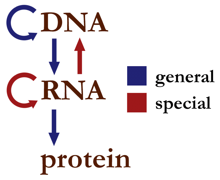
The special transfers matter for viruses. Retroviruses (HIV) use reverse transcriptase to copy their RNA genome into DNA, so their information path is RNA → DNA → RNA → protein; reverse transcription is the usual answer to "which process seems to violate the central dogma?" (Crick's version actually allowed it; the textbook one-way version does not). Influenza is a negative-sense RNA virus: its RNA-dependent RNA polymerase copies the genome into positive-sense mRNA and into new genomes, so its path is RNA → RNA → protein with no DNA at all (see The infection cycle). Prions are sometimes mentioned as an exception, but they pass on a misfolded shape, not a sequence, so they do not break the dogma.
Transcription and RNA processing
Transcription copies one strand of a gene into RNA. RNA polymerase binds a promoter, unwinds the DNA and reads the template (antisense) strand from 3′ to 5′, adding ribonucleotides to the new RNA's 3′ end, so the RNA grows 5′→3′ and is antiparallel to the template. Base pairing is G with C, and A in the template pairs with U in the RNA (RNA has uracil instead of thymine). The other DNA strand, the coding (sense) strand, has the same sequence as the mRNA except T in place of U.
In eukaryotes the primary transcript (pre-mRNA) is processed in the nucleus before translation: a 5′ cap (7-methylguanosine) is added, the 3′ end gets a poly(A) tail, and introns are spliced out so that exons join. Proteins are therefore not translated from unprocessed mRNA in eukaryotes. Alternative splicing joins different exon combinations, so one gene can make several protein isoforms. The exome is the set of all exons, the protein-coding part of the genome (only about 1–2% of human DNA). Bacteria have no nucleus, so their ribosomes start translating mRNA while it is still being transcribed.
Translation and the genetic code
Translation happens on ribosomes, in the cytoplasm or on the rough endoplasmic reticulum (for secreted and membrane proteins such as HA). Each codon of three mRNA nucleotides specifies one amino acid or a stop signal. A tRNA carries an amino acid at one end and an anticodon at the other that base-pairs antiparallel with the codon; aminoacyl-tRNA synthetases attach the correct amino acid to each tRNA. The ribosome links each new amino acid to the growing chain, so the protein grows from its N-terminus to its C-terminus as the mRNA is read 5′→3′.
| Feature | Fact |
|---|---|
| Codons | 4³ = 64: 61 sense codons plus 3 stops |
| Start codon | AUG (Met); the initiator tRNA's anticodon is 3′-UAC-5′ (written CAU 5′→3′). Bacteria start with formyl-Met |
| Stop codons | UAA, UAG, UGA; recognized by release factors, not tRNAs |
| Degeneracy | Most amino acids have 2–4 codons; Leu, Ser and Arg have 6; Met and Trp have only 1 |
| Wobble | The codon's third base can pair loosely with the anticodon's first base (G·U, or inosine with U, C or A), so one tRNA reads several codons. Wobble lets different codons give the same amino acid; it never lets one mRNA make different proteins |
| Reading frame | Fixed by the start codon; read in non-overlapping triplets with no gaps |
| Universality | Nearly universal; mitochondria and a few organisms have small variations. Selenocysteine (UGA) and pyrrolysine (UAG) are recoded stops (see Residues that get their own questions) |
| Ribosome start site | Eukaryotes: the cap is bound and the ribosome scans to the first AUG in a good Kozak context, consensus gccRccAUGG, where R (A or G) at −3 and G at +4 matter most. Bacteria: the Shine–Dalgarno sequence (AGGAGG) about 8 nucleotides upstream of AUG pairs with 16S rRNA |
Use the codon table below for any translation question. It is laid out by first base (blocks of four rows), then third base, so the four codons of a four-fold degenerate family sit on the same row.
ReferenceThe standard genetic code (all 64 codons)· 16 rows
Codons are mRNA triplets read 5′→3′. For DNA, replace U with T. Read a row to see how the third base usually matters least (wobble). Reverse lookup: the amino acid column tells you every codon that encodes a residue.
No rows match.
| Codon | Amino acid | Codon | Amino acid | Codon | Amino acid | Codon | Amino acid |
|---|---|---|---|---|---|---|---|
| UUU | Phe (F) | UCU | Ser (S) | UAU | Tyr (Y) | UGU | Cys (C) |
| UUC | Phe (F) | UCC | Ser (S) | UAC | Tyr (Y) | UGC | Cys (C) |
| UUA | Leu (L) | UCA | Ser (S) | UAA | STOP (ochre) | UGA | STOP (opal) |
| UUG | Leu (L) | UCG | Ser (S) | UAG | STOP (amber) | UGG | Trp (W) |
| CUU | Leu (L) | CCU | Pro (P) | CAU | His (H) | CGU | Arg (R) |
| CUC | Leu (L) | CCC | Pro (P) | CAC | His (H) | CGC | Arg (R) |
| CUA | Leu (L) | CCA | Pro (P) | CAA | Gln (Q) | CGA | Arg (R) |
| CUG | Leu (L) | CCG | Pro (P) | CAG | Gln (Q) | CGG | Arg (R) |
| AUU | Ile (I) | ACU | Thr (T) | AAU | Asn (N) | AGU | Ser (S) |
| AUC | Ile (I) | ACC | Thr (T) | AAC | Asn (N) | AGC | Ser (S) |
| AUA | Ile (I) | ACA | Thr (T) | AAA | Lys (K) | AGA | Arg (R) |
| AUG | Met (M) — START | ACG | Thr (T) | AAG | Lys (K) | AGG | Arg (R) |
| GUU | Val (V) | GCU | Ala (A) | GAU | Asp (D) | GGU | Gly (G) |
| GUC | Val (V) | GCC | Ala (A) | GAC | Asp (D) | GGC | Gly (G) |
| GUA | Val (V) | GCA | Ala (A) | GAA | Glu (E) | GGA | Gly (G) |
| GUG | Val (V) | GCG | Ala (A) | GAG | Glu (E) | GGG | Gly (G) |
Two habits make codon questions fast: look at the first two bases first, because they usually fix the amino acid, and check the strand's direction before reading anything. The table is your reference for codon questions; the worked example shows the method. Folding begins co-translationally: the N-terminal part of a chain can start folding while the rest is still being made, and chaperones assist.
Kinds of mutations
A mutation is a heritable change in DNA sequence. Classify it at the DNA level and then by its effect on the protein.
| Mutation | DNA change | Effect on protein | Typical severity |
|---|---|---|---|
| Silent (synonymous) | Base substitution that gives a codon for the same amino acid (often third position) | None in sequence | Usually none (rarely affects splicing or translation speed) |
| Missense, conservative | Substitution giving a chemically similar amino acid | One residue changes, properties kept | Often mild |
| Missense, non-conservative | Substitution giving a different kind of amino acid | One residue changes properties | Mild to severe, depending on location |
| Nonsense | Substitution creating a stop codon | Truncated protein (and often mRNA destroyed by nonsense-mediated decay) | Usually loss of function |
| Frameshift | Insertion or deletion of a number of bases not divisible by 3 | Every codon downstream changes, usually reaching a premature stop | Usually loss of function |
| In-frame insertion/deletion | Insertion or deletion of 3, 6, 9... bases | Amino acids added or removed, the rest unchanged | Variable; can be severe (CFTR ΔF508) |
| Splice-site | Change at an intron–exon boundary | An exon skipped or an intron retained | Often severe; skipping an exon removes the domain it encodes |
| Promoter / regulatory | Change outside the coding sequence | Same protein, made in different amounts | Variable |
Nonsense and frameshift mutations are the two classic ways to knock out a gene. A frameshift inside an intron is usually silent, because the intron is spliced out before translation (unless it hits a splice signal). Mutations are written as original residue, position, new residue: E6V (Glu6→Val, sickle cell), D614G (Asp614→Gly in the SARS-CoV-2 spike), ΔF508 (Phe508 deleted in CFTR).
Some mutation outcomes have names worth knowing. A loss-of-function mutation removes activity; a gain-of-function mutation adds or increases one. A dominant negative mutation makes a defective subunit that still assembles with normal ones and spoils the whole complex, so one bad copy is enough to block function. Multi-subunit proteins are vulnerable to this: a mutant subunit of a dimeric transcription factor that still dimerizes but cannot bind DNA, or an HA monomer that joins a trimer but cannot fold correctly, would poison the assembly.
Predicting the effect of a substitution
How to rank possible replacements for a residue: match polarity and charge first, then size and shape, and treat anything nonstandard as out of the running. For Thr, the best substitute is Ser (same –OH, one methyl smaller), then Val (same β-branched shape, but loses the –OH) or Asn; a charged or bulky aromatic residue ranks last. A substitution matrix such as BLOSUM62 encodes the same logic from how often each swap is seen between related proteins: Ile↔Leu↔Val, Ser↔Thr, Asp↔Glu, Lys↔Arg and Phe↔Tyr score highest.
Ask three questions about any amino-acid change: Where is the residue (surface, core, binding site, interface)? What changes (size, charge, polarity, hydrogen bonding, backbone flexibility)? What contact is lost or gained?
| Location | Typical effect of a non-conservative change | Example |
|---|---|---|
| Surface, away from function | Little effect; surface residues are the most variable | Antigenic-site changes on HA that alter only antibody binding |
| Hydrophobic core | Destabilizes folding: a charged or polar group buried in the core loses its water and has no partner; a larger residue does not fit, a smaller one leaves a cavity | Leu→Asp in the core; core Ile→Pro breaks a helix or strand |
| Active or binding site | Changes or removes binding or catalysis without necessarily affecting folding | Mutating a catalytic Ser, His or Cys kills activity; changes at HA positions 190 or 225 alter receptor preference |
| Subunit interface | Changes assembly or creates new sticky surfaces | Sickle-cell E6V on the hemoglobin surface |
| Salt bridge or disulfide | Removes a stabilizing interaction | Glu→Val breaks a salt bridge; Cys→Ser breaks a disulfide |
| Secondary structure | Pro inside a helix kinks or breaks it (no N–H, rigid ring); Gly adds flexibility and destabilizes helices | Ala→Pro in a helix |
A conservative substitution swaps an amino acid for one with similar size and chemistry, and usually has a small effect. Common conservative pairs: Lys↔Arg (both positive), Asp↔Glu (both negative), Leu↔Ile↔Val↔Met (hydrophobic aliphatic), Phe↔Tyr↔Trp (aromatic), Ser↔Thr (small hydroxyl), Asn↔Gln (amides), Asp↔Asn. A change that looks small in shape can still be non-conservative in chemistry: Thr→Val keeps the shape (Val has CH₃ where Thr has OH) but removes a hydroxyl that hydrogen-bonds, so it is polar→nonpolar; Asp→Gly (D614G) removes a charge and a whole side chain and makes the backbone more flexible.
Stability in energy terms. A stabilizing mutation makes the folded state lower in free energy relative to the unfolded state, so ΔG of unfolding becomes larger and positive (more energy needed to unfold) and the melting temperature rises. Two routes: add favorable contacts to the folded state (better packing, a new salt bridge or disulfide), or reduce the entropy of the unfolded state (for example a Gly→Ala or X→Pro change in a loop, or a disulfide, both of which restrict the unfolded chain). A destabilizing mutation does the reverse (see Why proteins fold, in energy terms).
Disease examples tests reuse
Three diseases recur as worked cases of single changes with large structural consequences. Each one illustrates a different row of the location table above: a surface change that creates a sticky patch, a deletion that blocks folding, and changes in a DNA-binding site.
| Disease | Change | Structural consequence |
|---|---|---|
| Sickle-cell anemia | β-globin Glu6→Val (GAG→GTG in DNA, a single A→T transversion) | Hydrophobic Val on the surface sticks to a hydrophobic pocket on another deoxy-Hb; fibers deform red cells |
| Cystic fibrosis (most common allele) | ΔF508: three bases deleted, Phe508 removed, in-frame | CFTR chloride channel misfolds and is degraded before reaching the membrane; changes primary, secondary and tertiary structure |
| Cancer (p53) | Missense mutations clustered in the central DNA-binding domain (e.g. Arg175, Arg248, Arg273) | The tumor suppressor can no longer bind DNA or unfolds; a dominant-negative effect occurs because p53 acts as a tetramer |
In each case the answer that earns credit names the level of structure affected: sickle cell changes how folded tetramers pack together (quaternary contacts between molecules), ΔF508 changes folding itself, and p53 hot-spot mutations remove DNA contacts or destabilize the domain that makes them.
Influenza: mutation in action
Influenza's RNA-dependent RNA polymerase has no proofreading, so it makes roughly one error per 10⁴–10⁵ nucleotides copied, about one new mutation per genome per replication. Most are harmful or neutral, but selection keeps the useful ones:
- Antigenic drift: substitutions in the antigenic sites around the HA head change the surface antibodies recognize, so last season's antibodies bind poorly.
- Receptor-specificity changes: substitutions in the pocket, such as at H3-numbered positions 190 and 225 (D222G in H1 numbering), shift which sialic-acid linkage the virus prefers (see What sets receptor preference in H1 HAs).
- Cleavage-site changes: inserting several basic residues at HA0's cleavage site lets furin-like proteases throughout the body activate HA, a hallmark of highly pathogenic avian strains.
- Drug resistance: the H275Y substitution in neuraminidase (N1 numbering) makes it resistant to oseltamivir.
Antigenic shift, the swap of a whole genome segment by reassortment, is a different process (see Influenza virus).
Conservation and sequence alignment
A residue is conserved if it stays the same (or nearly the same) at the matching position across related proteins or species. A sequence alignment lines up two or more sequences, inserting gaps where needed, so that matching positions sit in the same column; tools such as BLAST (for searching databases) and Clustal (for multiple alignments) do this. Identity is the percent of aligned positions that are the same amino acid; similarity also counts conservative substitutions. Homologous proteins share a common ancestor (an all-or-nothing statement); percent identity is the evidence for it.
Conservation is evidence of importance. Natural selection removes mutations that damage function, so residues that do essential jobs (catalytic residues, metal ligands, glycines in tight turns, prolines at kinks, disulfide cysteines, receptor-pocket residues) change least, while surface loops change most. In HA, the receptor-pocket floor residues (Tyr98, Trp153, His183, Tyr195 in H3 numbering) are conserved across subtypes, while the antigenic sites around them vary from year to year. Proteins with low sequence identity can still share the same fold, because structure is more conserved than sequence: as few as 25–30% identical residues usually means the same overall fold.
How mutation questions are asked
- Transcribe and translate a template or coding strand; find a start codon; spot a stop.
- Name the mutation type from a DNA change or from the protein outcome (truncated → nonsense or frameshift).
- Pick the most or least disruptive substitution (look for charge into core, Pro into helix, loss of Cys or catalytic residue; Lys→Arg, Leu→Ile, Asp→Glu are mild).
- Explain why a conserved residue matters, or why active-site residues are conserved.
- Central dogma statements, retroviral exceptions, and true/false items on wobble, introns and processing.
- Influenza links: drift, receptor-switching substitutions, cleavage site, resistance.
Viruses and how they infect cells
What a virus is, and what it is not
A virus is a set of genes packed in a protein shell (the capsid), sometimes wrapped in a lipid envelope. It has no metabolism of its own, so it must enter a host cell and use the cell's machinery — ribosomes, tRNAs, amino acids, ATP, membranes — to copy itself. Sizes run roughly 20–300 nm, one to two orders of magnitude smaller than a bacterium, and genomes run from about 2 kb to a few hundred kb.
What a virus lacks is the usual exam point: no ribosomes, no plasma membrane of its own making, no cytoplasm, no ATP synthesis, no growth, no binary fission. Virions do not get bigger and then split; they are assembled from parts. Outside a cell a virion is inert particles — which is why viruses are usually described as replicating rather than reproducing, and why "is a virus alive?" has no settled answer.
Some vocabulary that tests use interchangeably and should not:
| Term | Means |
|---|---|
| Virion | One complete, assembled virus particle outside a cell |
| Capsid | The protein shell; built from repeated subunits called capsomers |
| Nucleocapsid | Capsid plus the genome it encloses |
| Envelope | Lipid bilayer taken from a host membrane, studded with virus-encoded glycoproteins |
| Peplomer / spike | One of those envelope glycoproteins (HA and NA on influenza) |
| Tropism | Which species, tissues and cells a virus can infect — set mostly by receptor availability |
| Viroid / prion | Not viruses: a viroid is naked infectious RNA (plants), a prion is a misfolded protein with no nucleic acid |
Building a shell out of many copies of one small protein is forced on viruses by genome economy: a capsid large enough to hold the genome must be made from repeated subunits, because coding one protein per capsid position would need far more DNA than the capsid can hold. That constraint produces the two common symmetries — icosahedral (20 triangular faces, quasi-spherical) and helical (subunits spiralling around the nucleic acid). Influenza is a hybrid: helical nucleocapsids inside a roughly spherical (often pleomorphic or filamentous) envelope.

Enveloped versus naked viruses
The envelope is the single structural fact that predicts the most about a virus's behavior, so questions about it are common.
| Property | Enveloped (influenza, HIV, SARS-CoV-2, herpes) | Naked / non-enveloped (poliovirus, adenovirus, norovirus, papillomavirus) |
|---|---|---|
| Outer layer | Host-derived lipid bilayer with viral glycoproteins | Protein capsid only |
| Entry | Membrane fusion (at the surface or inside an endosome) | Endocytosis then capsid rearrangement/pore formation, or direct penetration |
| Exit | Budding (usually non-lytic; cell may survive a while) | Usually lysis of the cell |
| Environmental stability | Fragile: dried out, detergents (soap), alcohol and heat destroy the lipid | Tough: survive drying, detergents, stomach acid, many disinfectants |
| Typical transmission | Respiratory droplets, body fluids, close contact | Fecal–oral, fomites, surfaces |
| Antibody target | Envelope glycoproteins (HA on flu) | Capsid surface loops |
Soap works on influenza for a purely chemical reason worth stating: soap and alcohol dissolve the lipid envelope, so the spikes fall apart and the particle cannot attach or fuse. Naked viruses have no lipid to dissolve, which is why norovirus outbreaks need bleach rather than hand sanitizer.
The Baltimore classification
David Baltimore's scheme sorts viruses by the chemical nature of the genome and the number of steps between that genome and mRNA, because every virus must make mRNA the host ribosome can read. Learn it as "how many steps to (+) mRNA?"
| Class | Genome | Route to mRNA | Examples |
|---|---|---|---|
| I | dsDNA | Transcribe one strand, usually with host RNA polymerase in the nucleus | Herpesviruses, adenovirus, poxviruses (cytoplasmic, own polymerase) |
| II | ssDNA | Make dsDNA first, then transcribe | Parvovirus B19 |
| III | dsRNA | Viral RNA-dependent RNA polymerase (RdRp) copies the (−) strand | Rotavirus, reovirus |
| IV | (+) ssRNA | Genome is mRNA: ribosomes translate it directly | Poliovirus, hepatitis C, coronaviruses, dengue |
| V | (−) ssRNA | Genome is complementary to mRNA, so the virion must carry its own RdRp | Influenza A–D, measles, rabies, Ebola |
| VI | ssRNA with reverse transcriptase | RNA → DNA → integrate into host chromosome → transcribe | HIV and other retroviruses |
| VII | dsDNA with reverse transcriptase | DNA → RNA → DNA during replication | Hepatitis B |
Two consequences the bank asks about directly. First, a (+) ssRNA virus can be infectious as naked RNA, because injecting the genome alone is enough to start translation; a (−) ssRNA virus cannot, because the cell has no enzyme that reads RNA from RNA, so the polymerase must come packaged in the virion. Second, Class VI reverses the usual DNA → RNA direction of the central dogma: reverse transcriptase makes DNA from an RNA template, which is the exception every test cites (see Mutations and sequence changes for the dogma itself). Information flow for a retrovirus is RNA → DNA → RNA → protein.
The generic infection cycle
Every viral infection follows the same broad sequence, and exam questions often ask you to place an event in it:
- Attachment (adsorption): a viral surface protein binds a specific molecule (the receptor) on the host cell. Receptor choice sets which species and tissues the virus can infect (its tropism).
- Entry (penetration): the virus enters by fusing its envelope with the plasma membrane, or by being taken up in an endosome and escaping from it; naked viruses breach the endosome membrane or form a pore.
- Uncoating: the genome is released from the capsid.
- Replication and expression: viral genes are copied and translated by host ribosomes. Early proteins are usually enzymes; late proteins are usually structural.
- Assembly (maturation): new genomes and proteins come together, often with a proteolytic cleavage step that activates the particle.
- Release: by budding (enveloped viruses take their envelope from the host membrane) or by bursting the cell.
The interval between uncoating and the first new virions is the eclipse period, when no complete virus can be recovered from the cell — a classic "what is happening at point X on the one-step growth curve?" question. Burst size is the number of virions released per infected cell (hundreds to thousands for influenza).
Virologists count virus with a handful of standard measures, and a growth-curve or titer question assumes them:
| Measure | What it counts | How it is obtained |
|---|---|---|
| Plaque assay / PFU | Infectious particles, as plaque-forming units per mL | A dilution of virus is spread on a cell monolayer under an overlay; each infectious particle kills a spreading patch of cells, making one countable hole (plaque) |
| TCID₅₀ | The dilution that infects 50% of inoculated cell cultures | Serial dilutions in replicate wells, scored for cytopathic effect; roughly 0.7 PFU per TCID₅₀ |
| MOI (multiplicity of infection) | Infectious particles per cell | PFU added ÷ cells present. MOI ≫ 1 infects essentially every cell at the same moment, usually with several particles each (needed for a one-step growth curve); MOI ≪ 1 gives multiple rounds |
| HA titer | Total particles able to agglutinate red cells, infectious or not | The highest dilution that still clumps red blood cells |
| Particle-to-PFU ratio | How many physical virions it takes to score one infection | Electron-microscope or genome count ÷ PFU; usually far above 1, because most particles are defective |
On a one-step growth curve (high MOI, so every cell is infected at the same moment), the titer first falls or stays flat during the eclipse period, then rises steeply as progeny are released, then plateaus; the rise's height divided by the number of cells is the burst size.
Entry routes worth naming: direct fusion at the plasma membrane (HIV, pH-independent), receptor-mediated endocytosis followed by low-pH fusion (influenza), endosomal escape without fusion (adenovirus), and injection of the genome only (bacteriophages, which leave the capsid outside).
Lytic and lysogenic cycles
These two words come from bacteriophage biology but appear on protein-modeling tests as general virus vocabulary.
| Feature | Lytic cycle | Lysogenic cycle |
|---|---|---|
| What happens | Phage takes over at once, makes virions, lyses the cell | Phage genome integrates into the host chromosome as a prophage (or sits as a plasmid) and is copied with it |
| Host cell | Killed | Survives and divides, passing the prophage to daughters |
| New virions | Many, immediately | None until induction |
| Trigger to switch | — (already replicating) | Induction by stress such as UV or DNA damage switches a prophage into the lytic cycle |
| Animal-virus analogue | Acute infection (influenza) | Latency (herpesviruses) or provirus integration (HIV) |
Influenza has no lysogenic option: it is an acute, lytic-style infection with no integration step, because its genome is RNA and it encodes no integrase. Lysogeny also explains transduction, in which a phage carries host genes between bacteria, and why some bacterial toxins (diphtheria, cholera) are encoded on prophages.
Influenza virus
Structure of the virion
Influenza A is an enveloped, negative-sense single-stranded RNA virus (family Orthomyxoviridae). Its genome is split into eight segments, each wrapped with nucleoprotein (NP) and bound to its own polymerase heterotrimer (PB1, PB2 and PA) to form a ribonucleoprotein (vRNP), which folds back on itself as a double-helical hairpin. Under the envelope lies a layer of matrix protein (M1), the particle's structural scaffold. Three proteins sit in the envelope:
| Protein | Role |
|---|---|
| Hemagglutinin (HA) | Binds sialic acid receptors and drives membrane fusion; the main target of neutralizing antibodies; a homotrimer, roughly 300–500 trimers (spikes) per virion |
| Neuraminidase (NA) | An enzyme that cuts sialic acid off cell-surface glycans, freeing new virions and helping the virus move through mucus; a homotetramer with a mushroom-shaped head, roughly 40–100 tetramers per virion |
| M2 | A proton channel (a homotetramer) that acidifies the inside of the virion during entry; only on the order of tens of copies (published estimates range from about 15 to 60) |
Particles are pleomorphic: laboratory-adapted strains are roughly spherical at about 100 nm, while clinical isolates are often long filaments micrometers in length. That variability is why electron-microscope questions ask about "roughly spherical to filamentous" rather than a fixed shape, and why influenza cannot be given a crystallographic symmetry the way an icosahedral virus can.


A full strain name is written in a fixed order: type / host (omitted if human) / place of isolation / strain number / year (subtype). The 2027 protein comes from A/California/04/2009 (H1N1) — type A, human, isolated in California, isolate 04, in 2009, subtype H1N1. Tests love asking you to decode such a name or to say what is missing (the host, because it is human).
Influenza types A, B, C and D
| Type | Segments | Surface proteins | Natural reservoir | Human disease | Subtypes / lineages |
|---|---|---|---|---|---|
| A | 8 | HA + NA (separate) | Wild waterfowl and shorebirds; also pigs, horses, bats, poultry | Seasonal epidemics and all pandemics | H1–H18, N1–N11 (H17N10 and H18N11 only from bats) |
| B | 8 | HA + NA (separate) | Humans (also seals) | Seasonal epidemics, especially in children; no pandemics | No subtypes; two lineages, B/Victoria and B/Yamagata (Yamagata not detected in surveillance since March 2020) |
| C | 7 | One protein, HEF, does binding, esterase and fusion | Humans, pigs | Mild cold-like illness, mostly children | No subtypes |
| D | 7 | HEF | Cattle (also pigs) | No confirmed human disease | No subtypes |
The pattern to remember: only A has the animal reservoir wide enough and the subtype diversity needed for antigenic shift, so only influenza A causes pandemics. C and D get away with seven segments because HEF merges the jobs of HA and NA — and because their receptor is not Neu5Ac but 9-O-acetylated sialic acid, which the esterase removes.
The eight segments and what they encode
The segments are numbered by decreasing length. Every one of them must be packaged for a virion to be infectious, which is why co-infection so readily produces reassortants.
| Segment | Length (nt, approx.) | Protein(s) | Function |
|---|---|---|---|
| 1 | 2341 | PB2 | Polymerase basic 2: binds the host mRNA 5′ cap for cap-snatching. The residue 627 position (E627K) is a classic host-range marker for replication at mammalian airway temperature |
| 2 | 2341 | PB1, PB1-F2, PB1-N40 | PB1 is the catalytic RdRp subunit that adds nucleotides. PB1-F2 is a short accessory protein from a second reading frame that targets mitochondria and promotes apoptosis and secondary bacterial pneumonia; it is truncated and non-functional in 2009 pdm09 viruses |
| 3 | 2233 | PA, PA-X | PA carries the endonuclease that snips the snatched cap, and is the target of baloxavir. PA-X (a frameshift product) shuts off host gene expression |
| 4 | 1778 | HA | Receptor binding and membrane fusion; chief antigen (your model protein) |
| 5 | 1565 | NP | Coats the vRNA every ~24 nucleotides, carries nuclear localization signals, and is the main target of cross-reactive cytotoxic T cells |
| 6 | 1413 | NA | Sialidase: cleaves sialic acid to release progeny and penetrate mucus; target of oseltamivir and zanamivir |
| 7 | 1027 | M1, M2 (spliced mRNA) | M1 is the matrix scaffold and controls vRNP export; M2 is the proton channel and target of the adamantanes |
| 8 | 890 | NS1, NEP/NS2 (spliced mRNA) | NS1 is the interferon antagonist (it sequesters dsRNA and blocks RIG-I signalling and host mRNA processing); NEP (nuclear export protein) ferries vRNPs out of the nucleus with M1 |
Two structural tricks are on this list. Segments 7 and 8 each make two proteins by alternative splicing of the viral mRNA in the nucleus — an unusual move for an RNA virus and one of the reasons influenza must replicate there, since splicing happens only in the nucleus. Segments 2 and 3 each make an extra protein from a different reading frame of the same RNA. Both are genome-economy devices: eight segments, far more than eight proteins.
The infection cycle
- Attachment. HA on the virion binds sialic acid at the tips of glycans on airway epithelial cells. Each contact is weak; many HA–sialic acid contacts at once make the attachment stick (avidity, below).
- Endocytosis. The cell takes the bound virus into an endosome, mostly by clathrin-mediated endocytosis.
- Acidification. The endosome pumps in protons and its pH falls to about 5–6. M2 lets protons into the virion interior, which loosens M1 from the vRNPs so they can be released after fusion.
- Fusion. Low pH (about 5.0–5.5 for most human strains) triggers a large, irreversible change in HA's shape that drives the viral and endosomal membranes together (details below). The vRNPs are released into the cytoplasm.
- Nuclear import and replication. Unlike most RNA viruses, influenza copies its genome in the nucleus; NP's nuclear localization signals get the vRNPs through the nuclear pore. The viral polymerase makes mRNA, using 5′ caps snatched from host mRNAs to prime transcription (cap-snatching: PB2 grabs the cap, the PA endonuclease cuts 10–13 nucleotides downstream, and PB1 extends that fragment). Transcription makes capped, polyadenylated mRNA; replication instead makes a full-length positive-sense copy, the cRNA, which serves as template for new (−) vRNA.
- Translation and assembly. HA, NA and M2 are translated on ER-bound ribosomes, glycosylated, folded and sent through the Golgi to the apical plasma membrane; M1, NP, NS1 and the polymerase subunits are made on free ribosomes. New vRNPs leave the nucleus with M1 and NEP and gather under patches of membrane rich in viral proteins.
- Budding and release. New virions bud from the apical surface, taking host membrane as their envelope, with M2 helping to pinch off the particle. HA on the new virions would stick them right back onto sialic acid on the same cell, so NA cuts sialic acid to set them free.

The viral RdRp has no proofreading exonuclease, so it misincorporates roughly one nucleotide in – — about the length of a single segment. Every infected cell therefore produces a cloud of slightly different genomes (a quasispecies), which is the raw material for antigenic drift and for drug resistance.
Hemagglutinin, from precursor to fusion machine
HA is made as one chain, HA0, which folds and assembles into a homotrimer in the ER. A host protease must cut HA0 into HA1 and HA2, held together by a disulfide bond, before the virus can infect — HA0 binds sialic acid perfectly well but cannot fuse. The cut is therefore an activation step exactly like a zymogen's (see Post-translational modifications), and uncleaved particles are non-infectious.
| Feature | Seasonal / 2009 pandemic H1, H3 (low pathogenicity) | Highly pathogenic avian H5, H7 |
|---|---|---|
| Cleavage site sequence | A single basic residue (monobasic), typically …Q/E–R↓G… | A multibasic stretch, e.g. …R–X–K/R–R↓G… |
| Activating proteases | Airway proteases such as TMPRSS2 and HAT, plus bacterial and plasmin-like proteases | Furin and related proprotein convertases, present in essentially every cell |
| Where those proteases are | Respiratory epithelium (and gut epithelium in birds) | Ubiquitous, in the Golgi of most tissues |
| Consequence | Infection confined to the respiratory tract | Virus can be activated in many organs, giving systemic disease in poultry |
- HA1 forms the globular head on top. It carries the receptor binding site and most of the antibody targets. Your model segment, chain A residues 153–252, is part of HA1.
- HA2 forms most of the long stem (the triple-helical coiled coil) and anchors the trimer in the membrane. It holds the fusion peptide, a hydrophobic stretch of about 20 residues at the new N-terminus created by cleavage, buried in a charged pocket in the pre-fusion shape. Its sequence in H1 HAs begins GLFGAIAGFIE…: glycines and alanines for flexibility and small size, two phenylalanines and two isoleucines for bulk hydrophobicity, and almost no polar residues until the glutamate, which is exactly the composition that lets it bury itself in the hydrocarbon core of the target membrane. It is a favorite side-chain-properties question because every letter in it can be justified from the chemistry.
At endosomal pH the HA1 heads loosen their grip on each other, and HA2 refolds in what is the textbook example of a spring-loaded conformational change: a loop between two helices becomes helical, extending one long helix that throws the fusion peptide roughly 100 Å toward the endosomal membrane, where it inserts. The molecule then jackknifes, folding the C-terminal part of HA2 back alongside the long helix into a hairpin, which drags the viral membrane (held by the transmembrane anchor) against the endosomal membrane (held by the fusion peptide). Several trimers acting together bend the two bilayers into a hemifusion stalk and then a pore.
The trigger is protonation: as pH falls, histidine side chains (pKa ≈ 6) and carboxylates pick up protons, salt bridges that clamped the pre-fusion state break, and the molecule falls into the lower-energy post-fusion shape. The change is irreversible — the pre-fusion state is metastable, a loaded spring, and the energy released does the work of fusion. That is also why HA exposed to low pH outside a cell is permanently inactivated and non-infectious, and why acid-stability of HA is studied as a transmissibility trait.

Antigenic drift, shift and pandemic history
| Term | Mechanism | Rate | Result |
|---|---|---|---|
| Antigenic drift | Point mutations accumulate in HA and NA antigenic sites, selected by antibody pressure | Continuous, every season | Gradual change; reason flu vaccines are updated each year; causes epidemics |
| Antigenic shift | Reassortment brings a new HA (and sometimes NA) segment into a human-transmissible virus, or a whole animal virus adapts to humans | Rare, decades apart | A virus most people have no immunity to; can cause a pandemic |
Drift is mutation within a subtype (H3N2 of 2015 versus H3N2 of 2025); shift changes the subtype itself (H1N1 → H2N2). A mixing vessel host — classically the pig, whose airway carries both avian-type and human-type receptors — allows an avian and a human virus to meet in one cell and swap segments.
| Pandemic | Virus | Nickname | Notes |
|---|---|---|---|
| 1918–19 | H1N1 | "Spanish flu" | The deadliest: an estimated 50 million deaths worldwide and an estimated 675,000 in the US by current CDC figures. Unusual W-shaped mortality curve with a peak in healthy young adults |
| 1957–58 | H2N2 | "Asian flu" | Reassortant: avian HA, NA and PB1 on a human H1N1 backbone. An estimated 1.1 million deaths worldwide |
| 1968–69 | H3N2 | "Hong Kong flu" | Reassortant: avian HA and PB1 on the circulating H2N2 backbone; NA stayed N2, so partial immunity blunted it. An estimated 1 million deaths worldwide. H3N2 still circulates |
| 2009–10 | H1N1pdm09 | "swine flu" | Quadruple reassortant from swine lineages (below). CDC's modeled estimate is 151,700–575,400 deaths worldwide in the first year |
Notice the pattern in 1957 and 1968: the new virus kept some segments, so people with antibodies to the retained N2 were partly protected, and those pandemics were milder than 1918. Note too that H1N1 disappeared from human circulation after 1957, reappeared in 1977, and was then replaced by the 2009 virus — so "H1N1" on a test can mean three different viruses, and the year matters.
The 2009 H1N1 pandemic
The 2009 pandemic virus arose in pigs by reassortment, the swapping of whole genome segments between two influenza viruses that infect the same cell. Its segments came from two swine influenza lineages, which themselves carried genes of swine, avian and human origin — which is why it is called a quadruple reassortant:
| Segment(s) | Lineage of origin |
|---|---|
| HA, NP, NS | Classical swine H1N1, in North American pigs since about 1918 |
| PB2, PA | North American avian lineage, via the triple-reassortant swine viruses of the late 1990s |
| PB1 | Human seasonal H3N2, via the same triple-reassortant swine viruses |
| NA, M | Eurasian avian-like swine lineage, in European pigs since the late 1970s |
Its HA is an H1 from the classical swine lineage, which descends from the 1918 pandemic virus. That ancestry explains a striking feature of 2009: many older adults, exposed decades earlier to 1918-like H1 viruses, had antibodies that cross-reacted with the new HA, while children and young adults did not. The age distribution of severe cases was therefore inverted relative to a normal flu season — one of the clearest real-world demonstrations of immune imprinting.
The immune response to influenza
Innate immunity
The first defenses act within minutes to hours and do not depend on prior exposure. They are non-specific (they respond to classes of molecule, not to one strain), germline-encoded (not rearranged, so no new specificities can be made), and they have no memory — a second infection gets the same innate response as the first.
Barriers come first. The airway is lined by a mucus blanket moved by beating cilia (the mucociliary escalator), which traps virions and sweeps them to the throat to be swallowed. Mucus is itself rich in heavily sialylated glycoproteins (mucins), so it acts as a decoy receptor: HA sticks to mucus instead of to cells, and NA must cut the virus free. Lysozyme, defensins, surfactant proteins A and D (which bind HA's glycans), low skin pH and stomach acid round out the chemical barriers.
Pattern recognition. Infected and sentinel cells detect PAMPs (pathogen-associated molecular patterns) with PRRs (pattern recognition receptors). For influenza, four sensors matter — three that detect viral RNA and one that senses the damage the virus does:
| Sensor | Where it sits | What it detects | Output |
|---|---|---|---|
| TLR3 | Endosome membrane | Double-stranded RNA from replication intermediates | Type I interferon and inflammatory cytokines |
| TLR7 | Endosome membrane, especially in plasmacytoid dendritic cells | Single-stranded viral RNA | Large bursts of type I interferon |
| RIG-I | Cytoplasm | Short dsRNA and RNA bearing a 5′-triphosphate — the signature of viral, not host, RNA | Signals through MAVS to IRF3/IRF7 and NF-κB |
| NLRP3 inflammasome | Cytoplasm | Cell damage and M2 ion flux | Activates caspase-1, releasing IL-1β and IL-18 |
Interferon is the central antiviral signal. Infected cells secrete type I interferons (IFN-α and IFN-β). These act mostly on neighboring cells, binding the IFNAR receptor and switching on, through the JAK–STAT pathway, several hundred interferon-stimulated genes (ISGs) that put the cell into an antiviral state before it is infected. The ones tests name:
| ISG | What it does |
|---|---|
| Mx1 / MxA | A GTPase that traps incoming vRNPs and blocks nuclear import; a major anti-influenza factor (many laboratory mouse strains lack functional Mx1) |
| PKR | Phosphorylates eIF2α on sensing dsRNA, shutting down translation in the infected cell |
| OAS / RNase L | OAS makes 2′–5′ oligoadenylates that activate RNase L, which degrades viral and cellular RNA |
| IFITM3 | Sits in endosome membranes and blocks HA-mediated fusion, so the genome never reaches the cytoplasm |
Influenza fights back with NS1, which binds dsRNA so sensors cannot see it, blocks RIG-I signalling, and interferes with host mRNA processing and export. NS1 is the reason an otherwise healthy interferon system does not stop the infection outright, and deleting it is one way live attenuated vaccines are designed.
Cells and cascades. Alveolar macrophages and neutrophils phagocytose virions, debris and infected cells. Natural killer (NK) cells kill infected cells without needing MHC-restricted recognition: they read the balance of activating signals against inhibitory signals from MHC class I, so a cell that has lowered its MHC I to hide from T cells ("missing self") is killed by NK cells instead. NK cells also perform ADCC (antibody-dependent cellular cytotoxicity) by binding, through their CD16 Fc receptor, IgG that is already stuck to HA on the infected cell's surface — a bridge from the adaptive side back to the innate one.
Complement is a cascade of about 30 plasma proteins with three activation routes: the classical pathway (C1q binding antibody already attached to antigen), the lectin pathway (mannose-binding lectin recognizing the sugars on HA's glycan shield), and the alternative pathway (spontaneous C3 hydrolysis on any surface lacking host regulators). All three converge on C3 convertase, and the shared outputs are opsonization (C3b tagging particles for phagocytes), chemotaxis and inflammation (the anaphylatoxins C3a and C5a), and the membrane attack complex (C5b–C9), which punches pores in membranes. Note that complement can neutralize an enveloped virion directly, which naked viruses largely escape.
Inflammation and symptoms. Infected cells and macrophages release cytokines — IL-1, IL-6, TNF-α — and chemokines that recruit more cells. IL-1 and IL-6 act on the hypothalamus to raise the thermostat, producing fever; prostaglandin E₂ is the final mediator, which is why aspirin and ibuprofen (COX inhibitors) lower a fever. Fever, aches and fatigue largely come from these signals, not from the virus directly. When the response runs out of control — most often in severe influenza — massive cytokine release ("cytokine storm") damages the alveolar barrier and causes the acute respiratory distress that kills. This is the mechanistic answer to "why were healthy young adults at risk in 1918?": a vigorous immune system can itself do the damage.
Adaptive immunity
Adaptive immunity is slow the first time because it works by clonal selection: the body makes an enormous repertoire of lymphocytes, each with one randomly generated receptor, and an antigen selects the few that fit, which then divide. That takes about a week. The second time, the expanded memory clones are already there, so the response is faster, larger and of higher affinity.
| Arm | Cells and molecules | What it does against influenza |
|---|---|---|
| Humoral | B cells → plasma cells → antibodies | Neutralizing antibodies bind HA and block attachment or fusion; IgA protects the airway surface, IgG circulates in blood; anti-NA antibodies slow release |
| Cellular | CD8⁺ cytotoxic T cells | Recognize viral peptides (often from conserved internal proteins such as NP and M1) on MHC class I and kill infected cells |
| Helper | CD4⁺ T cells | Help B cells switch class and mature their antibodies; support CD8⁺ responses |
| Memory | Memory B and T cells | Faster, stronger response on reinfection with a similar strain |
Antigen presentation and MHC
T cells cannot see a whole virus. They see short peptides held in the groove of a major histocompatibility complex (MHC) molecule — in humans, HLA. There are two pathways, and the pairing with the T cell co-receptor is the fact most asked:
| Feature | MHC class I | MHC class II |
|---|---|---|
| Found on | All nucleated cells | Professional antigen-presenting cells: dendritic cells, macrophages, B cells |
| Peptide source | Endogenous: proteins made inside the cell, including viral ones | Exogenous: proteins taken in by phagocytosis or endocytosis |
| Processing | Proteasome chops the protein; TAP pumps peptides into the ER, where they load onto MHC I | Endosome/lysosome proteases chop the antigen; the invariant chain keeps the groove empty until then |
| Peptide length | About 8–10 residues, ends tucked into a closed groove | About 13–25 residues, groove open at both ends |
| Chains | One α chain plus β₂-microglobulin | α and β chains, both membrane-anchored |
| Read by | CD8⁺ cytotoxic T cells | CD4⁺ helper T cells |
| Message sent | "I am infected — kill me" | "Here is what I found outside — organize a response" |
Mnemonic: multiply to eight. CD8 × MHC I = 8; CD4 × MHC II = 8.

A CD8⁺ T cell that finds its peptide kills by releasing perforin (which lets granzymes in) and granzymes (proteases that trigger apoptosis), and by engaging Fas on the target. Killing the infected cell before it finishes making virions is how cell-mediated immunity limits an established influenza infection. Because the peptides come from whatever protein is being made, many of them come from conserved internal proteins (NP, M1) rather than from the variable HA head — so T-cell immunity cross-protects between strains and even subtypes far better than antibody immunity does, which is why previous infection often reduces severity even when it does not prevent infection.
B cells, antibodies and affinity maturation
A B cell's receptor is a membrane-bound antibody. When it binds its antigen and gets help from a CD4⁺ T cell recognizing the same antigen on the B cell's MHC II, the B cell proliferates in a germinal center, where two things happen:
- Somatic hypermutation, carried out by the enzyme AID, scatters point mutations through the variable-region genes. B cells whose mutated receptors bind antigen better capture more of it, get more T-cell help, and survive; the rest die. Over successive rounds, average antibody affinity climbs — affinity maturation.
- Class switch recombination swaps the heavy-chain constant region (IgM → IgG, IgA or IgE) while leaving the variable region, and therefore the specificity, unchanged. Cytokines from the helper T cell choose the class.
The output is plasma cells (antibody factories, no longer displaying surface receptor) and memory B cells.

An antibody is a protein-structure question in disguise, so expect it on this event's tests. The basic unit is two identical heavy chains and two identical light chains, held together by interchain disulfide bonds in a Y shape. Each chain is a string of immunoglobulin folds — compact β-sandwich domains, two antiparallel sheets with Greek-key topology pinned face to face by an internal disulfide (a sandwich, not a barrel: the sheets stack, they do not close into a cylinder). Papain digestion cuts above the hinge into two Fab fragments (one binding site each) and one Fc; pepsin cuts below it, giving a bivalent F(ab′)₂. The actual contact surface, the paratope, is built from six hypervariable loops — the complementarity-determining regions (CDRs), three from VH and three from VL — and the patch of antigen it touches is the epitope. A conformational epitope, like most of those on HA, exists only in the folded protein, which is why denaturing the antigen can abolish antibody binding.
| Class | Structure | Where | Role against influenza |
|---|---|---|---|
| IgM | Pentamer (10 binding sites) with a J chain | Blood | First antibody made in a primary response; low affinity per site but high avidity; best complement activator; a rise in IgM means recent infection |
| IgG | Monomer | Blood and tissue; crosses the placenta | The main neutralizing and longest-lived class; what HAI and most serology measure; four subclasses |
| IgA | Dimer with a J chain and secretory component | Mucus, saliva, tears, breast milk | The front-line antibody on the airway surface, where influenza lands; induced strongly by live attenuated (nasal) vaccine |
| IgE | Monomer | Bound to mast cells and basophils | Allergy and parasites; not an antiviral class |
| IgD | Monomer | Mostly on naive B cell surfaces | Receptor for antigen; little known secreted role |

How antibodies actually stop influenza
"Neutralization" is not one mechanism. Against HA there are several, and a good test question asks which one a given antibody uses:
- Block receptor binding. An antibody whose footprint covers or overlaps the receptor binding site physically prevents HA from reaching sialic acid. This is the mechanism HAI measures.
- Block fusion. Stem antibodies let the virus bind and be endocytosed but clamp HA2 so it cannot do its spring-loaded refolding; some also prevent HA0 cleavage.
- Aggregation. Multivalent antibodies, especially IgM, cross-link virions into clumps that cannot infect and are easily cleared.
- Opsonization and complement. Fc regions on bound antibody recruit phagocytes (via Fcγ receptors) and C1q, destroying the particle or the infected cell.
- ADCC. IgG bound to HA on an infected cell's surface is read by NK cell CD16, which triggers killing.
Most neutralizing antibodies bind the HA head, around and over the receptor binding site, because it is exposed and immunodominant. On H1 heads these targets are called antigenic sites Sa, Sb, Ca1, Ca2 and Cb (on H3 they are labeled A–E). The head tolerates mutation, so these sites drift. The conserved pocket floor cannot change much without losing sialic acid binding, but it is a small, shallow pocket that most antibodies cannot reach deeply; HA also hides variable surfaces behind N-linked glycans, and adding a new glycosylation site is one of the cheapest ways to escape an antibody. The stem is far more conserved, and broadly neutralizing stem antibodies — which often cross-react within a phylogenetic group (group 1 includes H1, H2 and H5; group 2 includes H3 and H7) — are a major target of "universal" vaccine research.
The hemagglutination inhibition (HAI or HI) assay measures anti-HA antibodies: HA on virions crosslinks red blood cells by binding their sialic acids (that is where the name hemagglutinin comes from), and serum antibodies that block the binding site prevent the clumping. Un-agglutinated red cells roll down a round-bottomed well into a sharp button; agglutinated ones form a diffuse mat.
Immune memory and original antigenic sin
Memory is why adaptive immunity is worth its slow start. A secondary response has a shorter lag, a higher peak, mostly IgG rather than IgM, and higher average affinity thanks to earlier affinity maturation.
Memory also has a downside peculiar to influenza. Original antigenic sin, now usually called immune imprinting, is the tendency of the immune system to answer a new, related strain by recalling memory cells raised against the first strain it ever met, rather than by generating fresh responses to the new epitopes. The recalled antibodies may fit the new HA poorly. Consequences the exam cares about:
- Your birth year predicts which HA group you respond to best, because it determines the subtype of your first influenza infection.
- In 2009, people born before about 1957 had 1918-descended H1 memory that cross-reacted with the pandemic HA, so severe disease skewed young — the opposite of a normal season.
- Imprinting also partly explains why repeated annual vaccination with similar strains can give diminishing boosts.
How influenza spreads through a population
Protein Modeling's exam asks about "the viral infection process", and the easiest population-level questions follow directly from the biology above.
| Term | Meaning | Influenza values (typical, from CDC and the literature) |
|---|---|---|
| Transmission routes | How virus reaches the next host | Respiratory droplets and smaller aerosols from coughing, sneezing and talking; contact with contaminated surfaces then the face |
| Incubation period | Infection → first symptoms | About 1–4 days (average ~2) |
| Infectious period | When a person sheds enough virus to infect others | From about a day before symptoms until roughly 5–7 days after onset; longer in young children and immunocompromised people |
| Latent period | Infection → start of infectiousness | Slightly shorter than the incubation period, which is why pre-symptomatic spread happens |
| Serial interval | Symptom onset in a case → onset in someone they infect | About 2–4 days, which is why flu epidemics rise quickly |
| Basic reproduction number (R₀) | Average secondary cases from one case in a fully susceptible population | About 1.2–1.8 for seasonal influenza; higher estimates for some pandemic strains |
| Effective reproduction number (Rₜ) | The same number given current immunity and behavior | Epidemic grows when Rₜ > 1, shrinks when Rₜ < 1 |
| Herd immunity threshold | Immune fraction needed to push Rₜ below 1 | 1 − 1/R₀, so roughly 20–45% for seasonal influenza — but drift erodes it every year |
| Case fatality ratio (CFR) | Deaths ÷ diagnosed cases | Sensitive to how many mild cases are counted, so early-epidemic CFRs are usually overestimates |
| Infection fatality ratio (IFR) | Deaths ÷ all infections, including undiagnosed | Lower than CFR; needs serological surveys to estimate |
| Seasonality | Why temperate epidemics peak in winter | Drier, colder air stabilizes virions and impairs mucociliary clearance; more indoor crowding; less UV. Tropical regions see year-round or rainy-season transmission |
Prevention and treatment
Influenza vaccines mostly work by raising anti-HA antibody, so the whole enterprise turns on predicting HA. Strain selection is done by WHO consultations twice a year — about February for the Northern Hemisphere season and September for the Southern — using global surveillance data, which leaves roughly six months for manufacture. Vaccines have been trivalent since the 2024–25 season, after B/Yamagata dropped out of circulation: an H1N1, an H3N2 and a B/Victoria component. A mismatch happens when the circulating virus drifts after the strains are chosen, and that, rather than any failure of the immune response, is the usual cause of a low-effectiveness season.
| Platform | How the antigen is made | Points tests ask |
|---|---|---|
| Egg-based inactivated | Virus grown in embryonated hen eggs, then inactivated and split | The classic method; slow (months); viruses can acquire egg-adaptation mutations in HA's receptor binding site that reduce the match to circulating strains; residual egg protein matters for allergy |
| Cell-based inactivated | Grown in mammalian cell culture (MDCK) | Avoids egg adaptation and egg allergy; faster to scale |
| Recombinant HA | HA gene expressed in insect cells via a baculovirus; purified HA protein only | No virus and no eggs at all; fastest, and the sequence is exactly the chosen one; higher HA dose |
| Live attenuated (LAIV, nasal) | Cold-adapted, temperature-sensitive master strain carrying the season's HA and NA | Replicates in the cool upper airway but not the warm lower airway; induces mucosal IgA and T-cell responses; being a live virus, it is not for immunocompromised or pregnant recipients |
| Adjuvanted / high-dose | Inactivated antigen plus an adjuvant (e.g. MF59) or 4× the HA dose | For older adults, whose antibody responses are weaker (immunosenescence) |
Inactivated and recombinant vaccines are given intramuscularly and raise mostly serum IgG, which is good at preventing severe disease; LAIV raises mucosal IgA at the site of infection, which is better at blocking transmission. Vaccination cannot cause influenza: inactivated and recombinant vaccines contain no replicating virus, and the soreness and low fever some people get are the innate response to the injected antigen.
Antivirals are a second line, most useful when started within about 48 hours of symptom onset.
| Drug | Target | How it works | Resistance note |
|---|---|---|---|
| Oseltamivir (oral), zanamivir (inhaled), peramivir (IV) | NA | Transition-state analogues of sialic acid that occupy NA's active site, so progeny virions stay stuck to the cell by their own HA | The H275Y substitution in N1 blocks oseltamivir binding but leaves zanamivir active |
| Baloxavir marboxil | PA endonuclease | Blocks the endonuclease that cuts the snatched host cap, so no viral mRNA is primed; a single oral dose | PA I38T reduces susceptibility |
| Amantadine, rimantadine | M2 | Plug the proton channel, so the virion interior is never acidified and M1 does not release the vRNPs | Essentially all circulating A strains carry the S31N change, so these are no longer recommended; influenza B uses a different proton channel, BM2, and was never susceptible |
| Favipiravir (not US-approved for flu) | RdRp | Nucleoside analogue incorporated by the polymerase, causing lethal mutagenesis | — |
Hemagglutinin and sialic acid receptors
This is the heart of the 2027 model and, at State and Nationals, a named exam topic.
Sequence to infection, in one chain of cause and effect
This is the synthesis a State or National question is really asking for, and the one thing to be able to recite while pointing at your model.
| Link | What happens | Where to point on the model |
|---|---|---|
| Sequence | The order of residues 153–252 in HA1 fixes which side chains line up where. Trp153, His183 and Tyr195 are conserved across subtypes precisely because the pocket depends on them, while position 190 varies and sets receptor preference | The colored α-carbons, and any side chain you built |
| Fold | Backbone hydrogen bonds build the strands and the 190-helix; the hydrophobic effect packs them into a compact subdomain; loop contacts, plus the head-domain disulfide Cys52–Cys277 outside your segment, hold the 130-, 150-, 190- and 220-elements in a fixed arrangement | The folded wire itself: the one helix at 187–196, the long 220-loop, the strands around them |
| Binding pocket | That arrangement leaves a shallow depression whose floor is aromatic and whose rim carries Asp190, Lys222, Asp225 and Gln226. The geometry reads the shape of a sialylated glycan, not just its terminal sugar | The depression you can see from the top of the model, with a modeled sialic acid in it |
| Receptor preference | Asp190 and Asp225 reach past the sialic acid to the galactose and GlcNAc of an α2,6 chain. An α2,3 chain cannot satisfy them, so a human-adapted virus binds the upper airway and an avian one does not | Those two side chains, flagged in the legend |
| Infection | Avidity from hundreds of such contacts holds the virion on the cell; the cell endocytoses it; the endosome acidifies; HA refolds and fuses the membranes; the genome enters and the cycle starts | Nothing physical: say it out loud, because it is where the model's chemistry becomes a disease |
Sialic acid, the receptor
Sialic acids are nine-carbon acidic sugars that cap the ends of glycan chains on cell-surface glycoproteins and glycolipids. The most common in humans is N-acetylneuraminic acid (Neu5Ac). Its key chemical features are the ones HA reads:
- a carboxylate at C1, negatively charged at physiological pH (pKa about 2.6), which makes sialic acid an acidic sugar and gives cell surfaces part of their negative charge;
- an N-acetyl group at C5, whose methyl packs against a nonpolar surface;
- a glycerol side chain (C7–C9) with hydroxyls that hydrogen-bond;
- the C2 carbon, which links to the next sugar (usually galactose) through a glycosidic bond.
A detail worth knowing: many animals, including pigs and horses, also make Neu5Gc (N-glycolylneuraminic acid, with an extra oxygen); humans cannot, because the CMAH gene is inactivated in our lineage. Human airway sialic acid is therefore essentially all Neu5Ac, which is one more species-level filter on which viruses bind us well.

α2,3 versus α2,6 linkages
The bond from sialic acid's C2 to galactose can go to galactose carbon 3 (α2,3) or carbon 6 (α2,6). This one detail controls host range.
| Feature | α2,3-linked receptor | α2,6-linked receptor |
|---|---|---|
| Preferred by | Avian influenza HAs | Human-adapted influenza HAs, including 2009 H1N1 |
| Where in birds | Intestinal epithelium (avian flu is an enteric infection in waterfowl, spread by water) | Rare |
| Where in humans | Present on ciliated cells of the trachea and bronchi, and deeper in the lungs on type II pneumocytes and non-ciliated bronchiolar cells | Abundant on the upper airway epithelium — nose, throat, trachea |
| Shape near the binding site | Fairly straight, extended ("cone") | Can bend back over the HA surface ("umbrella") when the chain is long |
| Consequence | Bird viruses infect people poorly and spread poorly between them, but can cause severe deep-lung disease if they do reach the alveoli | Human viruses infect the nose and throat and spread by coughing and sneezing |
Pigs carry both linkages in their airways, which is why they can be infected by both bird and human strains and can act as a mixing vessel for reassortment, as happened in 2009.
The linkage is not the whole story. In the glycan topology model (Chandrasekaran et al., 2008), what matters is the three-dimensional shape the whole glycan adopts: short glycans of either linkage fold into a cone, whereas long α2,6 glycans can fold into an umbrella that drapes over a wider patch of the HA surface. The extra C5–C6 bond in an α2,6 linkage gives the junction more rotational freedom, so only α2,6 chains can make the umbrella. Human-adapted HAs bind the umbrella form of long α2,6 glycans; avian HAs, including H5, do not, which is offered as one reason avian viruses can bind some human α2,6 receptors in the lab yet still fail to transmit. The practical exam version: linkage sets the shape, and HA reads shape, not just the bond.

The receptor binding site
The binding site is a shallow pocket near the top of each HA1 head — three identical sites per trimer, one per protomer. Its floor is formed by a set of conserved residues, usually listed as Tyr98, Trp153, His183 and Tyr195, and its rim by four structural elements: the 130-loop, 150-loop, 190-helix and 220-loop.
In 3UBE the ligand is LSTc, a human-type α2,6-linked pentasaccharide, bound to the A/California/04/2009 HA at 2.15 Å resolution. The companion structures in the same study are 3UBN (6′-SLN, a short α2,6 trisaccharide), 3UBJ (LSTa, avian-type α2,3) and 3UBQ (3′-SLN). The construct carries an engineered G205C/R220C disulfide across the subunit interface, added purely to stabilize the trimer for crystallography — a detail to mention in a legend, because those two cysteines are not in the wild-type sequence. In the file, chain A is HA1 and chain B is HA2, and the segment you build is chain A residues 153–252.
The contacts measured in the 3UBE coordinates themselves, sialic acid first and then the galactose beyond it:
| Protein atom | Ligand atom | Distance (Å) | Type |
|---|---|---|---|
| Tyr98 OH | Sia O8 / O9 (glycerol) | 3.0 / 2.9 | Hydrogen bond, pocket floor |
| Val135 backbone O | Sia N5 (acetamido N) | 3.0 | Backbone hydrogen bond, 130-loop |
| Thr136 OG1 | Sia carboxylate | 2.5–3.1 | Hydrogen bond, 130-loop |
| Ala137 backbone N | Sia carboxylate | 2.6 | Backbone hydrogen bond, 130-loop |
| Lys145 NZ | Sia O4 | 3.0 | Charged hydrogen bond, 140-loop |
| His183 NE2 | Sia O9 | 3.3 | Hydrogen bond, pocket floor |
| Gln226 | Sia carboxylate and O8 | 2.7–3.2 | Hydrogen bonds, 220-loop |
| Lys222 NZ | Gal O3 | 2.9 | Hydrogen bond to the second sugar |
| Asp225 backbone O | Gal O4 | 2.8 | Backbone hydrogen bond to the second sugar |
| Trp153 ring face | Sia N-acetyl methyl | van der Waals contact | Hydrophobic floor beneath the sugar |
Read the pattern rather than memorizing every number. The carboxylate is gripped by the 130-loop (backbone N of 137, side chain of 136) and by Gln226; the glycerol hydroxyls hydrogen-bond to Tyr98 and His183 on the floor; the N-acetyl methyl sits on the flat face of Trp153; and only the residues of the 220-loop and 190-helix reach past sialic acid to the galactose and beyond. Note also how many of these are backbone contacts (Val135 O, Ala137 N, Asp225 O): backbone atoms cannot mutate away, which is part of why the core of the site is conserved while its rim drifts.
Water matters too. Several contacts in this site are water-mediated rather than direct, including bridges from Ser186 and Asp190 to the glycerol hydroxyl and from Lys133A to the 4-hydroxyl — the last of these reported as a feature unique to H1 HAs. A single ordered water molecule in a 2.15 Å structure is a real, modeled atom, which is why high resolution matters for this kind of analysis.
What sets receptor preference in H1 HAs
In human-adapted H1 viruses, including 1918 and 2009, two aspartates matter most: Asp190 (on the 190-helix) and Asp225 (in the 220-loop), in H3 numbering. Avian H1 viruses usually have glutamate at 190 and glycine at 225, so the human-adaptation pair is written E190D / G225D. Their job is to reach past the terminal sialic acid: Asp225 hydrogen-bonds the galactose, and Asp190 is the only residue that contacts the glycan beyond galactose at all, hydrogen-bonding the acetamido nitrogen of GlcNAc-3 and the 2-hydroxyl of Gal-4. An α2,6 glycan, folded compactly with a cis linkage between sialic acid and galactose, lays those sugars where the aspartates can reach them; an α2,3 glycan, with its different geometry, cannot satisfy them.
The crystallography shows the difference directly. With LSTc (α2,6) the sugar is well ordered in every binding site in the crystal and can be modeled out to the fourth sugar. With LSTa (α2,3) the density is weak in four of six sites and absent in the other two, and only the sialic acid and the galactose beyond it could be modeled. Disorder in a crystal is the structural signature of weak, non-specific binding.
A third residue completes the picture: Lys222 is held in a fixed orientation by a small charge network with Asp225 and Glu227, which positions its side chain to hydrogen-bond the galactose.
| HA subtype | Avian-type residues | Human-type substitutions | Comment |
|---|---|---|---|
| H1 (1918, 2009) | Glu190, Gly225 | E190D, G225D | Two aspartates make the extra contacts to Gal and GlcNAc |
| H2 (1957), H3 (1968) | Gln226, Gly228 | Q226L, G228S | Leucine makes the pocket more hydrophobic, favouring the bent α2,6 glycan; Ser228 repositions the 220-loop |
| H5 (avian) | Gln226, Gly228 | — (not established in humans) | Laboratory studies identify similar positions as relevant, which is why 226/228 are watched in surveillance |
The two solutions are different chemistry for the same problem, and tests like to pair them: 190/225 for H1, 226/228 for H2 and H3. Both change the 190-helix/220-loop rim so that an α2,6 glycan fits and an α2,3 glycan does not.
D225G (H1 numbering D222G) is the variant to know from 2009. It removes the favourable hydrogen bond to galactose, which lowers α2,6 avidity, and makes the 220-loop more flexible, which improves α2,3 binding. Viruses carrying it were reported more often in severe and fatal cases, consistent with better binding deep in the lung where α2,3 receptors are, and it tends to reduce transmissibility. Its effect in the 2009 HA appears milder than the same change in the 1918 HA — possibly because Glu227 can partly compensate for the lost aspartate and keep Lys222 and the 220-loop in place. That contrast, the same substitution behaving differently in two related HAs, is exactly the kind of structure–function reasoning a National-level question wants: a residue's effect depends on its whole network, not on its identity alone.
Avidity and the HA–NA balance
Each HA–sialic acid contact is weak. A single HA binding one sialoside has a dissociation constant in the millimolar range — about a million times weaker than a typical nanomolar antibody–antigen interaction — and the Xu study likewise found 2009 HA binding α2,6 disaccharides more than tenfold more weakly than related strains in ELISA, with α2,3 binding barely detectable. The virus sticks anyway through avidity: a virion carries hundreds of HA trimers, each with three sites, and dozens of them engage sialic acids on the cell at once. Many weak contacts in parallel give an effective attachment that is many orders of magnitude tighter than any one of them, because all of them must let go simultaneously for the virion to drift away.
Avidity is also why NA is essential rather than a luxury. If HA binds too tightly or NA cuts too slowly, new virions are recaptured by sialic acid on the cell they just left, or trapped in sialylated mucus; if NA cuts too fast or HA binds too weakly, virions cannot hold on long enough to be taken in. Viruses therefore evolve a matched HA–NA balance, and changing one forces compensatory change in the other. Two testable consequences: drifting HA to escape antibodies often requires a compensating NA change, and a virus made resistant to oseltamivir (weakened NA) frequently acquires HA mutations that lower receptor avidity to restore the balance.
Structures, prediction and the PDB
The Protein Data Bank
The Protein Data Bank (PDB), founded in 1971 at Brookhaven with seven structures, is the single worldwide archive of experimentally determined 3D structures of proteins and nucleic acids. It now holds more than 250,000 experimental entries and is run by the wwPDB partners (RCSB PDB in the US, PDBe in Europe, PDBj in Japan, plus the NMR and EM data banks). Each entry has a four-character ID such as 3UBE, its coordinates, the experimental data and metadata. The archive's standard format is now PDBx/mmCIF, because the older fixed-column .pdb format cannot hold more than 99,999 atoms; viewers such as JUDE read both.
A PDB file is a text file. The records you should recognize:
| Record | Contents | Why it matters to you |
|---|---|---|
| HEADER, TITLE, COMPND, SOURCE | Name, molecule, organism | 3UBE's title names the 2009 pandemic HA in complex with LSTc |
| REMARK 2 / 3 | Resolution and refinement statistics (R, R-free) | Judge the structure's quality |
| REMARK 465 | Residues missing from the model (no density) | Gaps in loops or termini are not errors in your viewer |
| SEQRES | The full sequence of each chain | Compare with what is actually modeled |
| HELIX, SHEET | First and last residue of each helix and strand | The list you need for your build plan |
| SSBOND, LINK | Disulfide bonds; covalent links such as glycosylation | Find cysteines and sugar attachments |
| ATOM | One line per protein or nucleic-acid atom: serial number, atom name (N, CA, C, O, CB...), residue name, chain ID, residue number, x, y, z in Å, occupancy, B-factor, element | The coordinates themselves |
| HETATM | Same format for everything else: ligands, sugars, ions, water (HOH) | Ligands and glycans for the "ligand" visualization questions |
| CONECT | Bonds for HETATM groups | Rarely needed; the viewer uses it to draw ligand bonds |
| MODEL / ENDMDL | Separate models in an NMR ensemble | NMR entries hold many models |
The table's two central rows, ATOM and HETATM, are what a viewer draws; everything else is context. Note that CA in the atom-name column means the alpha carbon, not calcium (calcium appears as a HETATM ion named CA with element Ca), a classic trap when selecting atoms.
X-ray crystallography
How it works.
- Express and purify milligrams of pure, homogeneous protein.
- Crystallize it. Proteins are slowly brought out of solution (by adding a precipitant such as polyethylene glycol or ammonium sulfate, often by vapor diffusion in a hanging or sitting drop) until molecules pack into an ordered three-dimensional lattice. The first step is nucleation, the formation of a tiny ordered seed, followed by growth. Crystals form because molecules can lower their free energy by making regular, repeated contacts (hydrogen bonds, salt bridges, van der Waals contacts) with neighbors; they are held only by these weak contacts and contain roughly 30–70% solvent, which is why protein crystals are fragile and the proteins inside remain in a near-native state. Finding conditions is trial and error and is often the slowest step; flexible or membrane proteins may never crystallize.
- Collect diffraction: a beam of X-rays (wavelength about 1 Å, similar to bond lengths) hits the crystal, usually at a synchrotron and with the crystal frozen to limit radiation damage. Electrons scatter the X-rays, and the regular lattice makes the scattered waves reinforce only in particular directions, giving a pattern of spots (reflections). Their positions give the lattice geometry; their intensities depend on the arrangement of electrons in the repeating unit.
- Solve the phase problem. Detectors record each spot's intensity (amplitude) but not its phase, and both are needed to calculate the image. Phases come from molecular replacement (using a similar known structure as a starting model), heavy-atom derivatives (isomorphous replacement), or anomalous scattering (SAD/MAD, often with selenomethionine in place of Met).
- Calculate an electron density map (a Fourier transform of amplitudes plus phases), build the model into it using the known sequence, and refine it against the data.
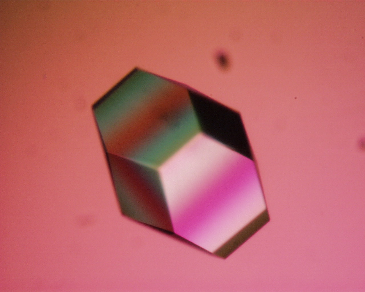

Diffraction obeys Bragg's law, the principle used to turn spot positions into lattice spacings.
Quality measures, and what each tells you:
| Measure | Definition | Good values | Meaning |
|---|---|---|---|
| Resolution | Smallest plane spacing (d) with measurable spots | Lower is better: < 1.5 Å excellent, ~2 Å good, > 3 Å limited | 3.5–4 Å: backbone and helices visible; ~2.5 Å: side chains placed; ~2 Å: waters and side-chain orientations clear; < 1.2 Å: individual atoms, some hydrogens |
| R-factor (R-work) | , the disagreement between observed and model-calculated amplitudes | About 0.20 or lower at 2 Å (rule of thumb: R ≈ resolution/10) | A random model gives about 0.59 |
| R-free | The same, computed on 5–10% of reflections left out of refinement | A few points (≤ ~0.05) above R-work | Guards against overfitting |
| B-factor (temperature factor) | in Ų, where ⟨u²⟩ is the mean-square displacement of the atom | Low (~10–30 Ų) in the ordered core; high (> 60 Ų) in flexible loops and surface side chains | High B means thermal motion or static disorder; the atom's position is less certain |
| Occupancy | Fraction of molecules in which the atom is at that position | 1.0 normally | Values below 1 for alternate conformations or partly bound ligands |
| Matthews coefficient V_M | Crystal volume per dalton of protein, | 1.6–3.5 ų/Da, typically ~2.2–2.7 | Estimates solvent content (~50%) and how many molecules are in the asymmetric unit |
The table moves from global quality (resolution, R, R-free) to atom-by-atom reliability (B-factor, occupancy). In a viewer, coloring by temperature (B-factor) shows rigid regions in blue and mobile regions in red; loops and the protein surface are usually hottest.
X-ray strengths and weaknesses. Strengths: atomic or near-atomic detail, no size limit in principle, high throughput once crystals exist, and clear views of bound ligands and water (most drug-design structures are crystallographic). Weaknesses: you must grow crystals, which can take months or fail; the result is a time-averaged static picture; crystal packing can distort surface loops; flexible regions and glycans may be missing; hydrogens are usually invisible; and the phase problem must be solved.
NMR spectroscopy
Nuclear magnetic resonance places a protein solution in a strong magnetic field and measures how nuclei with spin (¹H, and ¹⁵N and ¹³C introduced by isotope labeling) absorb radio waves. Each nucleus's chemical shift depends on its local environment, and NOE signals reveal pairs of hydrogens closer than about 5–6 Å. These distance and angle restraints are used to calculate a family of structures that all satisfy the data, so an NMR PDB entry contains an ensemble of models (often 20). Where the models agree the protein is rigid; where they spread out it is flexible.
- Advantages: no crystal needed; protein in solution near physiological conditions; directly measures dynamics and flexibility; can follow folding, binding and pH titrations.
- Limitations: practical mostly for small proteins (roughly under 30–50 kDa, larger with special labeling); needs concentrated, stable, isotope-labeled samples; generally lower precision than high-resolution X-ray; slow data analysis.
Two NMR effects show up in questions. In hydrogen–deuterium exchange, a protein dissolved in D₂O gradually loses the ¹H signals of its exchangeable amide protons as they are replaced by deuterium (which is not seen in ¹H NMR); amides on the surface exchange fast, while those buried in the core or locked in hydrogen bonds of helices and sheets exchange slowly, so the exchange rate maps structure and stability. And aromatic rings create ring-current shifts on nearby protons in a folded protein; those unusual shifts disappear when the protein unfolds and the environments average out.
Cryo-electron microscopy
In cryo-EM, a thin film of protein solution on a grid is plunged into liquid ethane so fast that water freezes as glass-like vitreous ice, trapping thousands of molecules in random orientations. A transmission electron microscope records many low-dose images (movies, called micrographs). Software then picks particles, sorts and averages particles with the same orientation into 2D class averages, combines the views into a 3D reconstruction (density map), and an atomic model is built into the map.

- Advantages: no crystals; tiny amounts of sample; ideal for large complexes, membrane proteins and flexible machines; can separate several conformations present in one sample. Resolution is now routinely 2–4 Å and better than 1.5 Å for the best samples.
- Limitations: small proteins (below roughly 50–100 kDa) give too little contrast; expensive microscopes; resolution often varies across one map (flexible edges are blurrier).
The 2017 Nobel Prize in Chemistry (Jacques Dubochet, Joachim Frank, Richard Henderson) recognized cryo-EM. Whole HA trimers and HA–antibody complexes are now routinely solved by cryo-EM, while HA ectodomains and heads with bound receptor analogs, like 3UBE (a full HA1/HA2 ectodomain), are mostly crystal structures.
Comparing the three methods
| Feature | X-ray crystallography | NMR | Cryo-EM |
|---|---|---|---|
| Sample | Crystal | Concentrated solution, isotope-labeled | Frozen thin film of solution |
| What is measured | Diffraction of X-rays by electrons | Radio-frequency absorption of nuclear spins | Electron images of single particles |
| Primary result | Electron density map | Distance and angle restraints → ensemble | Coulomb-potential density map |
| Size range | Any size that crystallizes | Small (roughly < 30–50 kDa) | Large (roughly > 50–100 kDa) |
| Typical resolution | 1–3 Å | Not stated as resolution; precision lower than good X-ray | 2–4 Å |
| Dynamics | Static average (B-factors hint at motion) | Yes, directly | Several states can be separated |
| Main bottleneck | Growing crystals; phase problem | Size limit; data analysis | Contrast for small proteins; cost |
| Share of PDB | About 80% of all entries | A few percent | Fast-growing; most new large complexes |
The table summarizes the trade-offs; questions usually give a protein's size or properties and ask which method fits. A 155 kDa soluble enzyme that crystallizes: X-ray (or cryo-EM). A 12 kDa flexible domain: NMR. A 1 MDa membrane complex: cryo-EM.
Other techniques measure structure without giving atomic coordinates: circular dichroism (CD) in the far-UV (about 190–250 nm) estimates secondary structure (α-helix shows minima at 208 and 222 nm; β-sheet a minimum near 218 nm) and follows unfolding; fluorescence of tryptophan reports whether Trp is buried or exposed; mass spectrometry gives mass, sequence and modifications; small-angle X-ray scattering (SAXS) gives overall shape in solution. Edman degradation sequences a protein from the N-terminus one residue at a time; historically, crystal structures were interpreted by combining diffraction with the separately determined amino-acid sequence.
A short history
| Year | Milestone |
|---|---|
| 1913 | W. H. and W. L. Bragg derive Bragg's law (Nobel 1915) |
| 1926 | James Sumner crystallizes urease, showing that enzymes are proteins (Nobel 1946) |
| 1951 | Linus Pauling and Robert Corey propose the α-helix and β-sheet from model building |
| 1958 | John Kendrew solves myoglobin, the first protein structure, by X-ray crystallography |
| 1959 | Max Perutz solves hemoglobin at low resolution (Kendrew and Perutz, Nobel 1962) |
| 1971 | Protein Data Bank founded with 7 structures |
| 1981 | First HA structure (H3) by Wilson, Skehel and Wiley |
| 1985 | First protein structure determined by NMR (Kurt Wüthrich; Nobel 2002) |
| 1994 | CASP, the blind structure-prediction competition, begins |
| 2017 | Nobel Prize for cryo-EM |
| 2020 | AlphaFold2 reaches near-experimental accuracy at CASP14 |
| 2024 | Nobel Prize in Chemistry: David Baker (computational protein design) and Demis Hassabis and John Jumper (AlphaFold structure prediction) |
The table is roughly the sequence tests draw from; the HA row ties the history to your protein.
Predicting and modeling structures
Anfinsen showed that a protein's sequence contains the information for its fold, so in principle structure can be computed from sequence. Methods, from most to least dependent on known structures:
- Homology (comparative) modeling: if the target's sequence is similar to a protein of known structure (a template, generally above about 30% identity), align the two sequences and build the target on the template's backbone, then model loops and side chains. Works because structure is conserved more than sequence. SWISS-MODEL is a common server.
- Threading (fold recognition): test the sequence against a library of known folds to find one it fits, even without obvious sequence similarity.
- Ab initio (de novo) prediction and molecular dynamics: start from the sequence or an extended chain and search for the lowest-energy structure using physics-based energy functions. Molecular dynamics (MD) simulates atomic motion over time using a force field (CHARMM, AMBER) whose terms include bond stretching, angle bending, torsion (dihedral) rotation, van der Waals interactions (Lennard-Jones potential) and electrostatics (Coulomb's law). MD is good for dynamics but too slow to fold most proteins from scratch.
- Deep learning (AlphaFold, RoseTTAFold): networks trained on the PDB and on multiple sequence alignments, which reveal pairs of positions that mutate together (co-evolution, a hint that they touch).
Most pipelines share four stages: backbone prediction, loop modeling, side-chain packing, and refinement.
Molecular replacement uses the same idea in crystallography: a known structure of a similar protein is placed in the new crystal's unit cell to supply initial phases, and the model is then rebuilt and refined against the new data. 3UBE was solved this way from another 2009 HA structure.
AlphaFold (Google DeepMind; John Jumper, Demis Hassabis and colleagues) predicts a 3D structure from an amino-acid sequence alone. AlphaFold2 achieved near-experimental accuracy for many proteins at CASP14 in 2020, and the AlphaFold Protein Structure Database (with EMBL-EBI) now holds over 200 million predicted structures, nearly every known protein sequence, freely available. AlphaFold 3 (2024) extends prediction to complexes with nucleic acids, ligands and ions.
| pLDDT (per-residue confidence) | Interpretation |
|---|---|
| > 90 | Very high: backbone and most side chains reliable |
| 70–90 | Confident: backbone generally correct |
| 50–70 | Low: treat with caution |
| < 50 | Very low: often an intrinsically disordered region rather than a failure |
pLDDT (predicted local distance difference test) reports local confidence for each residue; a separate PAE (predicted aligned error) plot reports confidence in the relative placement of domains. Limits: predictions are a single static model; they may be wrong for disordered regions, alternative conformations, the effect of a point mutation, or a protein's complex with a ligand; and they are not experimental evidence.
RMSD (root-mean-square deviation) is the standard single number for how similar two structures are. After the structures are optimally superimposed,
where is the distance between the i-th pair of matching atoms (usually Cα). Below about 1 Å the structures are nearly identical; 1–2 Å means the same fold with local differences; values above about 3–4 Å mean substantial differences. Because the differences are squared, a few large deviations dominate.
Distance matrices and contact maps represent a structure by the distances (or contacts below a cutoff such as 8 Å) between every pair of residues. They depend only on relative positions, so they are invariant to rotation and translation of the whole molecule, which makes them convenient inputs for prediction networks. The matrix is symmetric (entry i,j equals entry j,i) with zeros on the main diagonal; helices appear as bands next to the diagonal and sheets as lines perpendicular (antiparallel) or parallel to it. Distances are also unchanged by mirror reflection, so a distance matrix alone cannot tell a right-handed helix from a left-handed one.
De novo designed proteins ("designer proteins") are new sequences computed to fold into chosen shapes, pioneered by David Baker's group (Top7, 2003, a fold not seen in nature). Today they include designed binders that block HA's stem or receptor site, vaccine scaffolds and enzymes.
How structure-method questions are asked
- Advantage and disadvantage of X-ray, NMR or cryo-EM, or which method fits a given protein size.
- Steps of crystallography (crystallize → diffract → phase → map → model) or cryo-EM (freeze on grid → image → pick particles → 2D classes → 3D map → model).
- Definitions: resolution, R-factor, B-factor, Matthews coefficient, pLDDT, RMSD, molecular replacement, homology modeling.
- Bragg's law calculations.
- History: Sumner, Kendrew and Perutz, PDB founding, Nobel prizes (1962, 2017, 2024).
- PDB file contents: ATOM vs HETATM, HELIX and SHEET records, chain IDs.
Working in JUDE
JUDE is the browser version of Jmol that the 2027 rules name for both the build and the exam. It is free at the 3D Molecular Designs digital modeling hub, and it is the only website you may use during the event, so practice until the common operations are muscle memory. Under the menus, JUDE runs Jmol/JSmol, the open-source molecular viewer that reads PDB and mmCIF files, so anything you learn about Jmol commands carries over and questions are often written in Jmol syntax.
Molecular visualization matters for this event because a sequence or a flat picture hides the spatial relationships the exam asks about: which residues touch, which face the core, how a ligand sits in a pocket. JUDE lets you identify chains, residues, secondary structures, side chains and ligands and measure the distances between them.
| Task | What to do | Why it matters on the exam |
|---|---|---|
| Load a structure | Open the PDB file or ID | Every visualization question starts here |
| Restrict the view | Select a residue range in one chain | Your model is chain A, residues 153–252 |
| Change representation | Backbone or trace, cartoon, wireframe, ball-and-stick, spacefill | Trace and backbone show the fold; spacefill shows packing and surface; ball-and-stick shows individual side chains |
| Color by structure | Secondary-structure coloring | Fastest way to list helices and strands and the residues in them |
| Color by property | Charge, polarity, element | Shows which side chains point into the core and which face solvent |
| Show a side chain | Display the residue's side chain, label it | Needed for "name the residue contacting this ligand" questions |
| Measure | Distance between two atoms | Hydrogen bonds are roughly 2.7–3.2 Å between the heavy atoms; a contact of 8 Å is not a bond |
| Show ligands and water | Display non-protein molecules | The 2027 rules add protein–ligand interactions as a visualization topic |
The table lists the operations in the order you usually perform them. The command reference below gives the Jmol syntax behind each.
Representations
Each representation shows some things and hides others; tests ask for advantages and disadvantages.
| Representation (Jmol command) | What it draws | Advantage | Disadvantage |
|---|---|---|---|
Trace (trace) | A smooth tube through the α-carbons | Shows the chain path, exactly like your physical model | No side chains, no atoms |
Backbone (backbone) | Straight sticks joining consecutive α-carbons | Shows the fold and chain direction simply | Same as trace |
Cartoon / ribbon (cartoon, ribbon, rockets, strands) | Helices as coils or cylinders, strands as flat arrows (pointing N→C), loops as tubes | Secondary structure and chain direction obvious at a glance | Invented shapes, not atoms; hides side chains and packing |
Wireframe (wireframe) | Every bond as a thin line | All atoms and connectivity shown | Cluttered; secondary structure hard to see |
Sticks / ball-and-stick (wireframe 0.2; spacefill 0.4) | Bonds as sticks, atoms as small balls | Individual side chains and ligands clear; good for measuring | Cluttered for a whole protein |
Spacefill / CPK (spacefill) | Each atom as a sphere of its van der Waals radius | True volume, shape, packing and surface | Hides the interior and secondary structure |
Surface (isosurface molecular) | Molecular or solvent-accessible surface | Shows pockets and grooves where ligands bind | Hides everything underneath |
The rows run roughly from least to most atomic detail. A common combination for binding questions is cartoon for the protein plus ball-and-stick for the pocket side chains and the ligand.

Jmol command reference
Jmol commands work in two steps: select a set of atoms, then apply display, color, label or measurement commands, which act only on the current selection. Commands end at a line break or a semicolon. Selecting does not change the picture by itself.
ReferenceJmol commands for Protein Modeling· 25 rows
:A means chain A; 153-252 is a residue-number range; .CA names an atom. Commands are not case-sensitive.
Read the table by row when building a command and by the Notes column when a question asks why a command misbehaves.
No rows match.
| Purpose | Command | Notes |
|---|---|---|
| Select everything | select all (or select *) | Resets the selection before a new display command |
| Select a residue in one chain | select 190:A | Without :A, every chain's residue 190 is selected |
| Select a range in one chain | select 153-252:A | Your model's segment |
| Select a whole chain | select :A | Chain A is HA1 in 3UBE |
| Select by residue type | select TRP, select cys | Three-letter names |
| Select one atom | select 153:A.NE1 | Atom names as in the PDB file; CA = α-carbon |
| Predefined sets | select protein, ligand, hetero, water, solvent, helix, sheet, backbone, sidechain, acidic, basic, charged, polar, hydrophobic, aromatic, cystine | Combine with and, or, not. Jmol's hydrophobic set is Ala, Gly, Ile, Leu, Met, Phe, Pro, Trp, Tyr, Val; polar is every other amino acid |
| Combine conditions | select polar and sidechain, select hetero and not water | and, or, not; parentheses group them |
| Neighbors of a ligand | select within(5.0, ligand) | Everything within 5 Å (the distance in within() is always in Å) |
| Show only the selection | restrict 153-252:A | Hides everything else (selects and hides) |
| Show or hide | display selected, hide water | display and hide act without discarding the selection |
| Representations | trace, backbone 0.4, cartoon, ribbon, rockets, wireframe 0.15, spacefill, spacefill 50%, spacefill off | Apply to the current selection |
| Color by element | color cpk | C gray, O red, N blue, S yellow |
| Color by secondary structure | color structure | Helix magenta-pink, strand yellow, turn blue, other white |
| Color by chain | color chain | Each chain a different color |
| Color by residue type | color amino | Asp/Glu red, Lys/Arg blue, Cys/Met yellow, Ser/Thr orange, Phe/Tyr dark blue, Asn/Gln cyan, Leu/Ile/Val green, Gly white, Ala gray, Trp purple-pink, His pale blue, Pro salmon |
| Rainbow N→C | color group | Blue at the N-terminus to red at the C-terminus |
| Color by B-factor | color temperature | Blue = low B (rigid), red = high B (mobile) |
| Size by B-factor | spacefill temperature | Bigger spheres for more mobile atoms |
| Any color | color yellow, color [255,128,0] | Named colors or an RGB triple |
| Label | label %n%R | Residue name and number; label off |
| Measure distance | measure (153:A.NE1) (190:A.OD1) | Or double-click two atoms; result in Å |
| Hydrogen bonds | calculate hbonds then hbonds on | Backbone H-bonds are calculated from structure; ligand H-bonds may need measuring |
| Disulfides | ssbonds on | Draws S–S bonds; ssbonds 0.2 sets their thickness |
| View | center selected, zoom 200, spin on, reset | reset returns to the default orientation |
The two lines that cause the most trouble on tests:
- Integer versus decimal. Jmol inherited RasMol units from older software: an integer width or radius is in units of 1/250 Å, while a number with a decimal point is in ångströms.
backbone 1draws a thread 0.004 Å thick (almost invisible);backbone 1.0draws a 1 Å rod. Likewisebackbone 100equalsbackbone 0.4. The same rule applies towireframe,spacefillandtracewidths; distances insidewithin()are always in Å. - Chain ambiguity. A structure with several chains (3UBE has several copies of HA) has a residue 153 in each chain.
select 153grabs all of them;select 153:Agrabs only chain A's.
Colors to know
Element coloring follows the CPK convention (named for Corey, Pauling and Koltun's physical models): carbon gray or black, oxygen red, nitrogen blue, hydrogen white, sulfur yellow, phosphorus orange, iron orange-brown, chlorine green, sodium violet, calcium green, zinc slate blue-gray. Representation styles are often confused, so learn what each one hides: a cartoon invents ribbons that are not atoms at all, and a trace draws only α-carbons, exactly like your physical model.
| Coloring | Jmol default |
|---|---|
color structure | α-helix hot pink/magenta, 3₁₀ helix purple, π helix dark purple, β-strand yellow, turn light blue, other (coil) white |
color amino | Acidic red, basic blue, hydrophobic aliphatic green, aromatic Phe/Tyr dark blue, sulfur-containing yellow, hydroxyl orange, amides cyan, Gly white |
color temperature | Blue-white-red gradient by B-factor |
color chain | Chain A light blue, B light green, C pink, D light yellow... |
Model colors (your legend) and Jmol's default colors are different systems: your model might show hydrophobic side chains yellow while Jmol's color amino shows them green. Say which scheme you mean.
Ligands, water and other molecules
Ligands, sugars, ions and water are stored as HETATM records and are hidden or shown differently from the protein by default. select ligand (or select hetero and not water) finds them; select within(4.0, ligand) and sidechain lists the side chains that contact them. In 3UBE, look for N-acetylglucosamine (NAG) of the N-linked glycans attached to Asn residues (the LSTc chains also contain a NAG unit, so check whether a NAG is bonded to Asn), and the LSTc receptor analog (sialic acid, galactose and N-acetylglucosamine units) sitting in the receptor-binding pocket. Water appears as isolated red oxygen spheres (hydrogens are not in X-ray files).
Jmol computes backbone hydrogen bonds for secondary structure but does not automatically draw every protein–ligand hydrogen bond, so measure: if a donor N or O and an acceptor O or N are within about 3.5 Å (some tests use 3.4 Å), count it as a hydrogen bond; typical values are 2.7–3.2 Å. Other useful reference distances for measurements:
| Pair | Typical distance |
|---|---|
| Consecutive α-carbons (trans peptide) | 3.8 Å |
| Hydrogen bond, donor to acceptor heavy atoms | 2.7–3.2 Å (cutoff ~3.5 Å) |
| Salt bridge, charged N to charged O | ≤ 4 Å |
| Disulfide S–S | about 2.05 Å |
| Van der Waals contact between carbons | 3.4–4.0 Å |
| Zn²⁺ to Cys sulfur in a zinc site | about 2.3 Å |
These distances let you name an interaction from a measurement. Anything farther than about 5 Å is not a direct contact, though it may still be mediated by water.
Reading the chemical environment of a side chain
A common exam task gives you a residue and asks why it sits where it does. Work from the atoms outward:
- What is the side chain made of? Carbons only means hydrophobic; N or O means hydrogen bonding; charged groups mean salt bridges.
- What is within a few ångströms? Other side chains, backbone atoms, water, or a ligand.
- Name the interaction and say what it stabilizes: a buried hydrophobic cluster, a surface salt bridge, a disulfide bond tying two loops together, or a contact that holds a ligand.
- Predict a mutation's effect: putting a charged residue into a buried hydrophobic cluster destabilizes the fold; removing a cysteine breaks a disulfide; changing a pocket residue changes binding, not folding.
In JUDE, steps 1–3 translate into a short command sequence: select the residue and everything within about 4–5 Å (select within(4.5, 190:A)), show it as ball-and-stick colored by element, label the residues, and measure the closest N/O pairs. Spacefill of the same selection tells you whether the side chain is buried (no water can reach it) or exposed.
How visualization questions are asked
- Identify helices and strands in a residue range and list their first and last residues (use
color structureor the HELIX/SHEET records). - Name the residue(s) contacting a ligand, and the interaction type, from distances you measure.
- Write a command sequence that selects, displays and colors a feature.
- Compare representations: advantages and disadvantages of cartoon, wireframe, spacefill, trace.
- Explain a quirk: integer vs decimal units, chain ambiguity, why hydrogens are absent, what temperature coloring shows.
Strategy
- Start the model early. It must be finished before your first tournament and it travels to every level. Build it, then improve its features between tournaments as the rubric gets stricter.
- Split the preparation, not the knowledge. One partner can own the build and the JUDE operations while the other owns influenza biology and immunity, but both must be able to answer either half: the exam is 60% of the score and you sit it together.
- Put structure-specific facts on your sheets. The 3UBE segment's helix and strand boundaries, your legend's color key, the binding-site residues and loops, the α2,3 versus α2,6 contrast, the infection cycle in order, the innate and adaptive players, and the scale conversion (0.53 cm per Å).
- Rehearse JUDE under constraints. No search engine, one device, a borrowed network. Practice loading, selecting a residue range, measuring a distance and displaying a ligand without any notes.
- Protect the model in transit. It is impounded intact; a backbone that sags or a feature that falls off costs accuracy points no exam answer can win back. Build a stand and a box.
- Budget the 50 minutes. Leave a fixed block for the molecular exploration questions; they are the ones that need the device and are easiest to run out of time on.
Common pitfalls
One checklist of the mistakes that cost the most points, gathered from the pitfalls flagged through the guide plus the misreadings that show up most often in tournament answer keys. Read it the night before; each item is a complete answer on its own.
Modeling the wrong part of the protein
The build is chain A, residues 153–252, about 100 residues of the HA1 head. It is not the whole trimer, not HA1 plus HA2, and not every chain in the file.
Getting the scale or the backbone wrong
Two centimeters per amino acid, a mainly gray backbone 0.75–1.25 cm thick, built on the alpha-carbon display. A model at a different scale, in a different color scheme, or built as a cartoon ribbon does not match the specification.
3D printing the backbone
Printed parts are allowed for functional features only. The backbone has to be folded by hand from bendable material.
A full-page legend with the wrong columns
The Model Description Legend is a printed 8.5 × 5.5 in half sheet with three columns: what is displayed, how it is displayed, why it is displayed.
Adding decorative features
Features that do not explain structure or function earn nothing. A sticker, a logo or an unexplained bead is wasted effort and clutters the pocket you want judges to see.
Mixing up HA and NA
HA binds sialic acid; NA cuts it off. Antiviral drugs such as oseltamivir target NA's active site, not HA's binding pocket.
Confusing drift with shift
Drift is point mutation in HA and NA that accumulates year to year. Shift is reassortment that brings in a whole new segment, which is how the 2009 virus arose.
Calling a disulfide bond non-covalent
Disulfide bonds are covalent. The non-covalent list is hydrogen bonds, ionic interactions, the hydrophobic effect and van der Waals contacts.
Naming the structure level by the wrong count
One folded chain is tertiary structure; two or more chains assembled is quaternary. HA is a trimer, so the whole protein has quaternary structure even though your segment shows tertiary.
Saying all enzymes are proteins
Catalytic RNAs (ribozymes) are enzymes too.
Treating a zwitterion as a neutral molecule
A zwitterion carries charges that cancel. Lysine at pH 7 is net +1 and aspartate net −1, so not every free amino acid is a zwitterion at physiological pH.
Putting methionine in the disulfide club
Cysteine and methionine both contain sulfur, but only cysteine's thiol (–SH) can be oxidized to a disulfide. Methionine's sulfur sits inside a thioether with no hydrogen to lose.
Calling the hydrophobic effect "hydrophobic bonding"
Nothing bonds. Burying nonpolar side chains releases ordered water, so the gain is in the entropy of the solvent, not in an attraction between the side chains.
Mixing up the helix hydrogen-bond spacing
The α helix bonds C=O of residue i to N–H of i+4. The tighter 3₁₀ helix uses i+3 and the wider π helix i+5. A β turn spans four residues and bonds i to i+3.
Saying AlphaFold "determined" a structure
Prediction is not measurement. X-ray crystallography, NMR and cryo-EM determine structures from data; AlphaFold infers one from sequence and reports a per-residue confidence (pLDDT).
Reading a cartoon as if it were atoms
Cartoons and ribbons are drawn by the viewer, not measured: they show where the backbone runs, not where atoms are. Measure distances in a representation that shows atoms, and remember a trace shows only α-carbons, exactly like your physical model.
How tournaments test this
Three things matter about the question bank behind this guide. First, the archive's 1,962 Protein Modeling questions are dominated by the 2019-20 and 2020-21 seasons, when the event's protein was a CRISPR-associated nuclease. When we labeled the practice bank against the 2027 rules, only about three in five questions were still in scope; most of the rest are CRISPR, base editing, other retired target proteins or DNA-only molecular biology, and the October 2026 bank still tags 341 of its 1,874 questions CRISPR & Gene Editing. About 1,150 of those bank questions are exported from or match archive tests, and most of the rest come from older tournament tests the archive does not have. Practice on old tests, but skip those questions. Second, the question types are what the 2027 rules describe: about 54% free response, 32% multiple choice, 6% select-all, 4% true/false and 4% numeric, so expect to write sentences and expect select-all items with more than one correct option. Third, virus and immunity questions are almost absent from the old bank because the topic is new for 2027, while the rules give it half the exam's topic list: that is the gap between what you can practice and what you will be asked, and the influenza and immunity chapters here exist to fill it.
The table below shows roughly where the in-scope questions fell in that labeling, which is the best guide to where study time pays.
| Topic | Share of in-scope bank questions | Where this guide covers it |
|---|---|---|
| Helices, sheets, turns and their geometry | 13% | Secondary structure |
| Amino acid identity, codes and side-chain properties | 12% | Foundations: amino acids and side chains |
| Non-covalent interactions and disulfide bonds | 12% | Tertiary and quaternary structure |
| Folding thermodynamics, denaturation, chaperones | 12% | Why proteins fold, in energy terms |
| Structure levels, motifs and domains | 11% | Chemistry that drives folding |
| Protein function, enzymes, binding and inhibition | 9% | Protein function and binding |
| Mutations and sequence changes | 7% | Mutations and sequence changes |
| PDB files and structure determination | 6% | Structures, prediction and the PDB |
| Peptide bond, φ/ψ and Ramachandran plots | 6% | Chemistry that drives folding |
| Ionization, pKₐ, pI and net charge | 5% | Charge and pH |
| Post-translational modifications and targeting | 3% | Post-translational modifications |
| Jmol and JUDE operations | 3% | Working in JUDE |
| Model specifications and scale | 1% | The pre-built model |
| Viruses and immunity (rising sharply for 2027) | 1% | Viruses, influenza and immunity chapters |
Shares are rounded to the nearest percent, so they add to a little over 100.
The last line is the one to watch. Virus and immunity questions are almost absent from the old bank because the topic is new for 2027, yet the rules list it as a major exam area and give it the whole second half of the written test's topic list. Expect the 2027 crop of tests to ask far more about influenza and immunity than any archived test did, and expect hemagglutinin, sialic acid, receptor binding and influenza to join the archive's most frequent words (protein, amino, structure, acid, residues).
Most common correct answers. Keys most often name tertiary (16 questions), hydrogen bonds (14), glycine (13), proline (13), isoleucine and lysine (12 each), cysteine (11), tyrosine (11), arginine (10), quaternary and secondary (9 each). Structure-level names and residues with distinctive side chains dominate, which is a good hint about what to over-learn: the special residues (Gly, Pro, Cys, His), the four structure levels, and the bond types.
What a typical 2027 question looks like. Expect: identify a structure level or a secondary-structure element from a description or a JUDE view; pick the residue with a given property; explain how a named interaction stabilizes a fold; compute a net charge or a pI; place a step of the infection cycle in order; explain what innate or adaptive immunity does to the virus; and, at State and Nationals, explain how HA's grip on sialic acid starts an infection and what changes receptor preference.
Test yourself
Real questions from public tournament tests in our archive, limited to the parts of the bank that stay inside the 2027 scope. Click an answer to check it; each links to the full test.
- 1
Which of the following amino acids below is considered achiral?
From: University of Texas at Austin Regional, 2019-20, Division C
Practice more Protein Modeling questions →
- 1
The C terminus of a peptide chain has what charge at physiological pH?
From: University of Michigan Invitational, 2019-20, Division C
Practice more Protein Modeling questions →
- 1
Resonance causes peptide bonds in proteins to become
Practice more Protein Modeling questions →
- 1
Native proteins are generally far more stable than unfolded proteins, with the energy of these states often differing over a thousandfold.
From: University of Michigan Invitational - MI, 2025-26, Division C
1 question from this set is no longer available.
Practice more Protein Modeling questions →
- 1
A researcher proposes a model of an enzyme-catalyzed reaction in which a reactant is converted to a product. The model is based on the idea that the reactant passes through a transition state within the enzyme-substrate complex before the reactant is converted to the product. Which of the following statements best helps explain how the enzyme speeds up the reaction?
Practice more Protein Modeling questions →
- 1
Scientists analyze protein folding in both a normal water environment and a deuterated heavy water environment. They find that the mass of the native state in both of these environments is different, with the mass in the deuterated water being slightly higher. Which of the following could be a plausible explanation?
Practice more Protein Modeling questions →
Breadth reference
Tournament tests ask a long tail of one-line facts: a term you met once, a bond length, a pKₐ, the size of an antibody. This chapter collects those facts in two large lookup tables, a glossary and a table of numbers, so you can check a word or a value in seconds instead of hunting through the guide. Both tables are filterable: type in the filter box above a table (for example "helix", "Jmol" or "kDa") and only matching rows stay visible. When you build a notesheet, filter by topic and copy only the rows you keep forgetting; the earlier chapters explain the ideas, and these tables are the compact restatement.
Glossary of protein science
The glossary runs roughly in the order of the guide: amino acids and bonds, structure from secondary to quaternary, folding and stability, function and binding, protein classes and modifications, mutation, structure methods, visualization, and finally the virology and immunology vocabulary an influenza-themed exam uses. The third column says where the term earns its points: the kind of question it answers, or the link to hemagglutinin (HA) and the 3UBE model.
ReferenceGlossary of protein science· 474 rows
Terms are grouped by topic, not alphabetized; use the filter box. HA = hemagglutinin; 3UBE = the 2027 model structure (2009 H1N1 HA with receptor analog LSTc).
Filter for a single word first; many terms appear in several rows (for example "disulfide" finds the bond, cystine, reducing agents, PDI, keratin and HA0).
No rows match.
| Term | What it means | Where it matters |
|---|---|---|
| Amino acid | Molecule with an amino group and a carboxylic acid group; in proteins both sit on the same (α) carbon with an H and a side chain | Name and functional-group questions ("why are they called amino acids") |
| α-carbon (Cα) | The central carbon bonded to the amino N, the carboxyl C, an H and the side chain | The one point per residue your model shows; C2 in IUPAC numbering |
| IUPAC numbering | Carboxyl carbon is C1 and the α-carbon C2, so α-amino acids are 2-aminocarboxylic acids; side-chain carbons are β, γ, δ, ε | Naming questions; "ε-amino group of Lys" |
| Side chain (R group) | The variable group on Cα that gives each amino acid its identity and chemistry | Classifying residues; the part of a residue that differs |
| Residue | An amino acid unit inside a chain, i.e. the free amino acid minus the water lost when the peptide bond formed | Mass calculations (residue mass = free mass − 18.02 Da) |
| Proteinogenic amino acid | One of the amino acids built into proteins by ribosomes: the 20 standard ones plus selenocysteine and pyrrolysine | "How many amino acids" questions (20 standard, 22 genetically encoded) |
| Selenocysteine (Sec, U) | Cysteine with Se in place of S; inserted at a UGA codon read through by a special tRNA and an mRNA signal | "21st amino acid"; nonstandard amino acid lists |
| Pyrrolysine (Pyl, O) | Lysine derivative carrying a pyrroline ring, encoded by UAG in some archaea and bacteria | "22nd amino acid"; the only side chain with a pyrroline ring |
| Essential amino acid | One the body cannot make in sufficient amounts and must eat: His, Ile, Leu, Lys, Met, Phe, Thr, Trp, Val | "Name the essential amino acids"; mnemonic PVT TIM HaLL |
| Conditionally essential amino acid | Normally made by the body but needed in the diet in illness, growth or prematurity (e.g., Arg, Cys, Gln, Gly, Pro, Tyr) | Distractor in essential/nonessential questions |
| Zwitterion | A molecule with both + and − charges and net charge zero; a free amino acid near neutral pH (–NH₃⁺ and –COO⁻) | Definition questions; why amino acids are water-soluble crystals with high melting points |
| Amphoteric | Able to act as both an acid and a base | Amino acids and proteins; buffering |
| Chiral center | An atom (usually C) with four different substituents, giving non-superimposable mirror images | All standard amino acids except glycine are chiral |
| Enantiomers | Non-superimposable mirror-image stereoisomers (L and D forms) | Why ribosomes need L-amino acids; mirror-world questions |
| Diastereomers | Stereoisomers that are not mirror images; arise with two chiral centers | Ile and Thr have two chiral centers, so 2² = 4 stereoisomers each |
| L/D configuration | Fischer-projection naming relative to glyceraldehyde; protein amino acids are L (amino group on the left) | "Most protein amino acids are L"; L-amino acids give right-handed α helices |
| R/S configuration | Cahn–Ingold–Prelog naming by substituent priority; most L-amino acids are S | L-cysteine is R because sulfur raises the side chain's priority |
| CORN rule | Looking down the H–Cα bond, CO–R–N read clockwise identifies an L-amino acid | Quick chirality check from a 3D drawing |
| Optical activity | Rotation of plane-polarized light by a chiral compound | Glycine, being achiral, does not rotate light |
| Aliphatic | Nonaromatic hydrocarbon side chain (Ala, Val, Leu, Ile; Pro; Met often grouped here) | Hydrophobic-core residues; Met as an "honorary aliphatic" |
| Aromatic | Side chain with a planar, conjugated ring: Phe, Tyr, Trp (His is an aromatic heterocycle) | UV absorbance at 280 nm, π-stacking, cation–π |
| β-branched | Side chain branched at Cβ (Val, Ile, Thr) | Favor β strands; restrict helix formation |
| Thiol (sulfhydryl) | –SH group (cysteine) | Forms disulfides by oxidation; binds Zn²⁺ and Fe |
| Thioether | C–S–C group (methionine) | Met's sulfur cannot form disulfides |
| Imidazole | Five-membered aromatic ring with two N atoms (histidine) | Side-chain pKₐ ≈ 6, so His titrates near physiological pH; binds Ni²⁺ in His-tags |
| Guanidinium | Planar, resonance-stabilized group of arginine, pKₐ ≈ 12.5 | Arg is the most basic residue; binds phosphates and carboxylates |
| Indole | Benzene fused to a pyrrole (tryptophan) | Largest side chain; strongest 280 nm absorber; intrinsic fluorescence |
| Phenol | Aromatic ring bearing –OH (tyrosine) | Tyr phosphorylation; pKₐ ≈ 10 |
| Carboxamide (amide side chain) | –CONH₂ of Asn and Gln; polar, not ionizable | Asn is the N-glycosylation site; amides are not basic |
| Imino acid (secondary amine) | Proline's ring bonds back to the backbone N, so Pro has no backbone N–H | Helix breaker; cannot donate a backbone H-bond |
| Polar uncharged | Side chain with O or N that H-bonds but carries no net charge at pH 7 (Ser, Thr, Asn, Gln, Cys, Tyr) | Surface residues; classification questions |
| Acidic residue | Asp and Glu: side-chain carboxylate, negatively charged at pH 7 | Salt bridges; red in common color schemes |
| Basic residue | Lys, Arg, His: side chains that accept a proton; Lys and Arg positive at pH 7 | DNA and sialic-acid-carboxylate binding; blue in color schemes |
| Hydropathy index | A numeric scale of side-chain hydrophobicity; Kyte–Doolittle runs from Ile +4.5 to Arg −4.5 | Predicting buried residues and transmembrane helices |
| Amphipathic | Having a hydrophobic part and a hydrophilic part (a helix with a nonpolar face, a lipid) | Helical wheels; membrane-surface helices |
| Peptide bond | The amide C–N bond joining the carbonyl C of one residue to the amide N of the next | Primary structure; rigid, planar, almost always trans |
| Condensation (dehydration synthesis) | Reaction that joins two molecules with loss of water; forms each peptide bond | n residues are joined by n − 1 peptide bonds, releasing n − 1 waters |
| Hydrolysis | Splitting a bond by adding water; the reverse of condensation | Proteases; acid hydrolysis for composition analysis |
| Peptide, oligopeptide, polypeptide | Short chains (a few residues), up to about 20, and longer chains; "protein" usually means a folded, functional polypeptide (above about 50 residues) | Vocabulary questions |
| N-terminus | The chain end with the free α-amino group; sequences are written from it | Written first; +1 charge at pH 7; Edman degradation starts here |
| C-terminus | The chain end with the free α-carboxyl group | Written last; −1 charge at pH 7 |
| Backbone (main chain) | The repeating N–Cα–C(=O) atoms of every residue | Secondary-structure H-bonds; trace and backbone displays |
| Peptide plane | The six atoms Cα–C(=O)–N(H)–Cα that lie in one plane because of resonance | Only φ and ψ rotate, so the backbone is a chain of rigid plates |
| Resonance (peptide) | Delocalization of the N lone pair into the C=O, giving the C–N bond about 40% double-bond character | Explains planarity, shorter C–N bond, low reactivity |
| Cis / trans peptide | The two isomers about the peptide bond (ω ≈ 0° or 180°); trans is favored about 1000 : 1 except before Pro | Cis-Pro in turns; peptidyl-prolyl isomerases |
| Isopeptide bond | An amide bond involving a side-chain amino or carboxyl group (e.g., Lys ε-NH₂ to ubiquitin's C-terminus) | Ubiquitination; cross-linked proteins |
| pKₐ | The pH at which a group is half protonated; | Charge-at-pH questions; seven residues have an ionizable side chain |
| Henderson–Hasselbalch equation | Fraction protonated; one pH unit above pKₐ ≈ 91% deprotonated | |
| Isoelectric point (pI) | pH at which a molecule's average net charge is zero; the average of the two pKₐ values flanking the neutral form | Isoelectric focusing; solubility is lowest at the pI |
| Titration curve | Plot of pH against added base; flat buffering regions center on each pKₐ | Reading pKₐ and pI off a graph |
| Strecker synthesis | Laboratory synthesis of α-amino acids from an aldehyde, ammonia and cyanide via an α-aminonitrile, then hydrolysis | Gives a racemic (D + L) mixture; chemistry-themed questions |
| Covalent bond | Bond formed by shared electron pairs; peptide and disulfide bonds in proteins | Strongest bonds; primary structure and cross-links |
| Noncovalent interaction | Weak interaction without shared electron pairs: H-bonds, ionic, van der Waals, hydrophobic | Hold secondary, tertiary and quaternary structure |
| Hydrogen bond | Attraction between an H on an electronegative donor (N, O) and a lone pair on an acceptor (N, O) | Secondary structure; specificity of binding |
| H-bond donor / acceptor | Donor = N–H or O–H group supplying the H; acceptor = O or N lone pair receiving it | Backbone N–H donates, C=O accepts; Pro cannot donate |
| Electronegativity | An atom's pull on shared electrons (F > O > N > C ≈ S > H) | Why C=O oxygen is δ− and amide H is δ+ |
| Partial charge (δ) | Fractional charge on an atom from uneven electron sharing | Amide N–H and C=O dipoles; helix dipole |
| Salt bridge | Ionic interaction between oppositely charged side chains, often also H-bonded (Asp/Glu with Lys/Arg/His) | Tertiary and quaternary stability; mutations that remove a charge |
| Ionic (electrostatic) interaction | Coulomb attraction or repulsion between charges; weakened by high-dielectric water and salt | Charge-reversal mutations; electrostatic steering |
| Van der Waals interaction | Short-range attraction between all atoms at contact distance, with sharp repulsion if they overlap | Close packing of the core; contributes heavily in total |
| London dispersion force | The van der Waals attraction between temporary, induced dipoles | Nonpolar side-chain packing |
| Van der Waals radius | Effective radius of an atom's electron cloud for nonbonded contact | Spacefill sphere size; steric clashes |
| Steric clash (hindrance) | Atoms forced closer than their van der Waals radii allow | Forbidden Ramachandran regions; bulky residues |
| Hydrophobic effect | Nonpolar groups cluster away from water because ordered water around them is released, increasing solvent entropy | The main driving force of folding |
| Clathrate (ordered water shell) | Cage-like, more ordered water around a nonpolar surface | Its release on burial is the entropy gain of the hydrophobic effect |
| Dipole–dipole interaction | Attraction between permanent dipoles (e.g., aligned peptide groups) | Helix dipole; polar interactions |
| Ion–dipole interaction | Attraction between a charge and a polar group (e.g., Na⁺ and water, Lys⁺ and a C=O) | Hydration; helix capping |
| Cation–π interaction | Attraction between a cation (Lys, Arg, metal) and the electron-rich face of an aromatic ring | Binding pockets; a non-obvious noncovalent force |
| π-stacking | Face-to-face or edge-to-face packing of aromatic rings | Aromatic clusters; ligand binding |
| Disulfide bond | Covalent S–S bond between two cysteine thiols, formed by oxidation | Main covalent cross-link; extracellular proteins; HA1–HA2 link |
| Cystine | Two cysteines joined by a disulfide | Jmol select cystine; keratin of hair |
| Oxidation / reduction (thiol) | 2 R–SH → R–S–S–R + 2 H⁺ + 2 e⁻ is oxidation; adding electrons breaks it (reduction) | The reducing cytosol keeps disulfides rare inside cells |
| Reducing agent | Reagent that breaks disulfides: dithiothreitol (DTT), β-mercaptoethanol (BME), TCEP | Reducing SDS-PAGE; separates disulfide-linked subunits |
| Metal coordination | Dative bonds from side-chain atoms (Cys S, His N, Asp/Glu O) to a metal ion | Zinc fingers, heme iron, Ca²⁺ in EF hands |
| Dielectric constant | How strongly a medium weakens electrostatic forces (water ≈ 80, protein interior ≈ 2–20, hexane ≈ 2) | Ionic interactions are much stronger when buried |
| Desolvation penalty | Free-energy cost of stripping water from a polar or charged group | Why burying a charge destabilizes a protein |
| Lennard-Jones potential | Energy model for van der Waals contact: an attractive r⁻⁶ term and a repulsive r⁻¹² term | Force fields and molecular dynamics |
| Force field | Set of energy terms (bond stretch, angle bend, torsions, van der Waals, electrostatics) used to compute a structure's energy | Molecular dynamics; refinement |
| Primary structure | The amino acid sequence, N → C | Held by peptide bonds |
| Secondary structure | Regular local backbone conformation (helix, strand, turn) held by backbone C=O···H–N bonds | "Which level" questions; DSSP output |
| Supersecondary structure (motif) | A small, recurring combination of secondary elements (β-α-β, helix-turn-helix, hairpin, Greek key) | Between secondary and tertiary in some textbooks |
| Tertiary structure | Full three-dimensional fold of one chain, held by side-chain interactions, the hydrophobic core and disulfides | HA1 head domain; most of what denaturation destroys |
| Quaternary structure | Arrangement of two or more chains (subunits) | HA homotrimer; hemoglobin α₂β₂ |
| Quinary structure | Transient, weak contacts proteins make in the crowded cell | Distractor; not one of the four classical levels |
| α helix | Right-handed helix, 3.6 residues per turn, C=O(i) H-bonded to N–H(i + 4) | Most common secondary structure |
| 3₁₀ helix | Tighter helix with 3 residues per turn and i → i + 3 H-bonds (10-atom H-bonded ring) | Short helices at helix ends; DSSP "G" |
| π helix | Wider helix with about 4.4 residues per turn and i → i + 5 H-bonds | Usually a π-bulge in an α helix; DSSP "I" |
| π-bulge | Single residue inserted into an α helix, creating a short π-helical turn of about 7 residues | Often at functional sites |
| Left-handed α helix (αL) | Helix with positive φ and ψ; sterically strained for L-amino acids | Single Gly or Asn residues; rarely a full helix |
| Polyproline II (PPII) helix | Extended left-handed helix with 3 residues per turn and no internal H-bonds | Proline-rich segments; collagen chains; unfolded proteins |
| Collagen triple helix | Three left-handed PPII-like chains wound into a right-handed superhelix, Gly every third residue | Fibrous protein; Gly-X-Y repeat |
| Helix dipole | Net dipole from aligned peptide groups: partial + at the N-terminus, − at the C-terminus | Negative residues near the N-cap, positive near the C-cap stabilize the helix |
| Helix capping | Side chains (often Ser, Thr, Asp, Asn at the N-cap; Gly at the C-cap) that satisfy the unpaired backbone H-bonds at helix ends | Helix start and stop signals |
| Helical wheel | Projection of a helix down its axis, residues 100° apart | Finding the hydrophobic face of an amphipathic helix |
| Heptad repeat | Seven-residue repeat abcdefg in which a and d are hydrophobic | Coiled coils and leucine zippers |
| Helix propensity | Tendency of a residue to adopt helical φ/ψ; Ala, Leu, Glu, Met, Gln, Lys high; Pro and Gly low | Secondary-structure prediction; design questions |
| Helix breaker | Residue that terminates or kinks helices, chiefly Pro (no N–H, rigid ring) and to a lesser extent Gly | "Why is Pro rare inside helices" |
| Helix–coil cooperativity | Nucleating the first turn is costly, but each added residue then adds one H-bond, so helix formation is cooperative (sharp transitions) | Zimm–Bragg σ (nucleation) and s (propagation) parameters |
| β strand | Nearly fully extended segment of backbone; side chains alternate sides | Building block of sheets; arrows in cartoons |
| β sheet | Two or more strands H-bonded side by side (backbone C=O to N–H) | Main secondary structure of the HA1 head |
| Antiparallel sheet | Adjacent strands run in opposite directions; H-bonds nearly linear | Slightly more stable than parallel; hairpins |
| Parallel sheet | Adjacent strands run the same way; H-bonds angled; needs long crossover connections | β-α-β units, Rossmann folds, TIM barrels |
| Mixed sheet | A sheet with both parallel and antiparallel strand pairs | Many α/β proteins |
| Pleat | Zig-zag of a sheet as successive Cα atoms go slightly up and down | Origin of "β-pleated sheet" |
| Sheet twist | Strands are not flat; sheets have a right-handed twist viewed along the strands | Real sheets in cartoons look twisted |
| β bulge | Irregularity where one strand has an extra residue opposite its partner | Breaks the alternation of side chains |
| Edge strand | Strand at the edge of a sheet with unpaired H-bond groups | Aggregation hot spots; protected by charges or prolines |
| Strand register | Which residues pair across two strands | Hairpin design; amyloid structure |
| Turn (reverse turn) | Short segment that reverses chain direction | Connect strands and helices; usually on the surface |
| β turn | Four-residue turn with C=O(i) H-bonded to N–H(i + 3); types I, II, I′, II′ and others | Gly often at position 3 of type II; Pro at position 2 |
| γ turn | Three-residue turn with an i → i + 2 H-bond | Less common than β turns |
| β hairpin | Two antiparallel strands joined by a short turn | Simplest β motif; low contact order |
| Loop (coil) | Irregular, non-repeating backbone segment | Often flexible, surface-exposed, high B-factor |
| Ω loop | Loop whose ends are close together, shaped like the Greek letter omega | Surface loops in many proteins |
| Random coil | An ensemble of disordered conformations with no stable structure | Unfolded state; CD minimum near 198–200 nm |
| Torsion (dihedral) angle | Rotation about a bond, defined by four consecutive atoms | φ, ψ, ω and χ angles |
| φ (phi) | Backbone torsion about the N–Cα bond | Ramachandran x-axis |
| ψ (psi) | Backbone torsion about the Cα–C bond | Ramachandran y-axis |
| ω (omega) | Torsion about the peptide C–N bond, near 180° (trans) or 0° (cis) | Fixed by resonance; not a free variable |
| χ angles and rotamers | Side-chain torsions (χ1, χ2...) and their preferred discrete combinations (rotamers) | Side-chain modeling; alternate conformations |
| Ramachandran plot | Plot of ψ against φ showing sterically allowed backbone conformations | α (lower left), β (upper left), αL (upper right) |
| Ramachandran outlier | Residue with φ/ψ in a disallowed region | Flags errors in a structure model; Gly excepted |
| DSSP | Algorithm that assigns secondary structure from 3D coordinates using backbone H-bond patterns (H, G, I, E, B, T, S) | It assigns from a known structure; it does not predict from sequence |
| Secondary-structure prediction | Estimating helix/strand/coil per residue from sequence (Chou–Fasman propensities, GOR, neural networks trained on PDB data) | Contrast with DSSP assignment |
| β-α-β motif | Two parallel strands joined by a crossover helix; almost always right-handed | Rossmann fold building block |
| Rossmann fold | Alternating β-α-β-α-β units forming a parallel sheet that binds a nucleotide (NAD⁺, FAD) | Classic α/β fold; dehydrogenases |
| Helix-turn-helix (HTH) | Two helices joined by a short turn; the "recognition helix" sits in the DNA major groove | Bacterial repressors; homeodomains |
| Helix-loop-helix (bHLH) | Two helices joined by a longer loop, dimerizing; basic region binds DNA | Transcription factors such as Myc |
| EF hand | Helix–loop–helix that binds Ca²⁺ in a 12-residue loop | Calmodulin, troponin C |
| Zinc finger | Small domain whose fold is held by a Zn²⁺ coordinated by Cys and His (classic C2H2: ββα) | DNA binding; each classic finger reads about 3 bp |
| Leucine zipper (bZIP) | Coiled-coil dimerization helices with Leu at every seventh position, plus a basic DNA-binding region | Transcription factors (Fos/Jun, GCN4) |
| Coiled coil | Two to seven α helices wound around each other in a left-handed supercoil, held by a hydrophobic heptad seam | Keratin, leucine zippers, the HA2 stem |
| Greek key | Four antiparallel strands where strand 4 folds back next to strand 1 | Common β topology; immunoglobulin and jelly-roll folds |
| β meander | Antiparallel strands each joined to its neighbor by a hairpin | Simplest sheet topology |
| β sandwich | Two β sheets packed face to face with a hydrophobic core between | Immunoglobulin domains |
| Jelly roll | β sandwich of eight strands in which hairpins are "rolled" so strand pairs interleave | Many viral capsid proteins; HA1 head |
| β barrel | A sheet closed on itself into a cylinder | Porins, lipocalins, GFP |
| TIM barrel | Eight parallel β strands surrounded by eight α helices, (βα)₈ | The most common enzyme fold |
| β propeller | Four to eight twisted four-stranded "blades" arranged around a central axis | Influenza neuraminidase head (six blades) |
| Four-helix bundle | Four roughly parallel or antiparallel helices packed around a core | Cytochrome b562, growth hormone, ferritin |
| Immunoglobulin (Ig) fold | β sandwich of 7–9 strands in two sheets with a conserved disulfide | Antibody domains, MHC, many receptors |
| Flavodoxin-like fold | A central five-stranded parallel sheet flanked by helices on both sides | α/β fold example |
| Domain | A compact unit of a chain that can fold, and often function, on its own | HA1 head vs HA2 stem; multi-domain proteins |
| Fold | A specific arrangement and connectivity of secondary elements shared by many proteins | Fold libraries; threading |
| Topology | The order and direction of secondary elements along the chain, ignoring exact geometry | Topology diagrams of sheets |
| CATH / SCOP classes | Structure classifications; SCOP top classes all-α, all-β, α/β (alternating) and α+β (segregated); CATH uses mainly-α, mainly-β and α–β (both mixed kinds together) | Classifying a fold from a picture |
| Sequence motif | A short conserved sequence pattern linked to a function (e.g., P-loop GxxxxGK[ST], NPA in aquaporins, N-X-S/T glycosylation sequon) | Identifying function from sequence |
| SH2 / SH3 domain | Modules that bind phosphotyrosine peptides (SH2) or proline-rich peptides (SH3) | Signal transduction; PTM-reading domains |
| Transmembrane helix | A run of about 20 hydrophobic residues spanning the lipid bilayer as an α helix | Hydropathy plots; HA's membrane anchor |
| Positive-inside rule | Lys and Arg are enriched on the cytoplasmic side of membrane proteins | Predicting membrane topology |
| Hydrophobic core | The buried interior of a globular protein, packed with nonpolar side chains | Core mutations are the most destabilizing |
| Interface | Surface buried when two chains or a ligand meet | HA trimer interface; antibody footprints |
| Subunit (protomer) | One chain, or the repeating unit, of a multi-chain protein | HA protomer = HA1 + HA2 |
| Homo- / hetero-oligomer | Assembly of identical (homo) or different (hetero) subunits | HA is a homotrimer; hemoglobin a heterotetramer |
| Symmetry (Cₙ) | n-fold rotational symmetry of an oligomer | HA trimer has 3-fold (C3) symmetry |
| Solvent-accessible surface area (SASA) | Area traced by the center of a water-sized probe rolled over the protein | Buried vs exposed residues; hydrophobic effect estimates |
| Packing density | Fraction of space filled by atoms; protein interiors are about 0.7–0.75, like a packed solid | "Proteins are well packed, not hollow" |
| Contact order | Average sequence separation of residues in contact in the native state, normalized for length | Low contact order (hairpins, helices) → faster folding |
| Native state | The folded, functional conformation | Folding questions; "native conformation" |
| Denatured (unfolded) state | Ensemble of non-native, largely disordered conformations | Loss of function; still has intact peptide bonds |
| Denaturation | Loss of secondary, tertiary and quaternary structure without breaking peptide bonds | Heat, pH extremes, urea, detergents, organic solvents |
| Renaturation | Spontaneous refolding of a denatured protein when the denaturant is removed | Anfinsen's ribonuclease experiment |
| Anfinsen's dogma (thermodynamic hypothesis) | The native fold is the global free-energy minimum and is encoded in the sequence; it needs a unique, stable and kinetically accessible minimum | Sequence determines structure; prions are the famous exception |
| Levinthal's paradox | A chain could never find its fold by random search of all conformations, yet real proteins fold in microseconds to seconds | Folding is guided, not random |
| Folding funnel (energy landscape) | Picture of folding as downhill flow on a rugged, funnel-shaped free-energy surface toward the native state | Explains multiple routes and kinetic traps |
| Principle of minimal frustration | Evolved sequences have native contacts that are mutually consistent, giving smooth, funneled landscapes (Bryngelson and Wolynes) | Named-principle questions |
| Kinetic trap | A local free-energy minimum where a misfolded chain can get stuck | Misfolding; why chaperones help |
| Molten globule | Compact intermediate with native-like secondary structure but loosely packed side chains | Folding intermediates |
| Hydrophobic collapse | Early, rapid compaction of the chain driven by burying nonpolar groups | Folding mechanism models |
| Nucleation–condensation | Model in which a small folding nucleus forms and the rest of the structure condenses around it | Folding mechanism models |
| Framework model | Model in which secondary structure forms first and then docks into tertiary structure | Folding mechanism models |
| Folding transition state | The highest free-energy point on the folding pathway that every folding molecule must pass | Saddle point of the landscape; sets the rate |
| Two-state folding | Only native and unfolded states are significantly populated | Basis of ΔG and T_m calculations |
| Gibbs free energy (ΔG) | ; negative means spontaneous | Folding has ΔG < 0 |
| Enthalpy (ΔH) | Heat change, mostly from bonds and interactions made or broken | Folding enthalpy from H-bonds and van der Waals contacts |
| Entropy (ΔS) | Measure of disorder (number of accessible microstates) | Chain loses entropy on folding; water gains it |
| Conformational entropy | Entropy of the chain's own flexibility, lost on folding | The main force opposing folding |
| Melting temperature (T_m) | Temperature at which half the molecules are unfolded (ΔG_unfold = 0) | Stability comparisons; DSC and CD melts |
| ΔC_p (heat-capacity change) | Increase in heat capacity on unfolding, from newly exposed nonpolar surface | Curved stability vs temperature; cold denaturation |
| Cold denaturation | Unfolding at low temperature, predicted by the curved stability curve | Stability is maximal at an intermediate temperature |
| Marginal stability | Native proteins are only about 20–60 kJ/mol more stable than unfolded ones | Why one mutation or a fever can matter |
| Chaotrope | Solute that disrupts water structure and weakens the hydrophobic effect (urea, guanidinium, SCN⁻) | Chemical denaturation |
| Kosmotrope | Solute that strengthens water structure and stabilizes folded proteins (sulfate, phosphate) | Hofmeister series; salting out |
| Hofmeister series | Ranking of ions by how they stabilize (kosmotropes such as sulfate) or destabilize (chaotropes such as thiocyanate) proteins | Salt effects on stability and solubility |
| Salting out | Precipitating proteins at high salt (e.g., ammonium sulfate) | Purification and crystallization |
| Metastable protein | Protein held in a higher-energy state by a kinetic barrier and released by a trigger | HA's pre-fusion form; serpins |
| Cotranslational folding | Folding of the nascent chain while it is still being made on the ribosome | Why in-vitro refolding can be slower |
| Molecular chaperone | Protein that binds exposed hydrophobic surfaces of non-native chains to prevent aggregation (e.g., Hsp70) | Folding helpers; does not encode the fold |
| Chaperonin | Barrel-shaped chaperone that encloses a chain in a cavity to fold (GroEL/GroES in bacteria, TRiC/CCT in eukaryotes) | Chaperone vs chaperonin questions |
| Heat shock protein (Hsp) | Chaperones induced by heat or stress, named by mass in kDa (Hsp60, Hsp70, Hsp90) | Stress response |
| Protein disulfide isomerase (PDI) | ER enzyme that forms and shuffles disulfides | Correct disulfide pairing |
| Peptidyl-prolyl isomerase | Enzyme that speeds cis–trans isomerization of X–Pro bonds | Slow folding steps |
| Unfolded protein response | ER stress signaling that boosts chaperones and slows translation when misfolded proteins build up | Cell biology of misfolding |
| Ubiquitin–proteasome system | Ubiquitin chains (Lys48-linked) tag proteins for degradation by the 26S proteasome | Disposal of misfolded proteins |
| Aggregation | Nonspecific association of misfolded chains, often via exposed hydrophobic surfaces | Loss of function; disease |
| Amyloid | Insoluble fibrils with a cross-β structure | Alzheimer's (Aβ, tau), Parkinson's (α-synuclein), type 2 diabetes (amylin) |
| Cross-β structure | Strands run perpendicular to the fibril axis, sheets parallel to it | Diagnostic X-ray reflections at 4.7 Å and about 10 Å |
| Prion | Misfolded protein (PrPˢᶜ) that converts the normal form (PrPᶜ) into more of itself | CJD, kuru, BSE; infectious protein without nucleic acid |
| Intrinsically disordered protein (IDP) | Protein or region with no stable fold under physiological conditions, often rich in charged and polar residues | Exceptions to "structure determines function"; tau |
| Hydrogen–deuterium exchange | Exchange of amide H for D in D₂O, slowed where the H is H-bonded or buried | Probing folding and protection (NMR, mass spec) |
| Enzyme | Biological catalyst (usually protein, sometimes RNA) that speeds a reaction without being consumed | Function questions |
| Ribozyme | Catalytic RNA (e.g., the ribosome's peptidyl transferase center) | "Enzymes can also be made of RNA" |
| Active site | Pocket or cleft where substrate binds and catalysis occurs | Conserved residues; inhibitor target |
| Substrate | Molecule an enzyme acts on | Specificity questions |
| Activation energy (Eₐ) | Free-energy barrier between reactants and the transition state | Enzymes lower it; they do not change ΔG of the reaction |
| Transition-state stabilization | Enzymes bind the transition state more tightly than the substrate | Core of catalysis; transition-state analog inhibitors |
| Catalytic triad | Ser–His–Asp (or Cys–His) arrangement making a strong nucleophile in proteases | Serine and cysteine proteases |
| Lock and key | Model in which substrate fits a rigid, pre-formed site (Fischer) | Contrast with induced fit |
| Induced fit | Model in which binding reshapes the site to fit the substrate (Koshland) | Conformational change on binding |
| Conformational selection | Ligand selects and stabilizes a pre-existing conformation from an ensemble | Modern view alongside induced fit |
| Cofactor | Non-protein helper an enzyme needs: a metal ion or an organic coenzyme | Zn²⁺, Fe²⁺, Mg²⁺; NAD⁺, FAD |
| Coenzyme | Organic cofactor, often vitamin-derived (NAD⁺ from niacin, FAD from riboflavin) | Cofactor classification |
| Prosthetic group | Cofactor bound tightly or covalently (heme in hemoglobin) | Cofactor classification |
| Apoenzyme / holoenzyme | Enzyme without / with its cofactor | Vocabulary |
| Ligand | Any molecule or ion that binds specifically to a protein | Jmol select ligand; LSTc in 3UBE |
| Binding pocket | Region whose shape and chemistry complement a ligand | Receptor binding site of HA |
| Affinity | Strength of a single binding interaction, measured by the dissociation constant K_d (lower K_d = tighter) | Antibody and receptor binding |
| Avidity | Combined strength of several simultaneous binding interactions, far greater than any single one | IgM's 10 sites; trimeric HA binding many sialic acids |
| Specificity | Preference for one partner over others, from shape and chemical complementarity | Enzymes, antibodies, HA linkage preference |
| Competitive inhibition | Inhibitor binds the active site and competes with substrate | Raises apparent K_M; overcome by more substrate (oseltamivir) |
| Noncompetitive inhibition | Inhibitor binds elsewhere equally well to E and ES, lowering activity | Lowers V_max |
| Uncompetitive inhibition | Inhibitor binds only the enzyme–substrate complex | Lowers both V_max and K_M |
| Mixed inhibition | Inhibitor binds E and ES with different affinities | General case of the above |
| Allosteric regulation | Binding at one site changes activity at a distant site via a conformational change | Hemoglobin O₂ cooperativity; feedback inhibition |
| Cooperativity | Binding at one site changes affinity at others (positive in hemoglobin) | Sigmoidal binding curve |
| Irreversible (covalent) inhibitor | Inhibitor that bonds covalently to the enzyme, inactivating it permanently | Aspirin acetylates COX; penicillin |
| Feedback inhibition | A pathway's end product inhibits an early enzyme | Regulation questions |
| Zymogen (proenzyme) | Inactive precursor activated by cleavage (trypsinogen, pepsinogen) | Prevents self-digestion; compare HA0 activation |
| Diffusion limit | Maximum rate set by how fast substrate reaches enzyme, about 10⁸–10⁹ M⁻¹ s⁻¹ | "Catalytically perfect" enzymes |
| Electrostatic steering | Charged residues guide a charged substrate into the active site, letting rates approach or exceed simple diffusion estimates | Superoxide dismutase, acetylcholinesterase |
| Enzyme Commission (EC) classes | Seven classes: oxidoreductases, transferases, hydrolases, lyases, isomerases, ligases, translocases | Classifying a reaction |
| Protease | Enzyme that hydrolyzes peptide bonds (serine, cysteine, aspartic, metallo-) | HA0 cleavage by host trypsin-like proteases |
| Kinase / phosphatase | Enzymes that add / remove phosphate groups | Signaling; phosphorylation questions |
| Nuclease | Enzyme that cleaves DNA or RNA | PA endonuclease of influenza |
| Isozyme | Different forms of an enzyme (distinct sequences, often from different genes) catalyzing the same reaction | Vocabulary |
| Globular protein | Compact, roughly spherical, water-soluble protein with a hydrophobic core | Enzymes, antibodies, HA1 head |
| Fibrous protein | Elongated protein with repetitive structure, usually structural and insoluble | Collagen, keratin, silk fibroin |
| Membrane protein | Protein embedded in (integral) or attached to the surface of (peripheral) a lipid bilayer | Hydrophobic outer surface in the bilayer |
| Type I membrane protein | Single-pass membrane protein with its N-terminus outside the cell and C-terminus in the cytoplasm | HA is type I; NA is type II (N-terminal anchor, C-terminus outside); M2 is N-out like HA but has no cleaved signal peptide, so it is usually called type III |
| GPCR | G-protein-coupled receptor with 7 transmembrane helices | Rhodopsin, β-adrenergic receptor |
| Rhodopsin | GPCR in rod cells whose bound retinal isomerizes on absorbing light, activating the G protein transducin | Light → signal conversion |
| Ion channel | Membrane protein forming a gated pore for ions | Faster than GPCR signaling; M2 proton channel |
| Transporter | Membrane protein moving solutes by conformational change (uniporter, symporter, pump) | Vocabulary |
| Receptor | Protein that binds a signal (hormone, ligand) and triggers a response | Function categories |
| Transcription factor | Protein that binds DNA to regulate transcription | Zinc fingers, HTH, bZIP |
| Structural protein | Protein that provides support or shape | Collagen, keratin, actin |
| Motor protein | Protein that converts ATP energy into movement (myosin, kinesin, dynein) | Function categories |
| Collagen | Triple-helical fibrous protein of connective tissue, Gly-X-Y repeat, Pro/hydroxyproline-rich | Most abundant protein in mammals; scurvy |
| α-Keratin | Coiled-coil fibrous protein of hair and nails, cross-linked by disulfides | Perms break and reform disulfides |
| Silk fibroin | Stacked antiparallel β sheets of Gly-Ala-Gly-Ala-Gly-Ser repeats | Strong, flexible β-sheet fiber |
| Hemoglobin / myoglobin | O₂-binding heme proteins; myoglobin is one chain, hemoglobin α₂β₂ and cooperative | Allostery, Bohr effect, sickle cell |
| Bohr effect | Lower pH and higher CO₂ reduce hemoglobin's O₂ affinity, partly via His protonation and new salt bridges | pKₐ and salt-bridge questions |
| Heme | Protoporphyrin ring with a central Fe²⁺, bound to the proximal His | Prosthetic group |
| Lectin | Protein that binds specific carbohydrates | HA is a viral lectin for sialic acid |
| Glycoprotein | Protein carrying covalently attached sugars (glycans) | HA and NA; most secreted proteins |
| Post-translational modification (PTM) | Covalent change to a protein after translation | Expands chemistry beyond 20 amino acids |
| Phosphorylation | Addition of phosphate to Ser, Thr or Tyr (His in bacteria) by kinases | Signaling switches; about a third of human proteins |
| N-linked glycosylation | Glycan attached to the amide N of Asn in an Asn-X-Ser/Thr sequon (X not Pro), starting in the ER | HA glycan shield; masking antigenic sites |
| O-linked glycosylation | Glycan attached to the O of Ser or Thr, added in the Golgi | Mucins |
| Sequon | The N-X-S/T consensus that allows N-glycosylation | Glycan sites gained in antigenic drift |
| Acetylation | Addition of an acetyl group to the N-terminus or a Lys ε-amino group | Lys loses its + charge; histone acetylation opens chromatin |
| N-terminal acetylation | Acetyl added to the α-amino group, on most human proteins | Affects stability and degradation signals |
| Methylation | Addition of methyl groups to Lys or Arg (proteins) or cytosine (DNA) | Histone code; gene silencing |
| Ubiquitination | Attachment of ubiquitin to Lys via an isopeptide bond | Lys48 chains → proteasome; Lys63 chains → signaling |
| SUMOylation | Attachment of the small ubiquitin-like modifier to Lys | Nuclear transport; regulation |
| Palmitoylation (S-acylation) | Reversible attachment of palmitate (C16) to Cys by a thioester | Membrane anchoring; HA's cytoplasmic tail cysteines |
| Myristoylation | Attachment of myristate (C14) to an N-terminal Gly | Membrane anchoring |
| Prenylation | Attachment of a farnesyl or geranylgeranyl group to a C-terminal Cys | Ras membrane targeting |
| Hydroxylation | Addition of –OH, e.g., Pro → hydroxyproline in collagen (vitamin C-dependent) | Scurvy; collagen stability |
| γ-Carboxylation | Vitamin K-dependent conversion of Glu to γ-carboxyglutamate (Gla) for Ca²⁺ binding | Blood-clotting factors |
| GPI anchor | Glycolipid anchoring a protein's C-terminus to the outer leaflet of the membrane | Prion protein |
| Proteolytic processing | Activating or maturing a protein by cutting its chain | HA0 → HA1 + HA2; zymogens; insulin |
| Signal peptide | N-terminal sequence (hydrophobic core of about 7–15 residues) directing a protein into the ER | HA's 17-residue signal peptide is cleaved off |
| Signal recognition particle (SRP) | Ribonucleoprotein that binds a signal peptide and docks the ribosome on the ER | Secretory pathway |
| Codon | Three-nucleotide mRNA unit specifying an amino acid or stop | Translation questions |
| Anticodon | Complementary three-base sequence on a tRNA | Base pairing with codons |
| Start codon | AUG (Met), which also sets the reading frame | Why Met is first |
| Stop codon | UAA, UAG, UGA, which code for no amino acid in the standard code | Nonsense mutations |
| Degeneracy (redundancy) | Most amino acids have more than one codon | Silent mutations |
| Wobble | Non-standard pairing at the codon's third base lets one tRNA read several codons | It does not let one mRNA make different proteins |
| Reading frame / ORF | The grouping of bases into codons; an open reading frame runs from a start to a stop codon | Frameshifts |
| Exon / intron | Coding segments kept in mature mRNA / non-coding segments spliced out | Splicing mutations; influenza M and NS mRNAs are spliced |
| Exome | All exons of a genome; about 1–2% of human DNA | Vocabulary |
| Point mutation | A change of a single base | Substitution categories |
| Missense mutation | Point mutation that changes one amino acid | HA drift; sickle cell E6V |
| Nonsense mutation | Point mutation that creates a premature stop codon | Truncated protein |
| Silent (synonymous) mutation | Base change that leaves the amino acid unchanged | Usually third codon position |
| Frameshift | Insertion or deletion not a multiple of three, shifting all downstream codons | Usually a garbled, truncated protein |
| In-frame indel | Insertion or deletion of a multiple of three bases | CFTR ΔF508 deletes one Phe |
| Transition / transversion | Purine↔purine or pyrimidine↔pyrimidine change / purine↔pyrimidine change | C → T is a transition |
| Conservative substitution | Replacement by a chemically similar residue (Leu → Ile, Lys → Arg, Asp → Glu) | Usually tolerated |
| Nonconservative substitution | Replacement by a dissimilar residue (charge, size or polarity change) | Core Ile → Pro, Glu → Val |
| Mutation notation | Wild-type residue, position, new residue (D225G = Asp225 → Gly) | Reading HA and SARS-CoV-2 variant names |
| Loss / gain of function | Mutation reducing / adding activity; dominant negative = a mutant that blocks the normal copy | Effect-prediction questions |
| Site-directed mutagenesis | Deliberately changing chosen codons in a cloned gene | Testing the role of a residue |
| Alanine scanning | Mutating residues one by one to Ala to map which matter | Binding hot spots |
| Conserved residue | Residue unchanged across related sequences, usually because it is structurally or functionally essential | Active sites, receptor-binding-site residues |
| Sequence alignment | Lining up sequences to match equivalent residues, allowing gaps | Conservation; homology modeling |
| Identity vs similarity | Percent identical residues vs percent identical-or-conservatively-substituted | Homology thresholds |
| Substitution matrix | Scores for replacing one residue by another, from observed alignments (BLOSUM, PAM) | Scoring alignments |
| Homolog / ortholog / paralog | Shared ancestry / in different species (speciation) / in one genome (duplication) | Evolution vocabulary |
| Consensus sequence | The most common residue at each position of an alignment | Motifs; vaccine design |
| X-ray crystallography | Structure determination from the diffraction of X-rays by a protein crystal | About 80% of PDB entries; 3UBE method |
| Bragg's law | : condition for constructive reflection from lattice planes spaced d | Plane-spacing calculations |
| Unit cell | Smallest box that, repeated by translation, builds the crystal | Cell edges a, b, c and angles α, β, γ |
| Space group | The set of symmetry operations of the crystal (65 possible for chiral molecules such as proteins) | Crystallography vocabulary |
| Asymmetric unit | Smallest part of the crystal from which symmetry generates the whole unit cell; the coordinates actually deposited | May contain several copies or only part of the biological assembly |
| Biological assembly | The functional oligomer, which may differ from the asymmetric unit | HA trimer generated from chains in the file |
| Reflection | One diffraction spot, indexed by Miller indices (hkl) | Data collection |
| Structure factor | Wave scattered for one reflection, with an amplitude (measured) and a phase (not measured) | Phase problem |
| Phase problem | Detectors record intensities but not phases, which are needed to compute the electron density | Solved by molecular replacement or experimental phasing |
| Molecular replacement | Getting initial phases by placing a known similar structure in the new crystal | 3UBE was solved this way |
| Experimental phasing | Phases from heavy-atom or anomalous scatterers (MIR, SAD, MAD; often selenomethionine) | For structures with no similar model |
| Electron density map | 3D map of electron density computed from amplitudes and phases, into which the model is built | Mesh displays; quality at a given resolution |
| Resolution | The smallest plane spacing with measurable data; smaller numbers mean finer detail | Judging a structure |
| R-factor (R-work) | Disagreement between observed and model-calculated amplitudes; lower is better | Model quality |
| R-free | R computed on 5–10% of reflections excluded from refinement | Guards against overfitting |
| B-factor (temperature factor) | Measure of each atom's positional spread, | High in loops and surface side chains; color temperature |
| Occupancy | Fraction of molecules in which an atom is at that position (1.0 = always) | Alternate conformations; partial ligands |
| Alternate conformation | Two or more modeled positions for an atom or side chain (altloc A, B) | Flexible side chains |
| Matthews coefficient (V_M) | Crystal volume per dalton of protein in the unit cell | Estimates solvent content and copies per asymmetric unit |
| Vapor diffusion | Crystallization by slowly drawing water from a hanging or sitting drop toward a reservoir | Crystal growth; first step is nucleation |
| Synchrotron | Particle accelerator producing intense, tunable X-rays | Most modern data collection |
| Cryo-cooling | Flash-freezing crystals (about 100 K) to reduce radiation damage | Data collection |
| Structure validation | Checking a model's geometry and fit (Ramachandran outliers, clashscore, R-free) | wwPDB validation reports |
| NMR spectroscopy | Structure from nuclear spin signals of a protein in solution | Small proteins; dynamics; ensembles of models |
| Chemical shift | Resonance frequency of a nucleus relative to a reference (ppm), set by its environment | Folded vs unfolded spectra |
| NOE | Nuclear Overhauser effect: signal between nuclei closer than about 5–6 Å | Distance restraints for NMR structures |
| NMR ensemble | A set of models (often 10–20) all consistent with the data | Spread shows flexibility |
| Cryo-electron microscopy (cryo-EM) | Structure from many electron images of frozen, randomly oriented particles | Large complexes; membrane proteins |
| Vitreous ice | Non-crystalline ice formed by plunge-freezing very fast | Preserves particles for cryo-EM |
| Particle picking / 2D class averages | Locating particles in micrographs / averaging particles in similar orientations | Cryo-EM workflow steps |
| 3D reconstruction | Combining 2D views of known orientation into a 3D map | Cryo-EM workflow |
| Fourier shell correlation (FSC) | Agreement between two half-maps vs resolution; cryo-EM resolution is quoted where FSC = 0.143 | Cryo-EM resolution |
| Protein Data Bank (PDB) | The single global archive of experimentally determined 3D structures of macromolecules (wwPDB; RCSB in the US) | Source of 3UBE |
| PDB ID | Four-character identifier, digit first (e.g., 3UBE) | Loading a structure in JUDE |
| PDB / mmCIF file | Text files holding coordinates and metadata; mmCIF is the current standard format | What a structure file contains |
| ATOM / HETATM records | Lines for standard polymer atoms / for ligands, waters, ions and modified residues | Selecting ligands and water |
| Chain ID | Letter naming each chain in a file | select 190:A; 3UBE chain A = HA1 |
| RMSD | Root-mean-square deviation of matched atoms after superposition, in Å | Comparing structures or predictions |
| Superposition | Rotating and translating one structure onto another to minimize RMSD | Structure comparison |
| Homology (comparative) modeling | Building a model on the structure of a related template | Works well above about 30% identity |
| Template | Experimentally solved structure used to build a model | Homology modeling |
| Threading (fold recognition) | Testing a sequence against a library of known folds | Remote homologs |
| Ab initio (de novo) prediction | Predicting structure from physics or statistics without a template | Rosetta-style methods |
| AlphaFold | DeepMind's deep-learning structure predictor; AlphaFold2 earned the 2024 Chemistry Nobel (Hassabis, Jumper; shared with Baker for design) | Prediction questions |
| pLDDT | AlphaFold's per-residue confidence score (0–100) | Low scores often mark disordered regions |
| PAE | Predicted aligned error: AlphaFold's estimate (Å) of relative position error between residue pairs | Domain placement confidence |
| Molecular dynamics | Simulation of atomic motion over time using a force field | Flexibility; folding simulations |
| De novo protein design | Designing new sequences that fold into chosen structures ("designer proteins") | Baker lab; Nobel 2024 |
| Edman degradation | Stepwise N-terminal sequencing: PITC labels the N-terminal residue, acid cleaves it off, and it is identified as a PTH-amino acid | One residue per cycle; blocked by N-terminal acetylation |
| Phenyl isothiocyanate (PITC) | Edman's reagent; couples to the free α-amino group at mildly alkaline pH | Edman chemistry |
| Sanger's reagent (FDNB) | 1-Fluoro-2,4-dinitrobenzene, labels the N-terminal residue (destroying the rest of the chain on hydrolysis) | Insulin, the first protein sequenced |
| Mass spectrometry | Measures mass-to-charge ratio of ionized molecules (ESI, MALDI); tandem MS sequences peptides | Protein ID; PTM mapping |
| Peptide mass fingerprinting | Identifying a protein from the masses of its tryptic peptides | Proteomics |
| SDS-PAGE | Electrophoresis of SDS-denatured proteins, separating by size (mass) | Subunit mass; reducing vs non-reducing gels reveal disulfide-linked subunits |
| Native PAGE | Electrophoresis without denaturant; separates by size, shape and charge | Oligomeric state |
| Western blot | Gel-separated proteins transferred to a membrane and detected with antibodies | Detecting a specific protein |
| Isoelectric focusing (IEF) | Electrophoresis in a pH gradient; each protein stops at its pI | First dimension of 2D gels |
| 2D gel electrophoresis | IEF (by pI) then SDS-PAGE (by mass) at right angles | Proteome mapping |
| Size-exclusion chromatography | Separation by size; large molecules elute first because they skip the bead pores | Oligomeric state; gel filtration |
| Ion-exchange chromatography | Separation by charge on a charged resin | Purification; depends on pH vs pI |
| Affinity chromatography | Purification by specific binding to an immobilized partner | His-tag, antibody columns |
| His-tag | Run of about six His residues added to a protein so it binds Ni²⁺ (or Co²⁺) resin; eluted with imidazole | Recombinant purification |
| Circular dichroism (CD) | Differential absorption of left vs right circularly polarized UV light, reporting secondary structure | Helix minima at 208 and 222 nm; thermal melts |
| Fluorescence spectroscopy | Emission after excitation; Trp emission shifts when Trp becomes exposed on unfolding | Folding studies |
| UV absorbance (A₂₈₀) | Absorbance at 280 nm, mainly from Trp and Tyr | Measuring protein concentration |
| Differential scanning calorimetry (DSC) | Measures heat absorbed while heating, giving T_m and ΔH of unfolding | Stability |
| Isothermal titration calorimetry (ITC) | Measures heat released or absorbed as a ligand is titrated in, giving K_d, ΔH and stoichiometry | Binding thermodynamics |
| Surface plasmon resonance (SPR) | Label-free, real-time binding on a sensor surface, giving on and off rates and K_d | Antibody and receptor kinetics |
| Dialysis | Exchanging buffer through a semipermeable membrane | Removing denaturant to refold |
| Recombinant expression | Producing a protein from a cloned gene in a host (E. coli, yeast, insect or mammalian cells) | Proteins from other species may misfold |
| JUDE | 3D Molecular Designs' browser-based Jmol viewer used in the event | Exam visualization tasks |
| Jmol / JSmol | Open-source molecule viewer (JSmol is the JavaScript version) driven by commands | Command questions |
| Representation | The drawing style of the molecule | Pros and cons of each style |
| Wireframe | Every bond drawn as a thin line | All atoms visible, cluttered |
| Sticks | Bonds drawn as cylinders | Side chains and ligands |
| Ball and stick | Small atom spheres joined by stick bonds | Measuring and showing pockets |
| Spacefill (CPK) | Atoms as van der Waals spheres | True shape and packing; hides interior |
| Cartoon / ribbon | Helices as spirals or cylinders, strands as arrows (N → C) | Secondary structure at a glance |
| Trace / backbone | A line or tube through the α-carbons | Matches the physical model |
| Molecular surface | Surface touched by a rolling probe (Connolly surface) | Pockets and grooves |
| CPK coloring | Element colors: C gray, O red, N blue, S yellow, H white, P orange | color cpk |
| Color structure | Helix magenta, strand yellow, turn blue, coil white | color structure |
| Color temperature | Coloring by B-factor, blue low to red high | Finding flexible regions |
| Color group | Rainbow along the chain, blue at the N-terminus to red at the C-terminus | Following chain direction |
| Selection expression | Jmol syntax that picks atoms (residue numbers, chain, sets, and/or/not, within) | Every command acts on the current selection |
| Restrict | Jmol command that selects a set and hides everything else | Showing only 153–252:A |
| Water (HOH) | Crystallographic water molecules listed as HETATM | select water, hide water |
| Alpha-carbon backbone model | Physical model showing one point per residue joined at 3.8 Å spacing | The 2027 model type |
| Legend | The model's printed key explaining what, how and why each feature is shown | Scored with the model |
| Virus | Obligate intracellular parasite: nucleic acid in a protein coat, sometimes with a lipid envelope | Not cellular; cannot replicate alone |
| Virion | One complete, infectious virus particle | Counting particles; morphology |
| Capsid | The protein shell surrounding the genome | Naked vs enveloped viruses |
| Capsomere | Protein subunit cluster from which a capsid is built | Icosahedral capsids |
| Nucleocapsid | Genome plus its tightly bound capsid or nucleoprotein | Influenza vRNP (helical) |
| Envelope | Lipid bilayer taken from host membrane, studded with viral glycoproteins | Influenza is enveloped; sensitive to soap and alcohol |
| Spike (envelope glycoprotein) | Surface protein projecting from the envelope | HA and NA spikes |
| Matrix protein | Protein lining the inside of the envelope, linking it to the nucleocapsid | Influenza M1 |
| Tropism | Which hosts, tissues or cells a virus can infect, often set by receptor distribution | Upper vs lower airway infection |
| Host range | The set of species a virus can infect | Avian vs human influenza |
| Viral receptor | Host molecule the virus attaches to | Sialic acid for influenza HA |
| Sialic acid | Nine-carbon acidic sugar (Neu5Ac in humans) at the tips of cell-surface glycans | HA receptor; NA substrate |
| α2,3 / α2,6 linkage | How sialic acid joins the next galactose; birds' gut has α2,3, the human upper airway α2,6 | Host specificity of HA |
| Receptor binding site (RBS) | Shallow pocket at the top of the HA1 head formed by the 130-loop, 150-loop, 190-helix and 220-loop | The 150-loop, 190-helix and 220-loop lie inside the 153–252 model segment; the 130-loop (about 133–138, H3 numbering as in 3UBE) lies just before it |
| Endocytosis | Uptake of a bound virion into an endosome | Influenza entry route |
| Membrane fusion | Merging of viral and host membranes to release the genome | HA2-driven, triggered by low pH |
| Fusion peptide | Hydrophobic N-terminal segment of HA2 that inserts into the target membrane | Exposed by HA0 cleavage |
| Spring-loaded mechanism | Low-pH refolding of HA2 in which a loop becomes helix, firing the fusion peptide toward the target membrane | Metastable proteins |
| HA0 cleavage | Host protease cuts the HA0 precursor into disulfide-linked HA1 and HA2, priming it for fusion | Required for infectivity |
| Polybasic cleavage site | Multiple Arg/Lys at the HA0 cleavage site, cut by ubiquitous furin-like proteases | Highly pathogenic avian H5 and H7 |
| Uncoating | Release of the genome from the capsid or vRNP into the cell | M2 acidifies the virion interior to help it |
| Negative-sense RNA | Genome complementary to mRNA, which must be copied by a viral polymerase before translation | Influenza (Baltimore Group V) |
| Segmented genome | Genome split into separate molecules (eight for influenza A and B) | Enables reassortment |
| RNA-dependent RNA polymerase (RdRp) | Viral enzyme copying RNA from an RNA template, without proofreading | PB1, PB2, PA heterotrimer |
| Cap-snatching | Influenza polymerase steals 5′ caps from host mRNAs to prime its own transcription | PB2 binds caps; PA cuts them |
| vRNP | Viral ribonucleoprotein: one RNA segment coated with NP plus the polymerase | Unit packaged into virions |
| Budding | Release of enveloped virions by wrapping in host membrane | Influenza buds from the apical plasma membrane |
| Reassortment | Swapping of whole segments between two viruses infecting the same cell | Antigenic shift; 2009 pandemic origin |
| Antigenic drift | Gradual accumulation of point mutations in antigenic sites | Annual vaccine updates |
| Antigenic shift | Abrupt acquisition of a new HA (± NA) subtype, by reassortment or direct jump | Pandemics |
| Subtype | Influenza A classification by HA and NA (H1–H18, N1–N11) | Strain names such as H1N1 |
| Clade / lineage | A group of related viruses descended from a common ancestor | Vaccine strain selection; B/Victoria and B/Yamagata |
| Quasispecies | Swarm of closely related variant genomes within one host | Error-prone RdRp |
| Zoonosis (spillover) | Infection that passes from animals to humans | Avian and swine influenza |
| Reservoir | The host population in which a pathogen persists | Wild aquatic birds for influenza A |
| Mixing vessel | Host infectable by both avian and human viruses, allowing reassortment | Classically the pig |
| Epidemic / pandemic / endemic | Unusual rise in one region / worldwide spread of a new strain / constant baseline presence | Vocabulary |
| Baltimore classification | Seven virus groups by genome type and mRNA route | Influenza is Group V, (−)ssRNA |
| Lytic / lysogenic cycle | Phage replicates and bursts the cell / phage genome integrates and is carried silently | Virus-cycle vocabulary |
| Antigenic site | Region on a protein surface recognized by antibodies; H1 sites are Sa, Sb, Ca1, Ca2 and Cb (H3: A–E) | Mostly on the HA1 head near the RBS |
| Glycan shield | Glycans covering a protein surface that hide epitopes from antibodies | HA glycosylation sites |
| HA stem (stalk) | Membrane-proximal HA region, mostly HA2, more conserved than the head | Target of broadly neutralizing antibodies and universal-vaccine designs |
| Neuraminidase inhibitor | Antiviral that blocks NA so progeny stay stuck to cells (oseltamivir, zanamivir, peramivir) | Treatment questions |
| Antigen | Any molecule recognized by an antibody or T-cell receptor | Immunology vocabulary |
| Immunogen | Antigen that actually provokes an immune response | Vaccine design |
| Epitope | The specific part of an antigen contacted by an antibody or T-cell receptor | HA antigenic sites |
| Linear vs conformational epitope | Continuous stretch of sequence vs residues brought together only by folding | Most HA-neutralizing epitopes are conformational |
| Paratope | The antigen-binding surface of an antibody | Formed by the CDR loops |
| Antibody (immunoglobulin) | Y-shaped protein of two heavy and two light chains joined by disulfides | Neutralization |
| Fab / Fc | Antigen-binding arm / constant stem that sets class and effector functions | Antibody structure |
| CDR | Complementarity-determining region: three hypervariable loops per chain, six per binding site | Antibody specificity |
| Isotype (class) | Antibody class set by the heavy-chain constant region: IgM, IgG, IgA, IgE, IgD | Function of each class |
| Class switching | B cell changes its antibody class (IgM → IgG/IgA/IgE) while keeping specificity | Requires T-cell help |
| Affinity maturation | Somatic hypermutation plus selection in germinal centers raises antibody affinity | Better antibodies over time |
| Neutralization | Antibody binding that prevents infection (blocking attachment, fusion or release) | Main protective mechanism against influenza |
| Broadly neutralizing antibody | Antibody that neutralizes many strains or subtypes, often by binding the conserved HA stem | Universal-vaccine research |
| Titer | Highest dilution of serum still showing activity, written as a reciprocal (1:40 = 40) | HAI and neutralization assays |
| Hemagglutination | Clumping of red blood cells by virus binding sialic acid on several cells | Basis of HA assays; gave HA its name |
| HAI (hemagglutination inhibition) assay | Measures antibodies that stop virus from agglutinating red blood cells | Standard flu-immunity measure |
| Seroconversion | A significant rise in specific antibody, for HAI usually a fourfold or greater increase | Vaccine trial endpoint |
| Seroprotection | Antibody level linked to protection, conventionally HAI titer ≥ 40 | Vaccine trial endpoint |
| Memory cell | Long-lived B or T cell that makes a faster, stronger secondary response | Basis of vaccination |
| Original antigenic sin (imprinting) | Lifelong bias of the antibody response toward the first influenza strains a person met | Birth-year effects on susceptibility |
| Innate immunity | Fast, non-specific first-line defense (barriers, interferon, phagocytes, NK cells, complement) | First hours to days of infection |
| Adaptive immunity | Slower, antigen-specific response with memory (B and T cells) | Vaccines rely on it |
| Pattern-recognition receptor (PRR) | Sensor of pathogen-associated molecular patterns (PAMPs), e.g., TLR3, TLR7, RIG-I | Innate detection of viral RNA |
| Interferon | Cytokine signal that puts nearby cells into an antiviral state (type I α/β, type II γ, type III λ) | Countered by influenza NS1 |
| Cytokine | Small signaling protein of the immune system (interleukins, TNF, interferons) | Fever, inflammation |
| Cytokine storm | Excessive, damaging cytokine release | Severe 1918 and H5N1 disease |
| MHC class I / II | Molecules that present peptides from inside the cell to CD8⁺ T cells / from engulfed antigen to CD4⁺ T cells | Rule of 8: CD8 × I, CD4 × II |
| HLA | Human leukocyte antigen, the human MHC (class I: HLA-A, -B, -C; class II: DP, DQ, DR) | Transplant and T-cell vocabulary |
| Antigen-presenting cell (APC) | Dendritic cells, macrophages and B cells that present antigen on MHC II | Activating helper T cells |
| CD4⁺ helper T cell | T cell that recognizes MHC II and helps B cells and CD8⁺ cells | Class switching, affinity maturation |
| CD8⁺ cytotoxic T cell | T cell that recognizes MHC I and kills infected cells (perforin, granzymes) | Cross-reactive anti-NP immunity |
| NK cell | Innate killer that attacks cells with low MHC I or antibody coating | Missing-self recognition; ADCC |
| Complement | About 30 plasma proteins forming a cascade that tags or lyses pathogens | Classical, lectin and alternative pathways |
| ADCC | Antibody-dependent cellular cytotoxicity: NK cells kill antibody-coated infected cells via Fc receptors | Non-neutralizing antibody protection |
| Adjuvant | Vaccine ingredient that boosts the immune response (aluminum salts, MF59, AS03) | Vaccine questions |
| Inactivated vaccine | Killed or split virus, or purified HA, given by injection | Most flu shots |
| Live attenuated vaccine (LAIV) | Weakened, cold-adapted virus given as a nasal spray | Mucosal IgA and T-cell responses |
| Recombinant HA vaccine | HA made in insect cells without eggs | Egg-free vaccine |
| Herd immunity | Indirect protection when enough of a population is immune that transmission falters | Public-health vocabulary |
| Secretory IgA | Dimeric IgA secreted onto mucosal surfaces | First antibody barrier in the airway |
The glossary defines; the earlier chapters explain. If a definition here prompts a "why", the section named in the third column has the mechanism and the pitfalls.
Numbers worth memorizing
Numeric questions reward recall more than reasoning: a bond length, the residues per helical turn, a pKₐ, the HAI titer that counts as protective. The table below collects the values tests ask for, grouped by topic. Where textbooks disagree, a range is given; quote the range, or the value your own source uses, and say "about".
ReferenceNumbers, constants and typical values· 177 rows
Lengths in Å (1 Å = 0.1 nm = 10⁻¹⁰ m); energies in kJ/mol with kcal/mol in parentheses (1 kcal = 4.184 kJ). pKₐ and pI values are Lehninger's free-amino-acid values unless noted.
Filter by unit ("Å", "kDa", "°") to see one kind of number at a time.
No rows match.
| Quantity | Value | Note |
|---|---|---|
| Ångström | 1 Å = 0.1 nm = 10⁻¹⁰ m | Atomic-scale unit used for all structure distances |
| Dalton | 1 Da = 1 g/mol = 1.66 × 10⁻²⁴ g | 1 kDa = 1000 Da |
| Gas constant R | 8.314 J mol⁻¹ K⁻¹ = 1.987 cal mol⁻¹ K⁻¹ | Thermodynamics problems |
| RT at 25 °C (298 K) | 2.48 kJ/mol (0.59 kcal/mol) | Thermal energy scale |
| 2.303 RT at 298 K | 5.7 kJ/mol (1.36 kcal/mol) | Each 5.7 kJ/mol of ΔG = a factor of 10 in K |
| Water concentration | 55.5 M | Hydrophobic-effect and binding calculations |
| Dielectric constant | Water ≈ 78–80; protein interior ≈ 2–20 (often 4); hexane ≈ 2 | Buried charges interact far more strongly |
| C–C single bond | 1.53–1.54 Å | Freely rotating |
| C=C double bond | 1.34 Å | No rotation |
| C–H / N–H / O–H | 1.09 / 1.01 / 0.96 Å | Hydrogens are not seen in most X-ray structures |
| C=O (peptide carbonyl) | 1.23–1.24 Å | Slightly long because of resonance |
| Peptide C–N | 1.32–1.33 Å | About 40% double-bond character |
| Amine C–N single bond | 1.47–1.49 Å | Compare with the shorter peptide bond |
| N–Cα / Cα–C (backbone) | 1.46 / 1.52 Å | The φ and ψ bonds |
| C–S bond | 1.81–1.82 Å | Cys and Met side chains |
| S–S (disulfide) bond | 2.03–2.05 Å | Longest common covalent bond in proteins |
| Disulfide Cα–Cα distance | About 4.4–6.8 Å (typically 5.5–6) | Cβ–Cβ about 3.4–4.3 Å; used to test whether two Cys can link |
| Disulfide C–S–S angle and χ3 | About 104°; χ3 ≈ ±90° | Disulfide geometry |
| Tetrahedral (sp³) angle | 109.5° | Cα geometry |
| Trigonal (sp²) angle | About 120° | Peptide C and N |
| N–Cα–C angle (τ) | About 111° | Backbone geometry |
| Cα(i)–Cα(i+1), trans peptide | 3.8 Å | Basis of the 2 cm per residue scale |
| Cα(i)–Cα(i+1), cis peptide | About 2.9 Å | Cis peptides are rare (mostly before Pro) |
| Peptide-bond rotation barrier | About 80–90 kJ/mol (about 20 kcal/mol) | Why ω is fixed |
| Trans : cis ratio | About 1000 : 1 for non-Pro peptide bonds; about 4 : 1 in short unfolded peptides for X–Pro, but only about 5–6% cis in folded proteins | Cis-Pro in turns |
| Van der Waals radii (Bondi) | H 1.20, C 1.70, N 1.55, O 1.52, S 1.80 Å | Spacefill sphere sizes |
| Van der Waals contact distance | About 3.3–4.0 Å between heavy atoms | Closer = clash |
| Water-probe radius | 1.4 Å | Solvent-accessible surface calculations |
| Hydrogen bond, donor to acceptor (N···O, O···O) | 2.7–3.2 Å (cutoff about 3.5 Å) | Measure in Jmol to confirm an H-bond |
| Hydrogen bond, H to acceptor | About 1.8–2.2 Å | Near-linear N–H···O is strongest |
| Salt bridge distance | Charged N to O within about 4 Å | Standard cutoff for calling a salt bridge |
| Aromatic stacking distance | About 3.4–3.8 Å between ring planes | π-stacking |
| Zn–S (Cys) / Zn–N (His) in zinc fingers | About 2.3 Å / about 2.0–2.1 Å | Tetrahedral Zn²⁺ site |
| Covalent C–C bond energy | About 350 kJ/mol (83 kcal/mol) | Covalent ≫ noncovalent |
| Disulfide S–S bond energy | About 250 kJ/mol (60 kcal/mol) | Covalent cross-link, far stronger than any noncovalent interaction |
| Hydrogen bond energy | About 4–20 kJ/mol (1–5 kcal/mol) in isolation; net only about 2–8 kJ/mol (0.5–2 kcal/mol) in water | Water competes for H-bonds |
| Ionic interaction (salt bridge) | Large in vacuum; net in water usually only about 0–12 kJ/mol (0–3 kcal/mol), and a buried salt bridge can even be net destabilizing | Desolvation cancels much of it |
| Van der Waals interaction | About 0.4–4 kJ/mol (0.1–1 kcal/mol) per atom pair | Weak each, large in total |
| Hydrophobic burial | About 0.1 kJ/mol per Ų buried (about 25 cal/mol/Ų) | Rough rule for the hydrophobic effect |
| α helix: residues per turn | 3.6 | 100° per residue |
| α helix: rise per residue / pitch | 1.5 Å / 5.4 Å | Pitch = 3.6 × 1.5 Å |
| α helix: H-bond and ring | C=O(i) → N–H(i + 4); 13-atom ring (3.6₁₃) | Every backbone N–H and C=O inside the helix is used |
| α helix: φ, ψ | About −57°, −47° (often −60°, −45°) | Lower-left Ramachandran region |
| α helix: Cα radius / diameter with side chains | About 2.3 Å / about 10–12 Å | Interior filled by backbone atoms in contact (no hole) |
| Typical α-helix length | About 10–12 residues (about 3 turns, about 15–18 Å); range 4 to more than 40 | Globular proteins |
| Helix dipole | About +0.5 charge unit at the N-terminus, −0.5 at the C-terminus (effective) | Capping preferences |
| 3₁₀ helix | 3.0 residues/turn, 2.0 Å rise, 6.0 Å pitch, i → i + 3, φ/ψ ≈ −49°/−26° | Usually 1–2 turns |
| π helix | About 4.1–4.4 residues/turn, about 1.15 Å rise, about 5 Å pitch, i → i + 5, φ/ψ ≈ −57°/−70° | Usually a π-bulge |
| Polyproline II helix | 3.0 residues/turn, 3.1 Å rise, 9.3 Å pitch, left-handed, φ/ψ ≈ −75°/+145° | No internal H-bonds |
| Collagen chain | About 3.3 residues/turn, about 2.9 Å rise per residue; three chains in a right-handed superhelix | Gly every 3rd residue |
| Collagen molecule | About 300 nm long, 1.5 nm wide; about 1000 residues per chain | Fibrous protein |
| Coiled coil | 3.5 residues per turn of the helix relative to the supercoil (heptad = 2 turns) | Leu at every 7th residue in leucine zippers |
| β strand: rise per residue | About 3.2–3.5 Å (antiparallel ≈ 3.4–3.5, parallel ≈ 3.2) | Nearly fully extended (max ≈ 3.6–3.8 Å) |
| β strand: two-residue repeat | Antiparallel ≈ 7.0 Å; parallel ≈ 6.5 Å | Side chains alternate faces |
| β strand: φ, ψ | Antiparallel ≈ −139°, +135°; parallel ≈ −119°, +113° | Upper-left Ramachandran region |
| Distance between adjacent strands | About 4.8 Å (4.7–5.0) | Also the 4.7 Å amyloid reflection |
| Distance between stacked sheets | About 10 Å | β sandwiches; amyloid 10 Å reflection |
| Typical strand length | About 5–10 residues | Globular proteins |
| β turn | 4 residues, C=O(i) → N–H(i + 3), Cα(i)–Cα(i + 3) < 7 Å | Type II: Gly at i + 2 |
| Type I β turn φ/ψ | i + 1: −60°, −30°; i + 2: −90°, 0° | Most common turn type |
| Type II β turn φ/ψ | i + 1: −60°, +120°; i + 2: +80°, 0° | i + 2 positive φ, usually Gly |
| γ turn | 3 residues, i → i + 2 H-bond | Rare |
| Proline φ | About −63° ± 15° | Ring locks φ |
| Left-handed helix (αL) φ, ψ | About +57°, +47° | Mostly Gly |
| Secondary-structure content of globular proteins | Roughly 30–35% helix, 20–25% strand, the rest turns and coil | Averages over the PDB |
| Transmembrane helix | About 20 residues (18–25), spanning about 30 Å of hydrocarbon core | Hydropathy plot window about 19 residues |
| Side-chain pKₐ (Lehninger, free amino acid) | Asp 3.65, Glu 4.25, His 6.00, Cys 8.18, Tyr 10.07, Lys 10.53, Arg 12.48 | Seven ionizable side chains |
| Side-chain pKₐ (typical in proteins, Berg) | Asp 4.1, Glu 4.1, His 6.0, Cys 8.3, Tyr 10.9, Lys 10.8, Arg 12.5 | Textbooks differ by a few tenths |
| α-COOH / α-NH₃⁺ pKₐ (free amino acids) | About 1.8–2.4 / about 8.8–11.0 (Pro 10.96) | Every amino acid |
| C-terminus / N-terminus pKₐ in peptides | About 3.1–3.7 / about 7.7–8.0 | Lower than in free amino acids |
| pKₐ shifts inside proteins | Often 1–3 units, occasionally more | Buried or paired groups |
| pI of amino acids | Gly 5.97; Asp 2.77 (lowest); Glu 3.22; His 7.59; Lys 9.74; Arg 10.76 (highest) | Most neutral residues fall about 5.0–6.3 (Cys 5.07 lowest, Pro 6.30 highest) |
| pI of example proteins | Pepsin ≈ 1; serum albumin ≈ 4.7–4.9; hemoglobin ≈ 6.8–7.1; cytochrome c ≈ 10; lysozyme ≈ 11 | Acidic vs basic proteins |
| Charge at pH = pKₐ ± 1 | About 91% / 9% deprotonated | Henderson–Hasselbalch rule of thumb |
| Charge at pH = pKₐ ± 2 | About 99% / 1% deprotonated | Treat as fully (de)protonated |
| Physiological pH | Blood 7.35–7.45; cytosol about 7.2 | Charges "at pH 7" |
| Endosome / lysosome pH | Early endosome about 6–6.5, late endosome about 5–5.5; lysosome about 4.5–5 | HA fusion is triggered in the endosome |
| HA fusion pH | About 5.0–6.0, depending on strain | Lower-stability HAs fuse at higher pH |
| Average residue mass | About 110 Da (abundance-weighted) | Residues ≈ mass ÷ 110 |
| Water lost per peptide bond | 18.02 Da | Mass of chain = sum of free amino acids − 18.02 × (n − 1) |
| Smallest / largest residue | Gly 57.05 Da / Trp 186.21 Da | Mass extremes |
| Protein density / partial specific volume | About 1.35 g/cm³ / about 0.73 cm³/g | About 1.2 ų per Da |
| Radius of a compact protein | nm (M in Da): 50 kDa ≈ 2.4 nm | Sphere estimate |
| Typical domain size | About 50–250 residues, most often about 100–150 | Domain definition questions |
| Typical protein length | Median human protein about 375–415 residues (about 40–45 kDa) | "Normal-sized protein" |
| Insulin | 51 residues (A chain 21, B chain 30), 5.8 kDa, 3 disulfides | First protein sequenced (Sanger) |
| Ubiquitin | 76 residues, 8.6 kDa | Small, very stable |
| Cytochrome c (human/horse) | 104 residues, about 12 kDa | Mass ÷ 110 ≈ 109 residues |
| Hen egg-white lysozyme | 129 residues, 14.3 kDa, 4 disulfides | Classic X-ray structure |
| Ribonuclease A | 124 residues, 4 disulfides; 105 ways to pair 8 Cys | Anfinsen's refolding experiment (random pairing ≈ 1% active) |
| Myoglobin | 153 residues (sperm whale), about 17 kDa, 1 heme | First protein crystal structure (Kendrew, 1958) |
| Hemoglobin | About 64.5 kDa; α chain 141, β chain 146 residues; 4 hemes | Sickle cell: β Glu6 → Val |
| GFP | 238 residues, about 27 kDa, 11-strand β barrel | β-barrel example |
| Titin | About 34,000 residues, about 3.8 MDa | Largest known human protein |
| ΔG of unfolding (net stability) | About 20–60 kJ/mol (5–15 kcal/mol) | Marginal stability |
| Folding time | Microseconds to seconds (fastest small domains ≈ 1 μs) | Kinetics questions |
| Levinthal estimate | 100 residues × about 3 states per residue gives about 3¹⁰⁰ ≈ 5 × 10⁴⁷ conformations; Levinthal's own figure for 150 residues was about 10³⁰⁰ | Random search would take longer than the age of the universe |
| ΔC_p of unfolding | About 50–60 J mol⁻¹ K⁻¹ per residue (12–14 cal) | Larger for bigger proteins |
| Typical T_m | Most mesophilic proteins about 45–75 °C; thermophile proteins often > 80 °C | Body temperature 37 °C |
| Urea / guanidinium chloride for full denaturation | About 6–8 M / about 6 M | Midpoints usually 2–5 M |
| SDS binding | About 1.4 g SDS per g protein (about 1 SDS per 2 residues) | Uniform charge-to-mass ratio |
| First protein structures | Myoglobin 1958 (Kendrew), hemoglobin 1959–60 (Perutz); Nobel 1962 | History questions |
| Other milestone dates | α helix and β sheet proposed 1951 (Pauling, Corey); Ramachandran plot 1963; Hodgkin Nobel 1964; Anfinsen Nobel 1972; PDB founded 1971; AlphaFold2 Nobel 2024 | History questions |
| PDB size | About 250,000–260,000 experimental entries (2026), about 80% X-ray overall | Grows by about 15,000–17,000 per year; cryo-EM is now about 40% of new entries |
| AlphaFold Database | More than 200 million predicted structures | Prediction questions |
| Resolution bands | > 4 Å: domain shapes, helices as rods; 3–3.5 Å: chain traceable, bulky side chains; about 2.5 Å: most side chains placed; about 2 Å: waters, side-chain orientation; < 1.2 Å: individual atoms, some H | Smaller number = more detail |
| 3UBE resolution | 2.15 Å | Good-quality X-ray structure |
| R-work | About 0.15–0.25 for good structures (rule of thumb R ≈ resolution ÷ 10) | A random model gives about 0.59 |
| R-free | Usually 0.02–0.07 above R-work; 5–10% of reflections set aside | Large gap suggests overfitting |
| B-factor | Core about 10–30 Ų; loops and surface often > 50–60 Ų | B = 20 Ų ↔ rms displacement ≈ 0.5 Å; 80 Ų ↔ ≈ 1 Å |
| Matthews coefficient | About 1.6–3.5 ų/Da, typically about 2–3 (mean about 2.5) | Solvent fraction ≈ 1 − 1.23/V_M |
| Crystal solvent content | About 30–70%, typically about 50% | Proteins in crystals are hydrated, nearly native |
| X-ray wavelength | Cu Kα 1.54 Å (home source); synchrotron often about 0.9–1.0 Å | E (keV) ≈ 12.4 ÷ λ (Å) |
| Crystal temperature during data collection | About 100 K | Reduces radiation damage |
| NMR practical size limit | Up to about 30–50 kDa routinely | Larger with special labeling |
| Cryo-EM | Routinely 2–4 Å; best below 1.5 Å; practical lower size limit about 40–50 kDa; 10⁴–10⁶ particle images | Large complexes |
| pLDDT bands | > 90 very high; 70–90 confident; 50–70 low; < 50 very low (often disordered) | AlphaFold confidence |
| RMSD between structures | < 1 Å nearly identical; 1–2 Å same fold, local differences; > 3–4 Å substantially different | Usually on Cα atoms |
| Homology modeling identity | Reliable above about 30–40%; "twilight zone" about 20–35% | Template choice |
| Contact-map cutoff | Often 8 Å between Cα (or Cβ) atoms | Contact maps |
| NOE distance limit | About 5–6 Å | NMR restraints |
| CD signatures | α helix: minima 208 and 222 nm, maximum about 192 nm; β sheet: minimum about 216–218 nm, maximum about 195 nm; random coil: minimum about 198–200 nm | Far-UV CD (190–250 nm) |
| Molar absorptivity at 280 nm | Trp 5500, Tyr 1490, cystine 125 M⁻¹ cm⁻¹ | Protein concentration from A₂₈₀ |
| Peptide-bond absorbance | Strong below 230 nm (often read at 205–214 nm) | Detecting peptides without Trp/Tyr |
| Trp fluorescence | Excite at 280 or 295 nm; emission about 330 nm buried, about 350 nm exposed | Red shift on unfolding |
| His-tag | Usually 6 His (6–10); Ni²⁺–NTA resin; eluted with about 100–500 mM imidazole | Purification |
| Edman degradation | One residue per cycle; practical length about 30–50 residues | Sequence peptides, not whole proteins |
| Genetic code | 64 codons = 61 sense + 3 stop (UAA, UAG, UGA); 1 start (AUG) | Codon-counting questions |
| Codons per amino acid | 6: Leu, Ser, Arg; 4: Gly, Ala, Val, Pro, Thr; 3: Ile; 2: Phe, Tyr, His, Gln, Asn, Lys, Asp, Glu, Cys; 1: Met, Trp | Degeneracy |
| Amino acid counts | 20 standard; 22 genetically encoded (+ Sec at UGA, Pyl at UAG); 9 essential in humans | "How many" questions |
| Residues with an ionizable side chain | 7 (D, E, H, C, Y, K, R) → three pKₐ values; the other 13 have two | Titration questions |
| Possible sequences | 20ⁿ for n residues (a 100-mer: 20¹⁰⁰ ≈ 10¹³⁰) | Combinatorics questions |
| Peptide bonds in a chain | n − 1 for n residues | Water released = n − 1 |
| Human protein-coding genes | About 19,000–20,000 | Proteome size |
| Translation speed | About 15–20 residues/s in bacteria; about 5–6 residues/s in eukaryotes | Cotranslational folding |
| Phosphoproteome | About one third of human proteins are phosphorylated; about 500 human kinases | PTM prevalence |
| N-terminal acetylation | About 80–85% of human proteins | Most common PTM by protein count |
| Model scale | 2 cm per residue ÷ 3.8 Å ≈ 0.53 cm per Å (magnification ≈ 5 × 10⁷) | Converting any real distance to model cm |
| 3UBE model segment | Chain A residues 153–252 = 100 residues = 200 cm of backbone | Cut a little over 2 m of material |
| Model helix | About 0.8 cm rise per residue; about 2.8 cm per turn; about 2.4 cm Cα-cylinder diameter | 1.5, 5.4 and 4.6 Å scaled |
| Model strand | About 1.7–1.8 cm advance per residue | 3.3–3.4 Å scaled; nearly straight |
| Model specifications | Backbone diameter 0.75–1.25 cm; fits in 50.0 × 50.0 × 50.0 cm; legend 8.5 × 5.5 in | 2027 rules |
| Influenza A virion | About 80–120 nm when spherical; filaments can exceed several μm | Pleomorphic |
| Influenza genome | 8 segments of (−)ssRNA, about 13.5 kb in total (13,588 nt in A/PR/8/34) | Segment 1–2: 2341 nt; segment 8: 890 nt |
| Influenza A proteins | 10 core proteins from 8 segments, plus accessory proteins (PB1-F2, PA-X and others), up to about 17 | Segments 7 and 8 each give two proteins by splicing |
| Spikes per virion | HA about 300–500 trimers; NA about 40–100 tetramers; M2 only about 15–70 molecules (a few tetramers) | HA:NA roughly 4–10 : 1 |
| HA of A/California/04/2009 | 566-residue precursor: 17-residue signal peptide, HA1 = 327 residues, HA2 = 222 residues (H1 numbering) | Polypeptide about 63 kDa, more with glycans |
| HA trimer size | About 135 Å long from membrane to head | Spike length about 13–14 nm |
| HA fusion-peptide length | About 20 residues at the HA2 N-terminus | Exposed by HA0 cleavage |
| HA/NA subtypes | H1–H18 and N1–N11 (H17N10 and H18N11 only in bats) | Subtype naming; a candidate H19 from wild ducks was reported in 2023 |
| Influenza RdRp error rate | About 1 per 10⁴–10⁵ nucleotides copied | About one mutation per genome copy |
| Reassortant genotypes from 2 parents | 2⁸ = 256 (254 new) | Each segment is an independent choice |
| Sialic acid | 9 carbons (Neu5Ac) | Receptor sugar |
| H1 antigenic sites | 5 (Sa, Sb, Ca1, Ca2, Cb); H3 also 5 (A–E) | Mostly on HA1 |
| Incubation period | 1–4 days, average about 2 | CDC |
| Infectious period | From about 1 day before symptoms to 5–7 days after (longer in children) | CDC |
| Antiviral treatment window | Best within 48 h of symptom onset | Neuraminidase inhibitors |
| Vaccine response time | About 2 weeks after vaccination for protective antibodies | Vaccine timing |
| Vaccine HA dose | 15 μg HA per strain (standard); 60 μg (high-dose, 65+); 45 μg (recombinant) | Inactivated vaccines |
| Global seasonal burden (WHO) | About 1 billion cases, 3–5 million severe cases, 290,000–650,000 respiratory deaths per year | Public-health context |
| Pandemic years | 1918 H1N1, 1957 H2N2, 1968 H3N2, 2009 H1N1pdm09 | Antigenic shift |
| 1918 deaths | About 50 million worldwide (about 675,000 US) | Deadliest pandemic |
| 2009 pandemic deaths, first year | About 151,700–575,400 worldwide (CDC model) | Mild compared with 1918 |
| IgG | About 150 kDa: 2 heavy (about 50 kDa) + 2 light (about 25 kDa) chains; 12 Ig domains; 2 binding sites | Fab and Fc each about 50 kDa |
| Ig domain | About 100–110 residues, one conserved disulfide | Ig fold |
| CDRs | 6 per binding site (3 heavy, 3 light) | CDR-H3 most variable |
| IgM | Pentamer, about 970 kDa, 10 binding sites, with J chain | First antibody in a primary response |
| Antibody classes / human IgG subclasses | 5 classes (IgM, IgG, IgA, IgE, IgD) / 4 IgG subclasses | Isotypes |
| IgG share and half-life | About 75–80% of serum antibody; half-life about 3 weeks | Passive immunity duration |
| Antibody affinity | K_d typically 10⁻⁷–10⁻¹¹ M after affinity maturation | Lower K_d = tighter |
| Antibody–antigen footprint | About 15–22 residues per side, about 600–900 Ų buried | Conformational epitopes |
| MHC peptide length | Class I about 8–10 residues; class II about 13–25 | Closed vs open groove |
| Primary vs secondary response | Primary: detectable antibody after about 7–10 days, IgM first, IgG peaking in 2–4 weeks; secondary: faster (days) and higher, mainly IgG | Memory |
| Protective HAI titer | ≥ 1:40, about 50% protection | Seroprotection |
| Seroconversion | ≥ 4-fold rise in HAI titer to at least 1:40 (or < 1:10 before to ≥ 1:40 after) | Vaccine trials |
| Complement | About 30 proteins, 3 activation pathways | Innate immunity |
| Interferon types | 3 (I: α/β; II: γ; III: λ) | Antiviral signaling |
When a test question gives its own constants (a pKₐ, a ΔC_p, a wavelength), use the question's values, not these. These are the defaults to fall back on when it does not.
Named proteins tests reuse
Test writers draw on a small, recurring set of proteins: the ones that appear in every biochemistry textbook because each one was the first, the clearest or the most medically important example of some principle. Myoglobin was the first structure, insulin the first sequence, ribonuclease A the refolding experiment, lysozyme the first enzyme structure, chymotrypsin the catalytic triad, hemoglobin cooperativity, collagen the triple helix. When a question names one of these proteins, it is usually not testing that protein's details. It is using the protein as a handle for the principle it is famous for. Learn the "why tests use it" column as carefully as the "what it does" column.
When a question names a protein you do not recognize, work from its class. The name usually gives the class away: "-ase" is an enzyme, and the root names the substrate (lactase splits lactose, protease cuts proteins, kinase adds phosphate, phosphatase removes it, polymerase builds polymers). "-in" proteins are often structural, transport or signaling proteins (keratin, globin, tubulin). "Receptor", "channel", "pump" or "transporter" means a membrane protein with hydrophobic transmembrane helices. Fibrous proteins (long repeats, high Gly/Pro/Ala) are structural and insoluble; globular proteins are soluble and do the chemistry. From the class you can usually predict the fold, the location, what a mutation would break and which technique would study it. Rows marked context only describe proteins whose internal details fall outside the 2027 rules; know the one-line identity, not the mechanism.
ReferenceNamed proteins and what they illustrate· 123 rows
Rows are grouped roughly by theme: oxygen carriers and fibrous proteins, motors, classic enzymes, membrane proteins, blood and immune proteins, folding and misfolding, regulation and the genome, viral proteins, and influenza. "Context only" means the protein's internal details are outside the 2027 rules.
No rows match.
| Protein | Class / fold | What it does | Why tests use it |
|---|---|---|---|
| Hemoglobin | Globular, all-α globin fold; α₂β₂ tetramer, 4 hemes | Carries O₂ in red blood cells from lungs to tissues | Quaternary structure, cooperativity (sigmoidal curve), T/R states, Bohr effect, 2,3-BPG; sickle cell (β Glu6→Val) |
| Myoglobin | Globular, all-α globin fold (8 helices, A–H), one heme | Stores O₂ in muscle | First protein structure (Kendrew, 1958); hyperbolic binding curve; contrast with hemoglobin |
| Fetal hemoglobin (HbF) | α₂γ₂ tetramer | Carries O₂ in the fetus with higher affinity than adult Hb | Binds 2,3-BPG weakly, so it pulls O₂ from maternal blood |
| Collagen | Fibrous; three left-handed polyproline-II-like strands wound into a right-handed triple helix | Tensile strength in skin, tendon, bone, cartilage | Gly-X-Y repeat (Gly every third residue fits the crowded center), hydroxyproline needs vitamin C (scurvy), most abundant mammalian protein |
| α-Keratin | Fibrous; α-helical coiled coil (two right-handed helices in a left-handed supercoil) | Hair, nails, wool, outer skin | Coiled coil; many disulfide cross-links (hair perms break and re-form them) |
| β-Keratin | Fibrous; β-sheet | Feathers, scales, claws of birds and reptiles | Contrast with α-keratin: same use, different secondary structure |
| Elastin | Fibrous, largely disordered chains cross-linked through modified lysines (desmosine) | Stretch and recoil in arteries, lungs, skin | Elasticity is entropic: stretching orders the chains, release restores disorder |
| Silk fibroin | Fibrous; stacked antiparallel β-sheets | Silk of silkworms and spiders | Gly-Ala-Gly-Ala-Gly-Ser repeat: small side chains let sheets pack tightly; strong but not stretchy |
| Actin | Globular G-actin polymerizes to F-actin filaments (uses ATP) | Microfilaments: cell shape, movement, muscle thin filaments | Polymerization of a globular protein into a filament; partner of myosin |
| Myosin | Motor protein; globular heads plus long coiled-coil tail | Walks along actin using ATP; muscle contraction | Chemical energy (ATP hydrolysis) converted to mechanical work; coiled-coil tail |
| Tubulin | Globular α/β heterodimer (binds GTP) | Builds microtubules: spindle, cilia, intracellular tracks | Dynamic instability; target of colchicine and taxol |
| Kinesin and dynein | Motor proteins | Carry cargo along microtubules (kinesin mostly toward the plus end, dynein toward the minus end) | Motor proteins other than myosin; ATP-driven stepping |
| Titin | Giant chain of Ig-like and fibronectin-like domains | Molecular spring that positions myosin in the sarcomere | Largest known protein (about 34,000 residues, roughly 3–4 MDa) |
| Spectrin | Long α-helical repeats | Mesh under the red-cell membrane that keeps cells flexible | Membrane skeleton; mutations give misshapen red cells |
| Tropomyosin and troponin | Coiled coil (tropomyosin); Ca²⁺-binding (troponin) | Block and unblock myosin sites on actin in response to Ca²⁺ | Calcium switch for muscle contraction |
| Insulin | Small globular hormone; A chain (21 aa) and B chain (30 aa) joined by 2 interchain disulfides plus 1 intrachain disulfide | Lowers blood glucose by signaling cells to take it up | First protein sequenced (Sanger); made as proinsulin, C-peptide removed; first recombinant human drug (1982) |
| Proinsulin | Single chain: B chain, C-peptide, A chain | Precursor folded in the ER | Shows that processing after folding can leave a two-chain protein that will not refold well on its own |
| Glucagon | 29-residue peptide hormone | Raises blood glucose | Opposite of insulin; peptide hormone example |
| Oxytocin and vasopressin | 9-residue cyclic peptides (one disulfide) | Hormones (uterine contraction; water retention) | Very small peptides with a disulfide ring; first peptide hormones synthesized (du Vigneaud) |
| Ribonuclease A (RNase A) | Globular α+β, 124 aa, 4 disulfides | Cleaves RNA | Anfinsen's refolding experiment: sequence determines structure; His12 and His119 act as acid and base |
| Lysozyme (hen egg white) | Globular α+β, 129 aa, 4 disulfides | Cleaves the bond between NAM and NAG sugars in bacterial cell walls | First enzyme structure (Phillips, 1965); Glu35 and Asp52 catalysis; a standard crystal in teaching labs |
| Chymotrypsin | Serine protease; two β-barrel domains | Cuts after large hydrophobic residues (Phe, Tyr, Trp) | Catalytic triad Ser195–His57–Asp102, oxyanion hole, zymogen chymotrypsinogen |
| Trypsin | Serine protease (chymotrypsin fold) | Cuts after Lys and Arg | Asp189 at the bottom of the specificity pocket pairs with the + side chain; activates other pancreatic zymogens |
| Elastase | Serine protease (chymotrypsin fold) | Cuts after small residues (Ala, Gly, Val) | Shallow pocket blocked by Val and Thr: same fold, different specificity |
| Subtilisin | Bacterial serine protease, different fold | Digests proteins; used in detergents | Same Ser–His–Asp triad in an unrelated fold: convergent evolution |
| Thrombin | Serine protease of blood clotting | Cuts fibrinogen to fibrin; activates platelets | Protease cascade; zymogen prothrombin; anticoagulant target |
| Fibrinogen and fibrin | Fibrous, long coiled-coil arms | Fibrinogen is soluble; once cut, fibrin polymerizes into the clot mesh | Proteolytic activation turns a soluble protein into a polymer |
| Plasmin | Serine protease | Dissolves fibrin clots | Clot breakdown; activated by tPA (a clot-busting drug) |
| Pepsin | Aspartic protease (two Asp in the active site) | Digests proteins in the stomach | Works best near pH 2; pepsinogen self-activates in acid; contrast with serine proteases |
| Papain and caspases | Cysteine proteases (Cys–His dyad) | Papain digests proteins (meat tenderizer); caspases execute apoptosis | Cysteine thiol as the nucleophile instead of serine |
| Carboxypeptidase A | Zinc metalloprotease | Removes the C-terminal residue of a peptide | Zn²⁺ activates water; exopeptidase versus endopeptidase |
| Enteropeptidase (enterokinase) | Serine protease of the gut wall | Cuts trypsinogen to trypsin | Starts the zymogen activation cascade |
| α₁-Antitrypsin (a serpin) | Metastable serpin fold | Traps and inhibits elastase in the lungs | Metastable native state that springs into a more stable form; deficiency causes emphysema |
| Bovine pancreatic trypsin inhibitor (BPTI) | Small α+β, 58 aa, 3 disulfides | Blocks trypsin | Classic folding model (disulfide intermediates) and early NMR and simulation subject |
| Lipocalins | 8-stranded β-barrel with a cup for small molecules | Carry hydrophobic ligands; some act as protease inhibitors | β-barrel binding pocket |
| Carbonic anhydrase | Zinc enzyme, mostly β-sheet | CO₂ + H₂O ⇌ HCO₃⁻ + H⁺ | Among the fastest enzymes (about 10⁶ reactions per second); Zn²⁺ bound by three His activates water |
| Catalase | Heme enzyme, tetramer | 2 H₂O₂ → 2 H₂O + O₂ | One of the highest turnover numbers known (millions per second); the fizzing peroxide demo |
| Superoxide dismutase (SOD1) | Cu/Zn enzyme, β-barrel dimer | Converts superoxide to H₂O₂ and O₂ | Antioxidant defense; misfolding mutants cause familial ALS |
| Urease | Nickel enzyme | Urea → ammonia + CO₂ | First enzyme crystallized (Sumner, 1926): proved enzymes are proteins |
| Hexokinase | Two-lobed α/β enzyme | Phosphorylates glucose | Induced fit: the lobes clamp shut around glucose |
| Triose phosphate isomerase (TIM) | TIM barrel (8 α/β units) | Interconverts glycolysis three-carbon sugars | Named the TIM-barrel fold; a "catalytically perfect" (diffusion-limited) enzyme |
| Lactate dehydrogenase | Rossmann fold (NAD⁺-binding), tetramer | Pyruvate ⇌ lactate | Rossmann fold for nucleotide binding; isozymes |
| Aspartate transcarbamoylase (ATCase) | Allosteric enzyme with catalytic and regulatory subunits | First step of pyrimidine synthesis | Feedback inhibition by CTP; allostery and T/R states in an enzyme |
| Acetylcholinesterase | α/β hydrolase, serine triad | Breaks down acetylcholine at synapses | Very fast enzyme; irreversibly inhibited by nerve agents and organophosphates |
| Dihydrofolate reductase (DHFR) | α/β enzyme | Makes tetrahydrofolate for DNA building blocks | Competitive inhibition by methotrexate (drug design example) |
| Rubisco | Large multi-subunit enzyme | Fixes CO₂ in photosynthesis | Most abundant protein on Earth; slow and confuses O₂ for CO₂ |
| Lactase | β-galactosidase enzyme | Lactose → glucose + galactose | Lactose intolerance versus lactase persistence; specificity from the substrate name |
| Amylase | Glycoside hydrolase (TIM barrel) | Cuts α-1,4 links in starch (saliva and pancreas) | Digestive enzyme specific to one bond type; cannot cut cellulose's β-1,4 links |
| Lipase | α/β hydrolase, serine triad | Breaks fats (triglycerides) into fatty acids and glycerol | Works at an oil–water interface; serine triad outside the proteases |
| Sucrase-isomaltase | Brush-border glycosidase | Splits sucrose and maltose | Digestive enzymes named after substrates |
| Firefly luciferase | Enzyme (adenylate-forming) | Luciferin + ATP + O₂ → light | Reporter gene and ATP assay; bioluminescence is enzyme-made light |
| Green fluorescent protein (GFP) | 11-stranded β-barrel around a central helix | Fluoresces green under blue light | Chromophore forms by itself from Ser65–Tyr66–Gly67; fusion tag for tracking proteins (Nobel 2008) |
| Streptavidin | β-barrel tetramer | Binds biotin | One of the tightest non-covalent bonds known; used in labeling and detection |
| Rhodopsin | GPCR, 7 transmembrane helices | Absorbs light in rod cells and triggers a G-protein cascade | Retinal (11-cis → all-trans) bound to a Lys by a Schiff base; light becomes a nerve signal |
| Bacteriorhodopsin | 7 transmembrane helices (not a GPCR) | Light-driven proton pump in archaea | First membrane protein structure, by electron crystallography (Henderson and Unwin, 1975) |
| β₂-Adrenergic receptor | GPCR | Responds to adrenaline | First structure of a GPCR bound to its G protein (Kobilka, 2011); about a third of drugs target GPCRs |
| Heterotrimeric G protein (Gα, Gβ, Gγ) | GTPase switch | Relays a signal from a GPCR | GTP = on, GDP = off; hydrolysis turns itself off |
| Ras | Small GTPase | Relays growth signals | Molecular switch; oncogenic mutations (such as at Gly12) lock it "on" |
| Insulin receptor and EGFR | Receptor tyrosine kinases (one transmembrane helix) | Hormone binding activates the inside kinase | Dimerization and autophosphorylation; contrast with GPCRs |
| K⁺ channel (KcsA) | Tetrameric ion channel | Conducts K⁺ at least a thousand times more selectively than the smaller Na⁺ | Selectivity filter (TVGYG) lined with backbone carbonyls (MacKinnon, Nobel 2003) |
| Voltage-gated Na⁺ channel | Ion channel with voltage sensors | Fires the action potential | Gating by membrane voltage; blocked by tetrodotoxin and local anesthetics |
| Nicotinic acetylcholine receptor | Ligand-gated ion channel (pentamer) | Opens on acetylcholine binding at nerve–muscle junctions | Ligand-gated versus voltage-gated channels |
| Aquaporin | Tetramer, 6 transmembrane helices each | Water channel; excludes H⁺ | Two NPA motifs in the pore; Agre, Nobel 2003 |
| Na⁺/K⁺-ATPase | P-type ATPase pump | Pumps 3 Na⁺ out and 2 K⁺ in per ATP | Primary active transport; Skou, Nobel 1997 |
| GLUT1 | Major facilitator transporter, 12 transmembrane helices | Moves glucose down its gradient | Facilitated diffusion (uniporter); saturates, unlike simple diffusion |
| CFTR | ABC transporter family, but works as a Cl⁻ channel | Moves chloride across epithelial membranes | ΔF508 misfolds and is degraded in the ER (cystic fibrosis) |
| ATP synthase | Rotary F₀F₁ complex in membranes | Uses the proton gradient to make ATP | Rotary motor; binding-change mechanism (Boyer and Walker, Nobel 1997) |
| Cytochrome c | Small heme protein | Carries electrons in the respiratory chain; triggers apoptosis | Highly conserved: classic for sequence comparison and evolutionary trees |
| Antibody (IgG) | Ig fold (β-sandwich domains); 2 heavy + 2 light chains, about 150 kDa, Y shape | Binds antigens; recruits immune effectors | Fab (binding) versus Fc (effector); 6 CDR loops; disulfide-linked chains |
| IgM, IgA, IgE | Antibody classes (pentamer, dimer, monomer) | First response; mucosal; allergy and parasites | Class switching; secretory IgA in the airway matters for influenza |
| MHC class I and II | Peptide-binding groove of two helices on a β-sheet floor | Present peptides to CD8⁺ (class I) and CD4⁺ (class II) T cells | Antigen presentation; class I shows internal (viral) peptides |
| T-cell receptor | Ig-fold heterodimer | Recognizes peptide–MHC | Recognizes processed peptides, never free antigen |
| Interferons (type I) | Small helical cytokines | Put neighboring cells into an antiviral state | First innate response to influenza; NS1 blocks their induction |
| RIG-I and TLR7 | Pattern-recognition receptors | Sense viral RNA (cytosol and endosome) | How innate immunity detects an RNA virus |
| Mx proteins (MxA) | Dynamin-like GTPase | Interferon-induced block of influenza replication | Example of an interferon-stimulated antiviral protein |
| Serum albumin | All-α, three domains | Keeps blood osmotic pressure; carries fatty acids, drugs, bilirubin | Most abundant plasma protein (about 66 kDa); nonspecific hydrophobic pockets |
| Ferritin | 24-subunit hollow shell of 4-helix bundles | Stores iron inside cells (thousands of Fe³⁺ per shell) | Quaternary cage; iron storage versus transport |
| Transferrin | Two-lobed globular glycoprotein | Carries Fe³⁺ in the blood (2 sites) | Receptor-mediated endocytosis; pairs with ferritin |
| Lectins (for example concanavalin A) | Carbohydrate-binding proteins | Recognize specific sugars | HA is a lectin: binding a glycan, not an enzyme reaction |
| Mucins | Heavily glycosylated proteins | Form the mucus layer of the airway | Carry sialic acid decoys that trap influenza; NA cuts through them |
| Erythropoietin (EPO) | Glycoprotein hormone, 4-helix bundle | Stimulates red-cell production | Glycosylation and protein drugs (and doping) |
| Prion protein (PrP) | Normal PrP^C mostly α-helical; PrP^Sc β-rich amyloid | Normal role unclear; misfolded form is infectious | Templated misfolding; CJD, kuru, BSE, scrapie; Prusiner (Nobel 1997) |
| Amyloid-β (Aβ) | 40–42 residue peptide cut from APP by β- and γ-secretase | Forms extracellular plaques | Alzheimer's disease; cross-β amyloid |
| Tau | Intrinsically disordered microtubule-associated protein | Stabilizes microtubules in neurons | Hyperphosphorylated tau forms tangles (Alzheimer's) |
| α-Synuclein | Intrinsically disordered | Synaptic vesicle protein | Lewy bodies in Parkinson's disease |
| Huntingtin | Large protein with an N-terminal polyglutamine tract | Neuronal functions | Expanded CAG repeat (above about 36) → long polyQ → aggregation (Huntington's) |
| Amylin (IAPP) | 37-residue peptide | Co-secreted with insulin | Pancreatic amyloid in type 2 diabetes |
| Hsp70 (BiP in the ER) | ATP-driven chaperone | Binds exposed hydrophobic stretches of new chains | Chaperone that binds directly; prevents aggregation |
| Hsp60 / GroEL–GroES | Chaperonin: two 7-subunit rings plus a lid | Encloses one chain so it folds alone | Chaperonin encloses; chaperone binds (the classic contrast) |
| Hsp90 | ATP-driven chaperone | Late folding of signaling proteins and kinases | Heat-shock protein family; cancer drug target |
| Protein disulfide isomerase (PDI) | Thioredoxin fold | Forms and reshuffles disulfides in the ER | Folding enzyme that speeds Anfinsen-type refolding in cells |
| Calnexin and calreticulin | ER lectin chaperones | Hold new glycoproteins (including HA) until folded | Glycan-based quality control |
| Ubiquitin | Small β-grasp fold, 76 aa | Tagged onto Lys of target proteins by E1–E2–E3 enzymes | Lys48 chains → proteasome degradation; extremely conserved (Nobel 2004) |
| Proteasome (26S) | Barrel-shaped protease complex | Destroys ubiquitin-tagged proteins | Regulated protein turnover; also makes MHC class I peptides |
| Calmodulin | Dumbbell of two lobes with EF hands | Binds 4 Ca²⁺ and then grips and activates target proteins | EF-hand motif; calcium sensing; conformational change on binding |
| Dystrophin | Long spectrin-like rod | Links the muscle cytoskeleton to the membrane | Duchenne muscular dystrophy; encoded by the largest human gene |
| p53 | DNA-binding core domain flanked by disordered regions; tetramer | Tumor suppressor: halts the cell cycle or triggers apoptosis after DNA damage | "Guardian of the genome"; most cancer mutations hit the DNA-binding core (Arg175, Arg248, Arg273) |
| Histones (H2A, H2B, H3, H4; H1) | Small, Lys/Arg-rich; histone fold | Octamer spools DNA into nucleosomes; H1 links them | Positive tails bind negative DNA; Lys acetylation removes + charge and opens chromatin |
| Zinc finger transcription factors | ββα fold with Zn²⁺ held by Cys₂His₂ | Read DNA bases in the major groove | Metal-stabilized small domain (context only: earlier-season target class) |
| Leucine zipper (GCN4) | Coiled coil of two helices, Leu every 7th residue | Dimerizes bZIP transcription factors | Heptad repeat and coiled coils |
| Lac repressor | Helix-turn-helix DNA-binding protein, allosteric | Blocks transcription until lactose is present | Allostery and helix-turn-helix DNA binding |
| DNA polymerase | Hand-shaped (fingers, palm, thumb) | Copies DNA 5′→3′; many proofread 3′→5′ | Enzyme as a protein machine; Taq polymerase is heat-stable (PCR) |
| RNA polymerase | Multi-subunit enzyme | Transcribes DNA into RNA | Eukaryotic transcription structure (Kornberg, Nobel 2006) |
| Ribosomal proteins | Many small, mostly basic proteins | Scaffold rRNA in the ribosome | The ribosome is a ribozyme: rRNA, not protein, forms the peptide bond (Nobel 2009) |
| Protein kinases (PKA, CDKs) | Two-lobed kinase fold binding ATP | Add a phosphate from ATP to Ser, Thr or Tyr | Phosphorylation as an on/off switch; CDK1 with cyclin drives the cell cycle; about 500 human kinases |
| BCR-ABL kinase | Fusion tyrosine kinase | Constantly active in chronic myeloid leukemia | Imatinib: a rational, structure-based kinase inhibitor |
| Protein phosphatases | Several folds (PP1, PP2A, PTP1B) | Remove phosphate groups | Reverse of kinases; reversible phosphorylation (Krebs and Fischer, Nobel 1992) |
| Glycogen phosphorylase | Allosteric dimer | Releases glucose from glycogen | Controlled by both phosphorylation and allostery |
| SARS-CoV-2 spike | Class I fusion glycoprotein trimer, like HA | Binds ACE2 and fuses membranes | Context only: same fusion logic as HA (cleavage, prefusion → postfusion); D614G as a mutation-naming example |
| HIV gp120/gp41 (Env) | Class I fusion glycoprotein trimer | Binds CD4 and coreceptor; gp41 fuses membranes | Context only: heavy glycan shield, another HA-like fusion protein |
| HIV protease | Aspartic protease, homodimer (99 aa per chain) | Cuts the Gag and Gag-Pol polyproteins into working proteins | Each monomer contributes one Asp25; landmark structure-based drug design (protease inhibitors) |
| HIV reverse transcriptase | p66/p51 heterodimer with polymerase and RNase H domains | Copies RNA genome into DNA | Error-prone, so HIV mutates fast; AZT (nucleoside) and non-nucleoside inhibitors |
| HCV NS3/4A protease | Serine protease (chymotrypsin-like) | Cuts the HCV polyprotein | Context only: shallow active site defeated by reversible covalent inhibitors (boceprevir, telaprevir) |
| Furin and TMPRSS2 | Host proteases (subtilisin-like; serine protease) | Cleave viral fusion proteins | Furin cuts multibasic HA sites (highly pathogenic H5/H7); TMPRSS2 cuts seasonal HA0 in the airway |
| Influenza hemagglutinin (HA) | Class I fusion glycoprotein, homotrimer; HA1 head + HA2 stem | Binds sialic acid; fuses viral and endosomal membranes at low pH | The 2027 protein (3UBE): receptor binding site, cleavage of HA0, fusion peptide, antigenic sites |
| Influenza neuraminidase (NA) | Tetramer; each head a 6-bladed β-propeller | Cuts sialic acid to release new virions and get through mucus | Target of oseltamivir and zanamivir; H/N subtype naming; HA–NA balance |
| Influenza M1 | Matrix protein, helical domains | Lines the inside of the envelope; organizes budding and vRNP export | Most abundant virion protein; the shell under the membrane |
| Influenza M2 | Single-pass membrane protein, tetrameric proton channel | Acidifies the virion interior so vRNPs uncoat | Target of amantadine and rimantadine; S31N resistance |
| Influenza NP | Nucleoprotein, RNA-binding | Coats every genome segment into an RNP | Conserved T-cell target; defines types A versus B |
| Influenza NS1 | Dimeric RNA-binding and effector domains | Blocks interferon induction (RIG-I) and host mRNA processing | Viral immune evasion protein |
| Influenza NEP (NS2) | Small nuclear export protein | Exports vRNPs from the nucleus with M1 | Why influenza needs nuclear export signals |
| Influenza PB1, PB2, PA | Heterotrimeric RNA-dependent RNA polymerase | PB1 adds nucleotides; PB2 grabs host 5′ caps; PA's endonuclease cuts them (cap snatching) | Polymerase lacks proofreading (drift); PB2 E627K host adaptation; baloxavir targets PA |
| Influenza PB1-F2 | Short accessory protein from an alternative reading frame | Targets mitochondria, promotes cell death | Overlapping reading frames; truncated in 2009 pandemic viruses |
The table is long, but the pattern is short: every row pairs a protein with a principle. If a question names a protein and asks something you do not know, fall back on the principle in the last column, which is usually what the question is about. The influenza rows at the end are the ones that connect directly to 3UBE; for HA itself, see the hemagglutinin chapters rather than this summary.
Techniques and what they tell you
Tests ask about methods in three ways: which technique measures a given property, what a named reagent or step does, and which method fits a given sample (size, purity, whether it crystallizes). The table below is organized by what the method measures, from atomic structure down to simple concentration. Use the middle column to answer "which technique would you use" and the right column for the one detail most likely to be asked.
ReferenceLaboratory and computational techniques· 77 rows
Grouped as: structure determination, sequencing and mass, separation and purification, spectroscopy and concentration, binding and stability, function and immunological assays, and computational methods.
No rows match.
| Technique | What it measures | Key detail to remember |
|---|---|---|
| X-ray crystallography | Electron density of a crystal, hence atomic positions | Needs crystals; phase problem; Bragg's law; quality by resolution, R, R-free; about 80% of the PDB; 3UBE was solved this way |
| Molecular replacement | Initial phases for a new crystal | Places a similar known structure in the new unit cell (3UBE used 3LZG); works when sequences are similar |
| Isomorphous replacement (MIR) | Phases from heavy-atom soaks | Heavy atoms (Hg, Pt, U) change spot intensities; used for myoglobin and hemoglobin |
| Anomalous scattering (SAD/MAD) | Phases from atoms that absorb near the X-ray wavelength | Selenomethionine replaces Met; MAD needs a tunable synchrotron beam, SAD only one wavelength |
| Fiber diffraction | Repeat distances in aligned fibers | Photo 51 (DNA); α-keratin's 5.1 Å repeat was an early clue to coiled coils |
| Neutron crystallography | Nuclear positions, including hydrogen and deuterium | Sees H atoms and protonation states that X-rays miss; needs large crystals and a reactor or spallation source |
| NMR spectroscopy | Chemical shifts and through-space distances of nuclei (¹H, ¹⁵N, ¹³C) in solution | Ensemble of models; dynamics; best for proteins under about 30–50 kDa; isotope labeling |
| NOE / NOESY | Distances between hydrogens closer than about 5–6 Å | The main source of distance restraints for NMR structures |
| ¹H–¹⁵N HSQC | One peak per backbone amide (prolines have none) | "Fingerprint" of a folded protein; peaks shift when a ligand binds |
| Hydrogen–deuterium exchange (NMR or MS) | How fast amide hydrogens swap with D₂O | Buried and H-bonded amides exchange slowly; maps structure and stability |
| Cryo-electron microscopy (single particle) | Density map from many images of frozen particles | Vitreous ice; particle picking, 2D classes, 3D reconstruction; best for large complexes; Nobel 2017 |
| Cryo-electron tomography | 3D images of objects in place, such as whole virions | Tilt series of one sample; shows HA and NA spikes on intact flu particles |
| Electron crystallography and MicroED | Structures from 2D crystals or tiny 3D crystals by electron diffraction | Bacteriorhodopsin (1975, 1990); MicroED works on crystals too small for X-rays |
| Small-angle X-ray scattering (SAXS) | Overall size and shape in solution | Low resolution (envelope only); radius of gyration; checks flexibility and oligomer state |
| Negative-stain electron microscopy | Shape of particles stained with heavy metal | Quick, low resolution; classic images of influenza virions |
| Atomic force microscopy and optical tweezers | Forces needed to unfold single molecules | Single-molecule mechanics, such as titin domains unfolding one at a time |
| Edman degradation | N-terminal sequence, one residue per cycle | (1) Phenyl isothiocyanate (PITC) couples to the free N-terminal amine at mildly basic pH; (2) anhydrous trifluoroacetic acid cleaves off the first residue as a thiazolinone; (3) it is converted to a stable PTH-amino acid and identified by HPLC. The rest of the chain is untouched; about 30–50 residues per run; fails if the N-terminus is blocked (acetylated) |
| Sanger N-terminal labeling | Identity of the N-terminal residue | Fluorodinitrobenzene (FDNB, "Sanger's reagent") tags the N-terminus; hydrolysis then destroys the chain, so only one residue per experiment |
| Dansyl chloride labeling | Identity of the N-terminal residue | Fluorescent tag, more sensitive than FDNB; also destroys the chain |
| Amino acid analysis | Amino acid composition | Acid hydrolysis (6 M HCl, about 110 °C, 24 h) then chromatography and ninhydrin; destroys Trp and converts Asn/Gln to Asp/Glu |
| Chemical and enzymatic cleavage | Fragments for sequencing or mapping | Trypsin after Lys/Arg; chymotrypsin after Phe/Tyr/Trp; cyanogen bromide after Met; neither protease cuts when the next residue is Pro |
| Overlapping-fragment sequencing | Full sequence from two different digests | Overlaps between fragments from two different cleavage methods give the order (Sanger's insulin strategy) |
| Disulfide reduction and alkylation | Breaks and caps disulfide bonds | β-mercaptoethanol or DTT reduce S–S; iodoacetamide caps Cys so the bonds cannot re-form |
| Mass spectrometry (ESI, MALDI) | Mass-to-charge ratio of ionized molecules | Exact mass, sequence and modifications; ESI and MALDI ionize intact proteins (Nobel 2002) |
| Tandem MS (MS/MS) | Sequence from fragmentation of peptides | Breaking peptide bonds gives b- and y-ion ladders that spell the sequence |
| Peptide mapping and peptide mass fingerprinting | Pattern of masses after digestion | Compared against a database to identify a protein or find a mutation or modification |
| SDS-PAGE | Apparent molecular mass of denatured chains | SDS unfolds and coats chains with uniform negative charge so they separate by size; small chains run fastest; add reducing agent to break disulfides (Laemmli, 1970) |
| Native PAGE | Size, shape and charge of folded proteins | No SDS: subunits stay together, so it reports the intact complex; compare with SDS-PAGE to count subunits |
| Isoelectric focusing | Isoelectric point (pI) | Protein migrates in a pH gradient until net charge is zero and it stops |
| 2D gel electrophoresis | pI (first dimension) then mass (second) | Isoelectric focusing then SDS-PAGE; resolves thousands of spots (O'Farrell, 1975) |
| Western blot | Presence and size of one specific protein | SDS-PAGE, transfer to a membrane, probe with an antibody |
| Size-exclusion chromatography (gel filtration) | Size (hydrodynamic radius) | Large molecules elute first because they cannot enter the bead pores; checks oligomer state |
| Ion-exchange chromatography | Net charge at a chosen pH | Cation exchanger (negative beads) binds proteins below their pI; elute with salt or pH change |
| Affinity chromatography | Specific binding to an immobilized ligand | Antibody, substrate or tag on the beads; often the single most powerful purification step |
| His-tag purification (IMAC) | Binding of a run of histidines to a metal | Ni²⁺ (or Co²⁺) on the resin binds the His₆ tag; elute with imidazole |
| Hydrophobic interaction and reverse-phase chromatography | Surface or overall hydrophobicity | Reverse-phase HPLC (organic solvent) denatures proteins; good for peptides and PTH-amino acids |
| Salting out (ammonium sulfate precipitation) | Solubility | High salt competes for water, so proteins precipitate in order of solubility; usually the first crude step |
| Dialysis | Removes small molecules and changes buffer | Semi-permeable membrane holds the protein; small solutes diffuse out; used to remove urea in Anfinsen-style refolding |
| Ultracentrifugation | Sedimentation coefficient (svedberg units, S) and mass | Analytical runs give size and shape; S values are not additive (ribosome 70S = 50S + 30S) |
| Density-gradient centrifugation | Buoyant density | Sucrose or CsCl gradients; used to purify virus particles |
| Recombinant expression | Producing protein from a cloned gene | E. coli, yeast, insect or mammalian cells; HA needs eukaryotic cells (insect or mammalian) for glycosylation |
| Site-directed mutagenesis | Effect of changing one chosen residue | Swap one codon, then measure binding or stability; how roles of RBS residues are proven |
| UV absorbance at 280 nm | Protein concentration | Mostly Trp and Tyr (plus disulfides); Beer–Lambert A = εcl; nucleic acids absorb at 260 nm, so A260/A280 checks purity |
| Colorimetric assays (Bradford, BCA, biuret, Lowry) | Protein concentration | Bradford: Coomassie dye binds Arg and aromatic residues and shifts to blue; biuret detects peptide bonds with Cu²⁺ |
| Circular dichroism (far-UV) | Secondary structure content | α-helix: minima at 208 and 222 nm; β-sheet: minimum near 218 nm; follows unfolding with temperature or denaturant |
| Intrinsic fluorescence | Environment of Trp (and Tyr) | Buried Trp emits at shorter wavelength (blue-shifted); exposure on unfolding shifts emission to about 350 nm |
| FRET | Distance between two fluorophores (about 1–10 nm) | Energy transfer falls with the sixth power of distance; a "molecular ruler" for conformational change |
| Atomic absorption spectroscopy | Amount of a metal ion | Measures metal content, such as Zn²⁺ in a zinc-binding protein |
| Chemical denaturation curves | Stability (ΔG of unfolding) | Titrate urea or guanidinium chloride, follow CD or fluorescence; midpoint and slope give ΔG |
| Differential scanning calorimetry (DSC) | Heat absorbed on unfolding; melting temperature Tm | Peak at Tm; area gives the enthalpy of unfolding |
| Thermal shift assay (differential scanning fluorimetry) | Tm by dye fluorescence | Dye binds exposed hydrophobic core as protein melts; ligand binding raises Tm; high throughput |
| Isothermal titration calorimetry (ITC) | Heat of binding | Gives Kd, stoichiometry and ΔH in one experiment, and hence ΔG and TΔS |
| Surface plasmon resonance (SPR) | Binding in real time without labels | One partner on a gold chip; gives on-rate, off-rate and Kd |
| Equilibrium dialysis | Binding affinity of a small ligand | Ligand passes the membrane, protein cannot; excess ligand on the protein side measures binding |
| Enzyme activity assays | Rate of product formation | Often followed by a color or absorbance change (NADH at 340 nm); define units of activity |
| Hemagglutination (HA) assay | Amount of virus (HA titer) | Virus cross-links red cells into a lattice instead of a button; titer = highest dilution that still agglutinates (Hirst, 1941) |
| Hemagglutination inhibition (HAI) assay | Antibodies that block receptor binding | Serum + virus, then red cells: antibodies prevent agglutination; titer ≥ 40 is the usual correlate of protection |
| Microneutralization assay | Antibodies that stop infection of cells | Measures neutralization by any mechanism, including stem antibodies that HAI misses |
| Plaque assay and TCID₅₀ | Infectious virus titer | Counts plaques or infected wells; HA assay counts particles, not infectivity |
| ELISA | Amount of antigen or antibody | Antibody–enzyme conjugate makes a colored product; sandwich or indirect formats |
| Glycan arrays | Which glycans a protein binds | Hundreds of glycans printed on a slide; how α2,3 versus α2,6 preference is measured (used for 3UBE's virus) |
| Antigenic cartography | Antigenic distance between flu strains | Maps HAI data onto a 2D map; guides vaccine strain choice |
| Deep mutational scanning | Effect of every single mutation | Libraries of all variants selected for function or escape; maps HA's tolerance to mutation |
| DSSP | Secondary structure assigned from 3D coordinates | Uses backbone H-bond patterns and geometry; assigns, does not predict (Kabsch and Sander, 1983) |
| Sequence alignment and BLAST | Similarity between sequences | BLAST searches a database for similar sequences; scored with BLOSUM or PAM matrices; reports E-values |
| Multiple sequence alignment | Conserved and variable positions across many sequences | Conserved residues are structurally or functionally important; covarying pairs hint at contacts |
| Hydropathy plot (Kyte–Doolittle) | Hydrophobicity along a sequence | Windows of about 19–20 hydrophobic residues predict transmembrane helices |
| Chou–Fasman and later secondary-structure prediction | Helix, strand or coil per residue from sequence | Uses residue propensities (Ala, Leu, Glu, Met favor helix; Val, Ile, Tyr favor strand; Pro, Gly break) |
| Homology (comparative) modeling | 3D model built on a related known structure | Needs a template, roughly above 30% sequence identity; SWISS-MODEL |
| Threading (fold recognition) | Which known fold a sequence fits | Works when sequence identity is too low for homology modeling |
| Molecular dynamics | Atomic motion over time | Force field of bonds, angles, torsions, van der Waals and electrostatics; nanoseconds to microseconds |
| Docking | Likely pose and strength of ligand binding | Scores complementary shape and interactions; used for drug and glycan placement |
| Rosetta and de novo design | New sequences for a chosen shape or binder | Baker lab; designed HA-stem binders and nanoparticle vaccines |
| AlphaFold and pLDDT | Predicted structure from sequence; per-residue confidence | pLDDT above 90 very high, below 50 often disordered; PAE gives domain placement confidence; a prediction, not a measurement |
| Ramachandran validation | Whether φ/ψ angles are in allowed regions | Good structures have about 98% of residues in favored regions; outliers flag errors (MolProbity) |
| RMSD | Average distance between matched atoms after superposition | Under 1 Å nearly identical; 1–2 Å same fold; squares make large deviations dominate |
| Contact map and distance matrix | Which residue pairs are close | Symmetric; helices as bands by the diagonal, sheets as off-diagonal lines; rotation-invariant |
The single most common trap in this table is mixing up what each method separates by: SDS-PAGE separates by size alone, native PAGE by size, shape and charge, isoelectric focusing by pI, ion exchange by charge, size-exclusion by size (largest out first). The second is confusing assignment with prediction: DSSP assigns secondary structure from coordinates, while Chou–Fasman and AlphaFold predict from sequence.
People and milestones
History questions reward knowing a short list of names with one fact each. Most fall into three groups: those who solved the first structures and sequences, those who explained folding, and those who built the methods (often with a Nobel Prize). A few are specific to influenza and to 3UBE itself. The year given is the year of the discovery; the Nobel year, if any, is listed with the field.
ReferencePeople and milestones in protein science· 67 rows
Nobel fields: Chemistry or Physiology or Medicine ("Medicine" below), with the year awarded.
No rows match.
| Person or team | Year | Contribution | Why it is asked |
|---|---|---|---|
| Gerardus Mulder (name suggested by Jöns Jacob Berzelius) | 1838 | Coined "protein", from Greek "of first importance" | Origin of the word |
| Emil Fischer | 1894 (enzyme model); 1902 Nobel Chemistry (for sugars and purines) | Lock-and-key model of enzyme specificity; also showed proteins are chains of amino acids joined by peptide bonds | Lock and key versus induced fit |
| Max von Laue; W. H. and W. L. Bragg | 1912–1913; Nobel Physics 1914 (Laue), 1915 (Braggs) | X-ray diffraction by crystals; Bragg's law nλ = 2d sin θ | Foundation of crystallography |
| Frederick Banting and Charles Best (with J. J. R. Macleod) | 1921; Nobel Medicine 1923 (Banting and Macleod) | Isolated insulin and treated diabetes | First protein hormone used as a drug |
| James B. Sumner | 1926; Nobel Chemistry 1946 (shared with Northrop and Stanley) | Crystallized urease | Proved enzymes are proteins |
| The Svedberg | 1920s; Nobel Chemistry 1926 | Analytical ultracentrifuge | Showed proteins have defined masses; the svedberg unit (S) |
| Wilson Smith, Christopher Andrewes and Patrick Laidlaw | 1933 | Isolated human influenza virus by infecting ferrets | Influenza A shown to be a virus; ferrets remain the standard model |
| Max Bergmann and Leonidas Zervas | 1932–1936 | Carbobenzoxy protecting group; an early stepwise peptide degradation | Precursor to Edman's method, which replaced it |
| Arne Tiselius | 1937; Nobel Chemistry 1948 | Electrophoresis of serum proteins | Origin of separating proteins by charge in an electric field |
| George Hirst | 1941 | Discovered that influenza virus agglutinates red cells; built the HA and HAI assays | Why the protein is called hemagglutinin; HAI still measures flu immunity |
| Archer Martin and Richard Synge | 1941; Nobel Chemistry 1952 | Partition chromatography | Basis of amino acid analysis and HPLC |
| Dorothy Crowfoot Hodgkin | Penicillin 1945, vitamin B12 mid-1950s, insulin 1969; Nobel Chemistry 1964 | X-ray structures of important biomolecules | The 1964 Nobel was for penicillin and B12; insulin came five years after the prize |
| Linus Pauling (with Itano, Singer and Wells) | 1949 | Showed sickle-cell hemoglobin has an abnormal charge | First "molecular disease" |
| Frederick Sanger | Insulin sequence complete 1955; Nobel Chemistry 1958 and 1980 | First complete protein sequence (insulin; FDNB labeling of N-termini, overlapping fragments); later DNA sequencing | Proved a protein has one exact sequence; only person with two Chemistry Nobels |
| Pehr Edman | 1950; automated sequenator with Geoffrey Begg 1967 | Stepwise N-terminal degradation with phenyl isothiocyanate | Sequencing without destroying the rest of the chain |
| Linus Pauling and Robert Corey (with Herman Branson) | 1951; Pauling's Nobel Chemistry 1954 | Predicted the α helix and β pleated sheet from model building with planar peptide bonds | Secondary structure predicted before any protein structure was solved |
| Rosalind Franklin and Raymond Gosling | 1952 | Photo 51, the X-ray fiber diffraction pattern of B-DNA | Context for X-ray history; Watson, Crick and Wilkins received the 1962 Nobel Medicine (Franklin had died in 1958) |
| Frank Macfarlane Burnet | 1957; Nobel Medicine 1960 (with Medawar, for acquired tolerance) | Clonal selection theory | How each B cell makes one antibody and is selected by antigen |
| Alick Isaacs and Jean Lindenmann | 1957 | Discovered interferon, using influenza virus | Innate antiviral response; NS1 blocks it |
| John Kendrew | 1958 (6 Å), 1960 (2 Å); Nobel Chemistry 1962 | Myoglobin, the first protein structure | Mostly α helix; confirmed Pauling's helix |
| Max Perutz | 1959–1960; Nobel Chemistry 1962 | Hemoglobin structure; used heavy-atom isomorphous replacement to solve phases | Quaternary structure; how O₂ binding shifts subunits |
| Daniel Koshland | 1958 | Induced-fit model | Enzyme and binding site shape changes on binding |
| Walter Kauzmann | 1959 | Identified the hydrophobic effect as a main driving force of folding | Folding is driven by water entropy |
| Christian Anfinsen | Ribonuclease refolding late 1950s–1961; Nobel Chemistry 1972 (with Moore and Stein) | Denatured RNase A (urea + β-mercaptoethanol) refolds and regains activity when they are removed slowly | Anfinsen's dogma: sequence determines the native structure |
| Stanford Moore and William Stein | 1950s–1960s; Nobel Chemistry 1972 | Automated amino acid analysis; RNase A sequence and active-site chemistry | Shared Anfinsen's prize |
| G. N. Ramachandran (with Ramakrishnan and Sasisekharan) | 1963 | Plot of allowed backbone φ/ψ angles from steric clashes | Ramachandran plot; still used to validate structures |
| David Phillips | 1965 | Lysozyme, the first enzyme structure (2 Å) | First structure that explained catalysis |
| Margaret Dayhoff | 1965 | Atlas of Protein Sequence and Structure; one-letter amino acid code; PAM matrices | Origins of bioinformatics and sequence databases |
| Jacques Monod, Jeffries Wyman and Jean-Pierre Changeux | 1965 | Concerted (MWC) model of allostery | T and R states for hemoglobin and allosteric enzymes |
| Cyrus Levinthal | 1968–1969 | Levinthal's paradox: a random search of conformations would take longer than the age of the universe | Folding must follow pathways or a funnel, not random search |
| Bruce Merrifield | 1963; Nobel Chemistry 1984 | Solid-phase peptide synthesis | Chemical synthesis of peptides on beads |
| Rodney Porter and Gerald Edelman | 1959–1969; Nobel Medicine 1972 | Structure of antibodies (heavy and light chains, Fab and Fc) | Antibody architecture |
| Ulrich Laemmli | 1970 | Discontinuous SDS-PAGE system | The standard protein gel |
| Protein Data Bank founded | 1971 | Archive at Brookhaven with 7 structures | Now run by wwPDB; over 250,000 entries |
| Günter Blobel | Signal hypothesis 1971, shown 1975; Nobel Medicine 1999 | Proteins carry intrinsic signals that direct them to their location (signal peptides) | Why HA's signal peptide sends it into the ER |
| Patrick O'Farrell | 1975 | High-resolution 2D gel electrophoresis | IEF plus SDS-PAGE |
| Richard Henderson and Nigel Unwin | 1975 (7 Å); atomic model 1990 | Bacteriorhodopsin by electron crystallography; first view of transmembrane helices | Start of cryo-EM; Henderson shared the 2017 prize |
| Georges Köhler and César Milstein (with Niels Jerne) | 1975; Nobel Medicine 1984 | Hybridomas for monoclonal antibodies | Monoclonal antibody tools and drugs |
| Erwin Neher and Bert Sakmann | Patch clamp 1976; Nobel Medicine 1991 | Recorded currents through single ion channels | Channels open and close as single molecules |
| Susumu Tonegawa | 1976; Nobel Medicine 1987 | Gene rearrangement creates antibody diversity | V(D)J recombination |
| Ian Wilson, John Skehel and Don Wiley | 1981 | First HA structure (H3, 3 Å), from bromelain-cleaved HA trimer | Defined HA1 head, HA2 stem, receptor site and antigenic sites |
| Jane Richardson | 1981 | Ribbon diagrams and a taxonomy of protein folds | The cartoon representation used in JUDE |
| Stanley Prusiner | Named prions 1982; Nobel Medicine 1997 | Infectious proteins (PrP) cause scrapie and CJD | Protein-only infection; templated misfolding |
| Gary Rogers and James Paulson | 1983 | Human flu isolates prefer α2,6-linked sialic acid, avian ones α2,3 | Basis of receptor specificity and host range |
| Jack Kyte and Russell Doolittle | 1982 | Hydropathy scale and plot | Predicting transmembrane segments |
| Wolfgang Kabsch and Chris Sander | 1983 | DSSP algorithm | How viewers and the PDB assign helices and sheets |
| Kurt Wüthrich | First protein structure by NMR 1985; Nobel Chemistry 2002 | Method to determine protein structures in solution by NMR | Shared 2002 with John Fenn and Koichi Tanaka (ESI and MALDI mass spectrometry) |
| Joseph Bryngelson and Peter Wolynes | 1987 | Energy landscape theory and the principle of minimal frustration | Folding funnel |
| Edwin Krebs and Edmond Fischer | 1950s work; Nobel Medicine 1992 | Reversible protein phosphorylation (glycogen phosphorylase) | Kinases and phosphatases as switches |
| Stephen Altschul and colleagues | 1990 | BLAST | Fast database similarity searching |
| Peter Agre and Roderick MacKinnon | Aquaporin 1992; K⁺ channel structure 1998; Nobel Chemistry 2003 | Water channels; structure and selectivity of ion channels | Membrane channel structure and selectivity |
| Paul Boyer, John Walker and Jens Skou | Nobel Chemistry 1997 | ATP synthase mechanism and structure (Boyer, Walker); Na⁺/K⁺-ATPase (Skou) | Rotary catalysis; ion pumps |
| Per Bullough, Fred Hughson, John Skehel and Don Wiley | 1994 | Structure of HA at fusion pH | Huge refolding: fusion peptide moves about 100 Å; spring-loaded mechanism |
| John Moult and colleagues | 1994 | Started CASP, the blind prediction competition | AlphaFold2's 2020 result was judged at CASP14 |
| Osamu Shimomura, Martin Chalfie and Roger Tsien | GFP discovered 1962; Nobel Chemistry 2008 | Discovery and development of GFP | Fluorescent protein tags |
| Aaron Ciechanover, Avram Hershko and Irwin Rose | Nobel Chemistry 2004 | Ubiquitin-mediated protein degradation | Proteins are tagged for destruction |
| Brian Kuhlman, David Baker and colleagues | 2003 | Top7, a designed protein with a fold not seen in nature | Beginning of de novo protein design |
| Jeffery Taubenberger, Terrence Tumpey and colleagues | 2005 | Sequenced and reconstructed the 1918 pandemic virus | Pandemic history; HA receptor changes |
| Roger Kornberg | Nobel Chemistry 2006 | Structural basis of eukaryotic transcription (RNA polymerase II) | Protein machines that read DNA |
| Venkatraman Ramakrishnan, Thomas Steitz and Ada Yonath | Nobel Chemistry 2009 | Ribosome structure and function | The ribosome is a ribozyme |
| Robert Lefkowitz and Brian Kobilka | Nobel Chemistry 2012 | G-protein-coupled receptors | Rhodopsin family; 7 transmembrane helices |
| Rui Xu, Ryan McBride, Corwin Nycholat, James Paulson and Ian Wilson | 2012 (J. Virol. 86:982–990; entry released 2011) | Crystal structures of 2009 pandemic HA with receptor analogs, including 3UBE with LSTc | The structure named in the 2027 rules |
| Jacques Dubochet, Joachim Frank and Richard Henderson | Nobel Chemistry 2017 | Cryo-electron microscopy for high-resolution structures in solution | Vitrification (Dubochet), image averaging (Frank), atomic resolution (Henderson) |
| Frances Arnold; George Smith and Gregory Winter | Nobel Chemistry 2018 | Directed evolution of enzymes; phage display of peptides and antibodies | Evolution as a protein engineering tool |
| John Jumper, Demis Hassabis and the DeepMind team | AlphaFold2 at CASP14 2020; Nobel Chemistry 2024 | Protein structure prediction from sequence; database of over 200 million predictions | Half of the 2024 prize |
| David Baker | Nobel Chemistry 2024 | Computational protein design (Rosetta) | Other half of the 2024 prize; designed binders and vaccines |
| Katalin Karikó and Drew Weissman | Nobel Medicine 2023 | Nucleoside-modified mRNA, enabling mRNA vaccines | Vaccine platform now being tested for influenza |
Two attribution traps recur. The 2024 Chemistry prize was split: half to Baker for designing proteins, half to Hassabis and Jumper for predicting them. And Hodgkin's 1964 prize predates her insulin structure; Sanger, not Hodgkin, sequenced insulin. When a question asks who solved the "first" structure, the answer is Kendrew (myoglobin); the first enzyme structure is Phillips (lysozyme); the first NMR structure is Wüthrich's group; the first HA structure is Wilson, Skehel and Wiley.
Condensed facts
Everything below fits on one 8.5 × 11 in sheet, printed both sides. It deliberately repeats the guide's most quotable facts in compressed form: the chapters explain them, the Breadth reference tables hold the long tail, and this is what goes into the event with you. Print it, then add your own 3UBE helix and strand boundaries and your legend's color key.
Amino acids
| Property | Members |
|---|---|
| Nonpolar aliphatic | Gly (G), Ala (A), Val (V), Leu (L), Ile (I), Met (M), Pro (P) |
| Aromatic | Phe (F), Tyr (Y), Trp (W) |
| Polar uncharged | Ser (S), Thr (T), Cys (C), Asn (N), Gln (Q) |
| Basic (+ at pH 7) | Lys (K) pKₐ 10.5, Arg (R) 12.5, His (H) 6.0 |
| Acidic (− at pH 7) | Asp (D) 3.65, Glu (E) 4.25 |
| Other ionizable side chains | Cys 8.2, Tyr 10.1 |
| Sulfur | Cys (disulfides), Met (no disulfides) |
| Rings | Phe, Tyr, Trp, His (aromatic); Pro (not aromatic) |
| Aromatic heterocycles | His (imidazole), Trp (indole) |
| β-branched (favor strands) | Val, Ile, Thr |
| Achiral | Gly only (all others L; L-Cys is R) |
| Essential (9) | His, Ile, Leu, Lys, Met, Phe, Thr, Trp, Val |
| Six codons | Leu, Ser, Arg; one codon: Met (AUG, start), Trp (UGG) |
| Beyond the 20 | Selenocysteine (UGA), pyrrolysine (UAG) |
| Phosphorylated | Ser, Thr, Tyr (adds ≈ −2 charge) |
| Glycosylated | Asn (N-linked, sequon Asn-X-Ser/Thr), Ser/Thr (O-linked) |
| Acetylated / methylated / ubiquitinated | Lys |
| Absorb 280 nm | Trp ≫ Tyr |
| Model colors (rules example) | hydrophobic yellow, hydrophilic white, acidic red, basic blue, Cys green |
Charge math. Fraction protonated = 1/(1 + 10^(pH − pKₐ)). Acidic group: charge −1/(1 + 10^(pKₐ − pH)). Basic group: +1/(1 + 10^(pH − pKₐ)). pI = average of the two pKₐ values flanking the neutral form (acidic side chain → two lowest; basic → two highest). Below pI net +, above pI net −. Peptide: one N-terminus (+, pKₐ ≈ 9) and one C-terminus (−, pKₐ ≈ 2–3) plus ionizable side chains.
Mass. Residue ≈ 110 Da average; n ≈ M/110. Chain mass = Σ residue masses + 18 Da. Condensation releases one water per bond.
Structure and folding
| Element | Numbers |
|---|---|
| Peptide bond | Planar, ≈ 40% double character, C–N ≈ 1.32 Å, trans ≫ cis (X–Pro is the exception); only φ and ψ rotate; ω ≈ 180° |
| Ramachandran | Right-handed α: φ ≈ −57°, ψ ≈ −47° (lower left); β: φ ≈ −120°, ψ ≈ +130° (upper left); left-handed α upper right (mostly Gly); Pro and Gly are the outliers |
| α helix | Right-handed, 3.6 residues/turn, 1.5 Å rise/residue, 5.4 Å pitch, H-bond C=O(i)→N–H(i+4), ≈ 100° per residue, side chains point out, helix dipole + at N-terminus |
| 3₁₀ helix | 3.0 residues/turn, H-bond i→i+3; π helix ≈ 4.1–4.4/turn, i→i+5 |
| β strand | ≈ 3.3 Å/residue (≈ 3.2 parallel, 3.4 antiparallel), side chains alternate up/down |
| β sheet | Antiparallel = straight H-bonds (slightly stronger); parallel = slanted, needs crossover connections |
| β turn | 4 residues, H-bond i→i+3; Gly and Pro common |
| Helix formers / breakers | Ala, Leu, Glu, Met strong; Pro breaks (no N–H, rigid ring); Gly destabilizes (too flexible); β-branched and bulky residues disfavored |
| Model equivalents | Helix ≈ 2.4 cm wide, 2.8 cm per turn; 6-residue strand ≈ 10.5 cm vs 6-residue helix ≈ 4.8 cm |
| Interaction | Covalent? | Notes |
|---|---|---|
| Hydrophobic effect | No | Largest contributor; entropy gain of released water |
| Hydrogen bond | No | N/O donor–acceptor ≈ 2.7–3.2 Å (cutoff ≈ 3.5 Å); backbone bonds make 2° structure |
| Ionic / salt bridge | No | Opposite charges within ≈ 4 Å (Asp/Glu with Lys/Arg/His) |
| Van der Waals / London | No | Weak individually, huge in total in a packed core |
| π-stacking, cation–π | No | Aromatic rings; Arg/Lys over a ring face |
| Metal coordination | Dative | Cys, His, Asp, Glu to Zn²⁺, Fe, Ca²⁺ |
| Disulfide | Yes | Cys–Cys, oxidation; broken by DTT/BME; formed in the ER |
| Peptide bond | Yes | Primary structure only — never quaternary |
Levels. 1° sequence (peptide bonds) → 2° local backbone shape (backbone H-bonds) → 3° whole fold of one chain (side-chain forces, disulfides) → 4° several chains (HA = trimer). Denaturation destroys 2°/3°/4°, never 1°.
Thermodynamics. ΔG = ΔH − TΔS; folded state favored by only ≈ 20–60 kJ/mol. Tm = half unfolded, ΔG = 0. Anfinsen: sequence determines fold. Levinthal: folding cannot be a random search → funnel landscape. Chaperones (Hsp60, Hsp70) prevent wrong hydrophobic contacts. Denaturants: heat, extremes of pH (breaks ionic bonds), urea and guanidinium (H-bonds and hydrophobic core), detergents like SDS, organic solvents, heavy metals (bind thiols), reducing agents (disulfides only).
Motifs. β-α-β, Greek key, β-barrel, Rossmann fold, coiled coil (leucine zipper), helix-turn-helix, zinc finger (Cys₂His₂ + Zn²⁺), EF hand. Globular (soluble, hydrophobic core) vs fibrous (collagen, keratin) vs membrane (hydrophobic outside, ≈ 20-residue transmembrane helices).
Function, mutation and methods
- Enzymes lower activation energy by stabilizing the transition state; they do not change ΔG or Keq. Induced fit beats lock-and-key. Cofactors/coenzymes assist; some enzymes are RNA (ribozymes).
- Inhibition: competitive (binds the active site, overcome by more substrate), noncompetitive/allosteric (binds elsewhere, not overcome), uncompetitive (binds ES), irreversible (covalent).
- Mutations: silent, missense (conservative vs nonconservative), nonsense, frameshift (insertion/deletion not in threes). Surface change ≈ tolerated; core change destabilizes; active-site change alters binding. Sickle cell = Glu6 → Val in β-globin (polar → hydrophobic patch).
- Central dogma: DNA → RNA → protein; reverse transcription is the exception. Codon = 3 bases; 64 codons, 20 amino acids; AUG start; UAA/UAG/UGA stop.
- Structure methods: X-ray (needs crystals, atomic resolution, most PDB entries, gives electron density; R-factor and resolution in Å), NMR (solution, small proteins, gives an ensemble), cryo-EM (large complexes, no crystal needed), AlphaFold (prediction from sequence, pLDDT confidence — not a measurement). PDB file: ATOM, HETATM, HELIX, SHEET, SSBOND records; coordinates in Å.
JUDE / Jmol
| Need | Do |
|---|---|
| Show the fold | Backbone or trace (α-carbons only), or cartoon (drawn, not atoms) |
| Show atoms | Wireframe, ball-and-stick; spacefill for packing and surface |
| Secondary structure | Color by structure, then list helix and strand residue ranges |
| Side chains | Select residue → display side chain → label |
| Interactions | Measure heavy-atom distances: H-bond ≈ 2.6–3.2 Å, salt bridge ≈ 4 Å, hydrophobic contact ≈ 3.5–4.5 Å |
| Ligands and water | Display non-protein (HETATM) groups |
| CPK colors | C gray/black, O red, N blue, H white, S yellow, P orange |
Influenza and immunity
- Virion: enveloped, negative-sense ssRNA, 8 segments, Orthomyxoviridae; HA (binds sialic acid, fusion), NA (cuts sialic acid), M2 (proton channel), M1 matrix, NP, polymerase PB1/PB2/PA, NS1 (blocks interferon).
- Cycle: attachment to sialic acid → endocytosis → endosome acidifies (pH ≈ 5–6) → M2 lets protons in → HA refolds, fusion peptide inserted, membranes merge → vRNPs to the nucleus → cap-snatching transcription → new proteins and genomes → budding → NA frees progeny.
- HA0 must be cleaved to HA1 + HA2 (disulfide-linked) by a host protease before the virus is infectious; one basic residue (airway proteases) vs a polybasic site (furin, highly pathogenic avian strains).
- Drift = point mutations in HA/NA antigenic sites → annual vaccine updates. Shift = reassortment of whole segments → new HA most people lack immunity to.
- Receptors: α2,3 = avian-type (human lower airway), α2,6 = human-type (upper airway). Pigs carry both. H1 specificity switch: Asp190 + Asp225; H2/H3: Q226L + G228S. D225G (D222G in H1 numbering) shifts toward α2,3. Single contact is weak (mM); binding works by avidity; HA–NA balance must match.
- Binding site: floor Tyr98, Trp153, His183, Tyr195; rim 130-loop, 150-loop, 190-helix, 220-loop. Carboxylate held by the 130-loop and Gln226; glycerol by Tyr98/His183; N-acetyl methyl on Trp153.
- Innate: mucus and cilia, RIG-I and TLRs sensing viral RNA, type I interferon, ISGs, NK cells, macrophages, dendritic cells, inflammation (fever and aches come from cytokines).
- Adaptive: dendritic cells present antigen; MHC I → CD8⁺ cytotoxic T cells kill infected cells; MHC II → CD4⁺ helper T cells help B cells; B cells → plasma cells → antibodies (IgG in blood, IgA on mucosa, IgM first); class switching and affinity maturation; memory B and T cells.
- Antibodies to the HA head neutralize but drift; the stem is conserved (universal vaccine target). HAI assay measures antibodies that block HA from agglutinating red cells.
- Drugs: oseltamivir/zanamivir/peramivir → NA; baloxavir → polymerase (PA endonuclease, cap-snatching); amantadine/rimantadine → M2 (widespread resistance). Vaccines: inactivated, recombinant HA, live attenuated (nasal, mucosal IgA); strains chosen each year.
References and further reading
- Xu R et al., "Structural characterization of the hemagglutinin receptor specificity from the 2009 H1N1 influenza pandemic", Journal of Virology 2012, the article named in the 2027 rules: https://doi.org/10.1128/JVI.06322-11
- RCSB Protein Data Bank entry 3UBE (coordinates, ligands and structure summary): https://www.rcsb.org/structure/3UBE
- 3D Molecular Designs, Protein Modeling event page (the modified structure file and JUDE): https://learn.3dmoleculardesigns.com/science-olympiad-protein-modeling-event
- JUDE (Jmol User Design Environment), free in a browser: https://learn.3dmoleculardesigns.com/digital-modeling-hub/jude_online_jmol
- Xu R et al. 2012, free full text at PubMed Central (figures of the LSTc and LSTa complexes): https://pmc.ncbi.nlm.nih.gov/articles/PMC3255799/
- RCSB PDB "Molecule of the Month": Hemagglutinin, a plain-language tour of the protein you are modeling: https://pdb101.rcsb.org/motm/76
- Jmol documentation, the scripting commands JUDE runs underneath: https://chemapps.stolaf.edu/jmol/docs/
- Berg, Tymoczko and Stryer, Biochemistry, for side-chain chemistry, folding and enzyme mechanisms, latest edition
- CDC, influenza antiviral drug summary for clinicians: https://www.cdc.gov/flu/hcp/antivirals/summary-clinicians.html
- WHO, influenza vaccine composition recommendations (how each season's strains are chosen): https://www.who.int/teams/global-influenza-programme/vaccines/who-recommendations
- NCBI Bookshelf, Janeway's Immunobiology, for antigen presentation, antibodies and immune memory: https://www.ncbi.nlm.nih.gov/books/NBK27090/
- Skehel JJ and Wiley DC, "Receptor binding and membrane fusion in virus entry: the influenza hemagglutinin", Annual Review of Biochemistry 2000: https://doi.org/10.1146/annurev.biochem.69.1.531
- CDC, "How influenza viruses change: drift and shift": https://www.cdc.gov/flu/php/viruses/change.html
- NIAID, influenza overview and immune response: https://www.niaid.nih.gov/diseases-conditions/influenza
- Anfinsen CB, "Principles that govern the folding of protein chains", Science 1973: https://doi.org/10.1126/science.181.4096.223
- OpenStax, Biology 2e, chapters on biological macromolecules and on viruses and immunity: https://openstax.org/details/books/biology-2e
- Lehninger, Principles of Biochemistry, for amino-acid properties, pKₐ values and folding thermodynamics, latest edition
- Science Olympiad, Protein Modeling event page and rules: https://www.soinc.org
- SciOly Wiki, Protein Modeling: https://scioly.org/wiki/index.php/Protein_Modeling
- Practice more: the Scio.ly test archive has public Protein Modeling tests you can filter by subtopic.