Anatomy and Physiology - Immune System
On this page(19 sections)
The immune system is the body's defense network: barriers, white blood cells, signaling proteins and organs that find and remove pathogens, damaged cells and tumor cells. In 2027 it is one of the three systems Anatomy & Physiology tests, alongside respiratory and digestive. Almost every Immune question comes down to three ideas. Innate defenses respond fast and the same way every time. Adaptive defenses respond more slowly, target one specific antigen and remember it. And the same tools that protect you cause disease when they are aimed at the wrong target, too weak, or too strong, which is what hypersensitivity, autoimmunity, immunodeficiency and transplant rejection are. Keep those ideas in view and the cell names, antibody classes and disorders sort themselves out.
What the rules cover
Under the 2027 rules, teams of up to two take a written test or rotate through lab-practical stations (models, specimens, illustrations, data to analyze) in about 50 minutes. Each team may bring writing utensils, two Class II calculators and one 8.5" × 11" sheet of notes. The sheet may be written on both sides, laminated or kept in a sheet protector, but extra sheets and stuck-on labels are not allowed. The 2027 event covers the respiratory, digestive and immune systems only. The highest score wins, and preselected questions break ties.
The immune outline (Division B section 3.c, Division C section 3.c of the 2027 rules manual) is summarized below. Levels are cumulative: State tests everything Regional tests, and Nationals tests everything.
| Topic | Division B | Division C |
|---|---|---|
| Organs: bone marrow, thymus, spleen, lymph nodes, skin | Regional | Regional |
| Innate immunity: barriers, complement, Toll-like receptors, inflammation, innate cells | Regional | Regional (adds antimicrobial proteins and fever) |
| Adaptive immunity | Regional | Regional (B and T cells, antigen recognition, maturation, selection, tolerance, presentation, activation, memory) |
| The immune response and how innate and adaptive arms work together | Regional (with allergic reactions) | Regional |
| Active vs passive immunity, immunization | Not listed separately | Regional |
| The five antibody classes: structure and roles | Not listed separately | Regional |
| Cytokines, interferons, JAK-STAT and cGAS-STING; constant-region variation; IgG and IgA subclasses | Not listed | State & National |
| Transplant types and preventing rejection | State & National (allograft, autograft) | State & National (adds rejection types and graft-versus-host disease) |
| Cancer immunotherapies | State & National (antibodies, CAR-T) | National only, inside tumor immunology |
| Vaccine routes and adjuvants | Not listed | National only |
| Immunological imprinting and antibody-dependent enhancement | Not listed | National only |
| Treatment and prevention of every listed disorder | State & National | National only (with diagnosis) |
The disorder lists differ by division and level:
| Level | Division B | Division C |
|---|---|---|
| Regional | HIV/AIDS, CVID, SCID; multiple sclerosis, rheumatoid arthritis, Hashimoto thyroiditis, Graves' disease; anaphylaxis, urticaria, myasthenia gravis, Arthus reaction, serum sickness, contact dermatitis | The same, plus Sjögren's syndrome, and asthma and celiac disease as hypersensitivity examples |
| State & National | Systemic lupus erythematosus, dermatomyositis, scleroderma, psoriasis, psoriatic arthritis | The same, plus type 1 diabetes, erythroblastosis fetalis, DiGeorge syndrome, Bruton agammaglobulinemia, Job syndrome, Bloom syndrome, Wiskott-Aldrich syndrome, MHC class I and II deficiency |
| National only | None | Acute lymphoblastic, acute myeloid, chronic lymphocytic and chronic myeloid leukemia; severe congenital neutropenia; cyclic neutropenia; staph enterotoxin food poisoning and toxic shock syndrome (superantigens) |
Immune organs and tissues
The lymphatic system is made of lymph (the fluid drained from tissues), lymphatic vessels, lymph nodes and the lymphoid organs and tissues (bone marrow, thymus, spleen, tonsils, MALT); the 2027 rules test its immune half, the organs and the cells in them. Immune cells are made in one place, trained in another and put to work in a third, so the organs fall into groups by job. Primary (central) lymphoid organs are where lymphocytes are produced and become immunocompetent (able to recognize one antigen) and self-tolerant: the red bone marrow and the thymus. Secondary (peripheral) lymphoid organs are where mature, naive lymphocytes wait for antigen, get activated, proliferate (clonal expansion) and form memory cells: lymph nodes, spleen and mucosa-associated lymphoid tissue. Their design is the same idea repeated: a filter that slows fluid (lymph or blood) past dense crowds of B cells, T cells, macrophages and dendritic cells, so that the rare lymphocyte that fits an antigen gets to meet it. Tertiary lymphoid structures (ectopic lymphoid follicles, formed by lymphoid neogenesis) are organized follicles that appear in a chronically inflamed tissue (a joint in rheumatoid arthritis, a thyroid in Hashimoto thyroiditis, the edge of a tumor) and can regress when the inflammation ends.
Every organ has parenchyma (the working cells, here the lymphocytes and macrophages) and stroma (the supporting connective-tissue framework). Lymphoid tissue comes in two forms: diffuse lymphoid tissue, loosely scattered lymphocytes found in almost every mucous membrane, and lymphoid nodules (follicles), dense round clusters without a capsule. Larger organs (nodes, spleen, thymus) add a capsule. The supporting skeleton of the lymph nodes, spleen and bone marrow is reticular connective tissue: reticular cells (fibroblast-like stromal cells) secrete a mesh of reticular fibers made of type III collagen, and lymphocytes and macrophages sit in the mesh. The thymus is the exception; its framework is made of epithelial cells.

| Organ | Type | Capsule? | Afferent lymph vessels? | Follicles and germinal centers? | Filters or samples | Signature job |
|---|---|---|---|---|---|---|
| Red bone marrow | Primary | — | No | No | — | Makes all blood cells; B-cell maturation and selection; long-lived plasma cells live here |
| Thymus | Primary | Yes | No | No | — | T-cell maturation, positive and negative selection |
| Lymph node | Secondary | Yes | Yes (many) | Yes | Lymph | Meeting point for lymph-borne antigen and lymphocytes |
| Spleen | Secondary | Yes | No | Yes (white pulp) | Blood | Blood-borne antigens; removes old red cells and platelets |
| Tonsils | Secondary (MALT) | Partial | No | Yes | Samples air and food at the throat | Crypts trap microbes entering mouth and nose |
| Peyer's patches, appendix | Secondary (MALT) | No | No | Yes | Samples intestinal contents | M cells sample gut antigen; IgA production |
Red bone marrow
Red bone marrow is the site of hematopoiesis, the production of every blood cell from hematopoietic stem cells. In a child almost every bone contains red marrow; in an adult it is confined mainly to the flat bones and the axial skeleton (sternum, ribs, vertebrae, pelvis, skull) and the proximal ends of the femur and humerus. Red marrow fills the spaces of spongy bone; the medullary cavities of adult long-bone shafts hold fatty yellow marrow, which contains mesenchymal stem cells (able to form fat, bone and cartilage) and can convert back to red marrow after severe blood loss. A stem cell divides into a common myeloid progenitor (which gives red cells, platelets, granulocytes, monocytes, most dendritic cells and mast cells) or a common lymphoid progenitor (which gives B cells, T-cell precursors, NK cells and innate lymphoid cells). B cells finish maturing in the marrow; T-cell precursors leave for the thymus. Bone marrow is also where many long-lived plasma cells settle after a response and keep secreting antibody for years.
Thymus
The thymus is a two-lobed organ in the superior (and anterior) mediastinum, behind the sternum and in front of the great vessels above the heart. It develops from the endoderm of the third pharyngeal pouches (with neural-crest connective tissue), while the spleen and lymph nodes develop from mesoderm. It is largest relative to body size at birth and in early childhood, keeps growing in absolute size until about puberty, then slowly involutes: lymphoid tissue is replaced by fat and fibrous tissue, which is why an adult thymus looks small and yellow. Fewer new T cells is one reason immunity weakens with age.
Each lobule has two zones:
- Cortex (outer, dark-staining): densely packed immature T cells (thymocytes) and cortical epithelial cells. This is where T-cell receptor genes rearrange and positive selection happens. A blood-thymus barrier around the cortical capillaries keeps circulating antigens away from developing cells.
- Medulla (inner, pale): fewer, more mature thymocytes, medullary epithelial cells that display self-proteins (with the help of the transcription factor AIRE), dendritic cells and macrophages. Negative selection happens here. The medulla also contains Hassall's (thymic) corpuscles, concentric whorls of flattened, keratinized epithelial cells that stain pink (eosinophilic) on H&E and are the histological signature of the thymus. They are not just debris: their cells release cytokines (such as thymic stromal lymphopoietin) that condition medullary dendritic cells to promote regulatory T-cell development, and they help clear dying thymocytes.
The epithelial cells secrete hormones (thymosin, thymopoietin and thymulin) that promote T-cell maturation, which is why the thymus is sometimes listed with the endocrine glands. The thymus has no follicles, no germinal centers and no afferent lymphatic vessels; antigen is deliberately kept out, so it is the one lymphoid organ that does not fight infection directly.
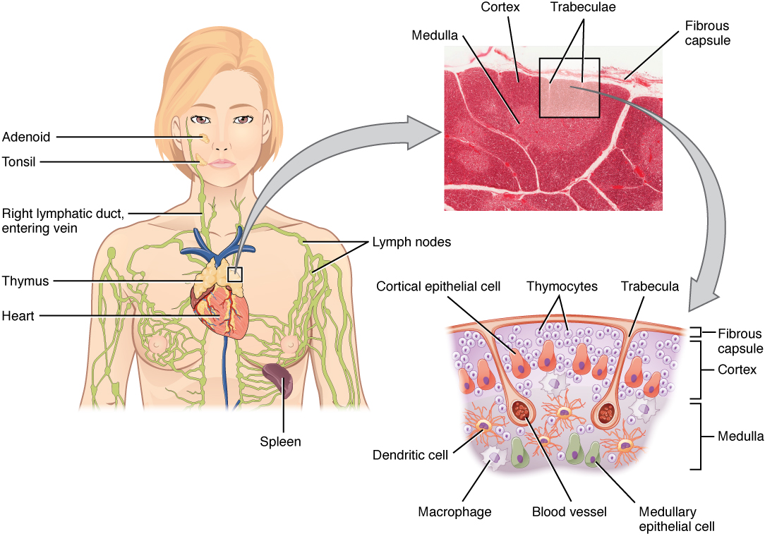
Lymph nodes
Lymph nodes are bean-shaped, encapsulated organs, usually 1–25 mm long, strung along lymphatic vessels. There are roughly 500–700 of them, clustered in the neck (cervical), armpit (axillary), groin (inguinal), chest (mediastinal and hilar) and abdomen (mesenteric and para-aortic). Each group drains a region, so a swollen node points to the region with the problem: an infected cut on the thigh enlarges the inguinal nodes; a sore throat enlarges the cervical nodes. The three groups you can feel easily (cervical, axillary, inguinal) are the largest superficial clusters.
| Node group | Location | Drains |
|---|---|---|
| Cervical (and submandibular) | Neck, along the jugular veins; under the jaw | Head, face, mouth, throat |
| Axillary | Armpit | Arm, breast, chest wall and upper back |
| Supratrochlear (cubital) | Just above the elbow, inner side | Hand and forearm (ulnar side) |
| Inguinal | Groin | Leg, lower abdominal wall, buttocks, external genitalia |
| Popliteal | Behind the knee | Lateral foot, back of the leg and deep tissues of the leg |
| Mediastinal and hilar (bronchopulmonary) | Chest, around the trachea and lung roots | Lungs and chest organs |
| Mesenteric | In the mesentery | Intestines |
| Iliac and para-aortic (lumbar) | Pelvis; along the aorta | Pelvic organs, kidneys, gonads |
Lymph follows one route through the node:
- Several afferent lymphatic vessels pierce the capsule on the convex surface.
- Lymph enters the subcapsular sinus just under the capsule.
- It flows inward through cortical (trabecular) sinuses alongside the trabeculae, connective-tissue partitions that extend in from the capsule.
- It reaches the medullary sinuses, lined by macrophages.
- It leaves through one or two efferent lymphatic vessels at the hilum, the indented side where the artery and vein also enter and leave.
Because there are more afferent than efferent vessels, lymph slows down in the node. Two jobs happen at once. Filtration: macrophages lining the sinuses phagocytose microbes, debris and cancer cells. Immune surveillance: naive B and T cells constantly recirculate from blood into nodes (through HEVs), patrol for their antigen for many hours (about a day), then leave in the efferent lymph and return to the blood; dendritic cells arriving in the lymph present antigen to them, so the rare lymphocyte that fits is found and activated.
Different cells live in different zones:
| Zone | Main cells | What happens there |
|---|---|---|
| Outer (superficial) cortex | B cells in follicles, follicular dendritic cells | Naive B cells wait in primary follicles. After activation a follicle becomes a secondary follicle with a pale germinal center, where B cells proliferate, switch class and improve their affinity. |
| Paracortex (deep cortex) | T cells, dendritic cells | Naive T cells enter from blood through high endothelial venules (HEVs), using the homing receptors CCR7 and L-selectin, and meet dendritic cells carrying antigen from the tissues. |
| Medulla | Medullary cords of plasma cells, B cells and macrophages; medullary sinuses | Plasma cells secrete antibody into the outgoing lymph. Lymph nodes, as a group, are the main antibody factories for antigens arriving from the tissues (the spleen does this for blood-borne antigens). |
A germinal center has two halves. In the dark zone, B cells (centroblasts) divide rapidly and undergo somatic hypermutation of their antibody genes. In the light zone, the mutated B cells (centrocytes) test their new receptors against antigen held on follicular dendritic cells and compete for help from follicular helper T (Tfh) cells; those with the best fit survive and become plasma cells or memory B cells. Follicular dendritic cells are not true dendritic cells: they hold intact antigen on their surface for B cells rather than presenting peptides to T cells.
A lymph node and a lymphoid nodule (follicle) are different things. A nodule is a small, unencapsulated cluster of lymphocytes in a mucous membrane or inside an organ; a node is a whole encapsulated organ with afferent and efferent vessels, cortex and medulla.
Lymphadenopathy means enlarged lymph nodes. A node swells when the lymphocytes in it are proliferating in response to infection (reactive), when it is inflamed (lymphadenitis), or when cancer has spread to it. Tender, soft, mobile nodes suggest infection; hard, painless, fixed nodes are more worrying.
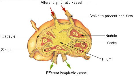

Spleen
The spleen is the largest lymphoid organ. It lies in the left upper quadrant of the abdomen, under the diaphragm, behind the stomach and in front of the left kidney, protected by ribs 9–11. A classic memory aid is "1 × 3 × 5 × 7 × 9 × 11": about 1 inch thick, 3 inches wide and 5 inches long, about 7 ounces, lying between the 9th and 11th ribs. Its smooth, convex diaphragmatic surface faces up and back against the diaphragm; its concave visceral surface carries impressions of the neighboring organs (gastric for the stomach, renal for the left kidney, colic for the left colic flexure), and the tail of the pancreas touches it at the hilum, where the splenic artery, splenic vein and nerves enter and leave. The spleen develops from mesoderm in the dorsal mesogastrium (the dorsal mesentery of the stomach) and stays intraperitoneal, wrapped in peritoneum except at the hilum, tethered by the gastrosplenic and splenorenal ligaments. A capsule sends trabeculae inward, and the tissue between them is the pulp.
- White pulp (about a quarter of the spleen) is lymphoid tissue wrapped around central arteries (arterioles). Its three parts are the periarteriolar lymphoid sheath (PALS), a sleeve of T cells hugging the artery; lymphoid follicles of B cells (with germinal centers after activation; the classic name for these splenic nodules is Malpighian corpuscles); and the marginal zone at the edge, where blood first percolates past macrophages and specialized marginal-zone B cells that respond quickly to bacterial polysaccharides. Activated B cells become plasma cells that collect at the edges of the white pulp and in the red-pulp cords, releasing antibody straight into the blood.
- Red pulp (most of the spleen) is made of splenic cords (cords of Billroth), loose tissue full of macrophages, red cells and other blood cells, and the venous sinusoids between them. Red cells must squeeze through slits in the sinusoid walls; old, stiff or damaged cells cannot and are eaten by cord macrophages.
The spleen's jobs, in the order tests ask them:
- Filters blood of microbes and starts immune responses to blood-borne antigens (white pulp and marginal zone).
- Destroys old and damaged red cells (the "red-cell graveyard") and platelets, and recycles their iron: macrophages break hemoglobin down, store iron as ferritin and hemosiderin, and pass heme on as bilirubin.
- Stores platelets (about a third of the body's supply) and a reserve of monocytes, and acts as a blood reservoir: after hemorrhage, sympathetic stimulation can squeeze some stored blood back into circulation. The reservoir is large in dogs and horses and modest in humans, but "blood reservoir" is a standard textbook answer.
- Finishes B-cell maturation: B cells leave the marrow as immature transitional B cells (T1, then T2 stages) and complete maturation into naive follicular or marginal-zone B cells in the spleen. The spleen is also a major site where lymphocytes meet blood-borne antigen and proliferate.
- Makes blood cells in the fetus. After birth it does not normally make red cells, though it can resume in severe marrow disease.
The spleen is not essential for life. After splenectomy a person survives but is more vulnerable to encapsulated bacteria (Streptococcus pneumoniae, Haemophilus influenzae type b, Neisseria meningitidis), because splenic macrophages and marginal-zone B cells (making IgM against capsule polysaccharides) are the main defense against those organisms in the blood. The risk is overwhelming post-splenectomy infection, so these patients get the pneumococcal, Hib and meningococcal vaccines. Other changes after splenectomy are hematologic: the platelet count rises (thrombocytosis, because the splenic platelet pool is gone), the white count rises, and old or damaged red cells that the spleen would have cleaned stay in circulation, carrying nuclear remnants called Howell-Jolly bodies and appearing as target cells. Sickle cell disease destroys the spleen without surgery: sickled cells repeatedly block its sinusoids, causing infarcts until the spleen shrinks to a fibrous remnant (autosplenectomy, usually by about age 5). These patients have functional asplenia and the same risk from encapsulated bacteria, which is why young children with sickle cell disease (to about age 5) take daily penicillin and all patients get the same vaccines. The spleen enlarges (hypertrophies) during infections and shrinks (atrophies) with age.

Mucosa-associated lymphoid tissue and the tonsils
Most pathogens enter through mucous membranes, so the body keeps unencapsulated lymphoid nodules in them: mucosa-associated lymphoid tissue (MALT). By location it is called GALT (gut-associated: Peyer's patches, the appendix and scattered follicles of the intestine), BALT (bronchus-associated), NALT (nasal, including the tonsils and adenoids), CALT (conjunctiva of the eye), and lymphoid tissue in the urogenital tract (the skin has a parallel system, SALT, but skin is not a mucosa, so SALT is not strictly part of MALT). Together MALT holds more lymphocytes than any other tissue, and its plasma cells make mostly dimeric IgA, which epithelial cells carry across into the lumen as secretory IgA. MALT also has to tolerate the trillions of harmless bacteria and food antigens in the gut. It does this by keeping them at a distance (a thick mucus layer, antimicrobial peptides and secretory IgA that coats bacteria in the lumen without inflaming the tissue) and by favoring regulatory T cells and IL-10 in the gut wall, so that food and commensals produce tolerance (oral tolerance) rather than an attack. A breakdown of this balance contributes to celiac disease and inflammatory bowel disease.
- Peyer's patches are aggregated lymphoid follicles in the wall of the small intestine, largest and most numerous in the ileum. Specialized epithelial M (microfold) cells over each patch grab antigen from the gut lumen and pass it to dendritic cells and lymphocytes underneath.
- The appendix is a blind pouch off the cecum whose wall is packed with lymphoid follicles (GALT). It samples gut bacteria, generates memory lymphocytes and IgA-producing cells that keep bacteria from invading the gut wall, and may act as a reservoir of helpful gut bacteria that can repopulate the colon after diarrheal illness.
- The tonsils are largest and most active in childhood and shrink by adolescence (the adenoids are usually small by the teenage years). They form Waldeyer's ring around the entrance to the throat: the single pharyngeal tonsil (adenoids) on the roof and back wall of the nasopharynx, the paired tubal tonsils at the openings of the auditory tubes, the paired palatine tonsils at the sides of the back of the mouth (the ones removed in a tonsillectomy), and the lingual tonsils at the base of the tongue. Palatine tonsils are covered by stratified squamous epithelium that dips into deep crypts; the crypts trap bacteria and debris so the germinal centers below can respond, which is also why they can become chronically infected.

Skin as an immune organ
The skin is listed as an immune organ because it is both barrier and sentinel. Keratinized epidermis blocks entry, sweat and sebum keep the surface acidic, and Langerhans cells (epidermal dendritic cells) carry antigen from the skin to the regional lymph nodes. Dermal dendritic cells, resident memory T cells, mast cells and macrophages in the dermis add a second layer. Together these make up skin-associated lymphoid tissue (SALT): antigen captured in the skin travels with Langerhans and dermal dendritic cells through lymphatics to the draining node group in the table above (an infected finger enlarges the axillary or supratrochlear nodes). This is why skin-applied haptens such as nickel cause contact dermatitis, and why the intradermal route works well for some vaccines.
Innate immunity
Textbooks often split defense into three lines. The first line is the external barriers (skin, mucous membranes and their secretions). The second line is the internal innate defenses: phagocytes, natural killer cells, antimicrobial proteins (complement, interferons), inflammation and fever. The third line is adaptive immunity (lymphocytes and antibodies). The first two lines are nonspecific and act the same way against every invader; only the third is specific and has memory. Tests ask you to sort items into these lines: tears and stomach acid are first line; neutrophils, NK cells, histamine and fever are second line; antibodies, plasma cells and cytotoxic T cells are third line.
Vocabulary that tests use without defining:
| Term | Meaning |
|---|---|
| Pathogen | An organism or agent that causes disease |
| Infection | Invasion and multiplication of a pathogen in the body |
| Resistance / susceptibility | The ability to ward off disease / the lack of that ability |
| Self vs nonself | The body's own molecules (normally tolerated) vs foreign ones (attacked) |
| Immunocompetence | A lymphocyte's ability to recognize and respond to one specific antigen, gained during maturation |
| Lymphopoiesis | Production of lymphocytes (part of hematopoiesis) |
| Thymic education | Maturation and selection of T cells in the thymus |
| Immunosenescence | The gradual decline of immune function with age (thymic involution, fewer naive T cells, weaker vaccine responses, more infections and cancers) |
| Opsonin | A molecule (IgG, C3b) that coats a microbe and makes it easier to phagocytose |
| Titer | The amount of a specific antibody in serum, measured by serial dilution |
Barriers and antimicrobial proteins
The first line of defense is physical, chemical and biological. Its job is to guard the portals of entry. The respiratory tract is the most common way pathogens get in, followed by the gastrointestinal tract; others enter through breaks in the skin (cuts, bites, needles), the urogenital tract, the conjunctiva of the eye, or across the placenta.
| Barrier | How it protects |
|---|---|
| Intact skin | Tightly packed, keratinized dead cells; constant shedding (desquamation) carries microbes away; dryness (desiccation) kills many. Sweat and sebum keep the surface acidic (pH about 4–5.5, the "acid mantle"), and sebum fatty acids and sweat peptides (dermcidin) kill microbes. |
| Nose and nasopharynx | Nasal hairs (vibrissae) filter large particles; the conchae make air swirl so particles hit the sticky mucus; sneezing expels irritants; swallowed mucus goes to the stomach acid. |
| Lower airways and other mucous membranes | Sticky mucus traps microbes; in the trachea and bronchi, cilia of the pseudostratified ciliated epithelium sweep it up to the throat (the mucociliary escalator); coughing clears the rest. Smoking paralyzes the cilia. |
| Ear canal | Cerumen (earwax) is acidic and contains lysozyme and fatty acids; it traps dust and microbes and carries them outward. |
| Tears and saliva | Wash surfaces and contain lysozyme, which digests peptidoglycan in bacterial (especially gram-positive) cell walls. |
| Stomach | Gastric acid (pH about 1.5–3.5) and pepsin kill most swallowed microbes. |
| Urine and vaginal secretions | Urine flushes the urethra; vaginal secretions are acidic. |
| Normal flora (microbiota) | Harmless resident bacteria use up space and nutrients and release inhibitory substances, crowding out pathogens (competitive exclusion). Antibiotics that kill them can allow C. difficile or yeast overgrowth. |
Many surfaces and cells add antimicrobial proteins and peptides: lysozyme; defensins and cathelicidin (LL-37), small positively charged peptides that punch holes in microbial membranes; lactoferrin, which binds the iron bacteria need; and collectins such as surfactant proteins A and D and mannose-binding lectin, which coat microbes. Inside the body, complement and interferons are the main antimicrobial proteins of the second line.
Pattern recognition and Toll-like receptors
The second line works inside the body. Its cells do not recognize one particular antigen; they recognize pathogen-associated molecular patterns (PAMPs), molecules that whole classes of microbes share and human cells lack (lipopolysaccharide, flagellin, peptidoglycan, double-stranded RNA, unmethylated CpG DNA), and damage-associated molecular patterns (DAMPs) released by stressed or dying cells (ATP, uric acid crystals, HMGB1). The receptors that see them, pattern-recognition receptors (PRRs), are encoded directly in the germline, so every macrophage carries the same set. The best-known family is the Toll-like receptors (TLRs), named after the Toll gene of fruit flies; humans have ten. TLRs are part of innate, not adaptive, immunity. Those on the cell surface detect microbial surfaces; those inside endosomes detect nucleic acids from microbes that have been engulfed.
| Receptor | Location | Detects |
|---|---|---|
| TLR4 | Cell surface | Lipopolysaccharide (LPS) of gram-negative bacteria |
| TLR2 (with TLR1 or TLR6) | Cell surface | Lipoproteins and peptidoglycan components of bacteria |
| TLR5 | Cell surface | Bacterial flagellin |
| TLR3 | Endosome | Double-stranded RNA (many viruses) |
| TLR7 and TLR8 | Endosome | Single-stranded viral RNA |
| TLR9 | Endosome | Unmethylated CpG DNA, common in bacteria and DNA viruses |
Binding a TLR sets off a chain inside the cell: most TLRs signal through the adaptor MyD88, which activates the transcription factor NF-κB → genes for inflammatory cytokines (IL-1, IL-6, TNF-α) switch on → inflammation and fever. TLR3 (and TLR4 in part) use a second adaptor, TRIF, that turns on type I interferons. TLR activation also makes dendritic cells mature and travel to lymph nodes, which is how innate recognition starts the adaptive response.
Some pattern-recognition molecules are soluble proteins in blood and tissue fluid rather than cell receptors: collectins (mannose-binding lectin, surfactant proteins A and D), ficolins and pentraxins (C-reactive protein, serum amyloid P). They coat microbes, act as opsonins and can start the lectin complement pathway. Other cell-associated PRR families include the C-type lectin receptors on the cell surface and, inside the cytoplasm, NOD-like receptors (some assemble the inflammasome, which activates caspase-1 to release active IL-1β), RIG-I-like receptors (cytosolic viral RNA → interferons), and cGAS (cytosolic DNA; see the signaling section).
Innate cells
All white blood cells (leukocytes) come from hematopoietic stem cells in the red marrow. A normal adult WBC count is about 4,500–11,000 cells/µL (4.5–11 × 10⁹/L). Above that range is leukocytosis, the normal response to infection, inflammation or stress; below about 4,000–4,500 is leukopenia. White cells are under 1% of blood volume (they sit in the thin buffy coat between plasma and packed red cells when blood is spun).
Leukocytes are classified two ways:
- By granules. Granulocytes (neutrophils, eosinophils, basophils) have conspicuous cytoplasmic granules and lobed nuclei, so they are also called polymorphonuclear leukocytes (PMNs). Agranulocytes (lymphocytes and monocytes) have no visible granules and a single unlobed nucleus (mononuclear cells).
- By lineage. Myeloid cells come from the common myeloid progenitor: neutrophils, eosinophils, basophils, mast cells, monocytes and macrophages, and most dendritic cells. Lymphoid cells come from the common lymphoid progenitor: B cells, T cells and NK cells. NK cells are lymphoid but innate.

The normal differential (percentage of each type among white cells) is learned with the mnemonic "Never Let Monkeys Eat Bananas", most to least abundant: Neutrophils > Lymphocytes > Monocytes > Eosinophils > Basophils.
ReferenceWhite blood cell reference· 10 rows
% = share of circulating leukocytes in a healthy adult (ranges differ slightly between labs). "Increased in" lists the classic causes tests use.
No rows match.
| Cell | % of WBCs | Appearance (Wright stain) | Lineage | Life span | Main jobs | Increased in |
|---|---|---|---|---|---|---|
| Neutrophil | 50–70% | 3–5 connected nuclear lobes; pale lilac fine granules; young form is a band with a horseshoe nucleus | Myeloid granulocyte | About 6–12 hours in blood, 1–2 days in tissue | First phagocyte to arrive at bacterial or fungal infection; respiratory burst; NETs; main cell of pus | Acute bacterial infection, tissue injury, stress, steroids |
| Lymphocyte | 20–40% | Round, dark nucleus filling most of the cell; thin rim of pale blue cytoplasm; small (most) or large (NK cells are large granular lymphocytes) | Lymphoid | Days (effector cells) to years (memory cells) | B cells: antibody; T cells: cell-mediated immunity; NK cells: kill infected and tumor cells | Viral infections, chronic infections, lymphocytic leukemias |
| Monocyte | 2–8% | Largest WBC; kidney- or horseshoe-shaped nucleus; abundant gray-blue cytoplasm | Myeloid agranulocyte | 1–3 days in blood, then months as a macrophage | Leaves blood and becomes a macrophage or dendritic cell in tissue | Chronic infections (tuberculosis), recovery phase of infection |
| Eosinophil | 1–4% | Usually bilobed nucleus; large red-orange granules (take up the acidic dye eosin) | Myeloid granulocyte | About 8–12 hours in blood, days in tissue | Kill parasitic worms (helminths) with major basic protein and other granule toxins; late phase of allergy and asthma; help limit allergic reactions | Parasitic worms, allergy, asthma |
| Basophil | 0.5–1% (least common) | Bilobed or S-shaped nucleus hidden by large dark blue-purple granules (take up basic dyes) | Myeloid granulocyte | Hours to days | Release histamine (vasodilator) and heparin (anticoagulant); IgE receptors make them part of allergy | Allergy, chronic myeloid leukemia |
| Mast cell | Not in blood | Tissue cell packed with histamine-containing granules | Myeloid (precursors leave marrow and mature in tissue) | Weeks to months | Fixed in connective tissue and mucosa near vessels; degranulate when IgE on their surface is cross-linked; start inflammation | Allergic tissue (mastocytosis is rare) |
| Macrophage | Not in blood | Large, irregular cell with many lysosomes | Myeloid (from monocytes) | Months | Phagocytosis, antigen presentation (MHC II), cytokines (IL-1, IL-6, TNF-α, IL-12) | Chronic inflammation, granulomas |
| Dendritic cell | Not in blood (precursors circulate) | Long branching processes | Mostly myeloid | Days in tissue | Most potent antigen-presenting cell; carries antigen from tissue to lymph nodes and activates naive T cells | — |
| Plasma cell | Rare in blood; found in lymph nodes, spleen, marrow and mucosa | Oval cell with an off-center (eccentric) round nucleus with "clock-face" chromatin, deep-blue cytoplasm and a pale halo (the Golgi) beside the nucleus | Lymphoid (from activated B cells) | Days to years (long-lived ones in marrow) | Secretes antibody | Multiple myeloma (not on the list), chronic infection |
| NK cell | 5–15% of lymphocytes | Large granular lymphocyte | Lymphoid, but innate | Weeks | Kill virus-infected and tumor cells that lack MHC I; ADCC | Some viral infections |

Tests name an abnormal count with the cell plus -philia/-cytosis (too many) or -penia (too few):
| Finding | Typical causes |
|---|---|
| Neutrophilia (with a left shift) | Acute bacterial infection, tissue injury or burns, stress, corticosteroids |
| Lymphocytosis | Viral infections; atypical (reactive) lymphocytes, large with abundant cytoplasm, are typical of infectious mononucleosis; chronic lymphocytic leukemia |
| Monocytosis | Chronic infections such as tuberculosis, recovery from infection, some autoimmune disease |
| Eosinophilia | Parasitic worms, allergy and asthma, some drug reactions |
| Basophilia | Allergy, chronic myeloid leukemia |
| Neutropenia | Chemotherapy, some viral infections, severe congenital or cyclic neutropenia |
| Lymphopenia | HIV/AIDS, corticosteroids, SCID |
Neutrophils
Neutrophils are the most numerous white cells and the first responders: they arrive at a bacterial infection within hours, phagocytose, kill and die, and their dead bodies with tissue debris and microbes make pus (a walled-off pocket of pus is an abscess). They kill in two ways. Oxygen-dependent killing is the respiratory (oxidative) burst: the enzyme NADPH oxidase turns oxygen into superoxide, which becomes hydrogen peroxide, and myeloperoxidase (the enzyme that makes pus greenish) converts that into hypochlorous acid (bleach). Oxygen-independent killing uses granule contents: lysozyme, defensins, lactoferrin and proteases such as elastase. Neutrophils can also cast neutrophil extracellular traps (NETs), webs of their own DNA studded with histones and granule enzymes that trap and kill microbes outside the cell; making NETs (NETosis) usually kills the neutrophil, because it has expelled its own chromatin. Neutrophils are short-lived and do not form memory.
Monocytes and macrophages
Monocytes circulate for one to three days, then leave the blood and become macrophages ("big eaters") or dendritic cells. Macrophages are long-lived, phagocytose microbes, dead cells and debris, present antigen on MHC class II to helper T cells, and release cytokines that start inflammation and fever (IL-1, IL-6, TNF-α) and steer T cells (IL-12). Free (wandering) macrophages move through tissues; fixed macrophages stay in one organ and have their own names. Together, monocytes and tissue macrophages form the mononuclear phagocyte system (older name: reticuloendothelial system).
| Tissue macrophage | Location |
|---|---|
| Kupffer cells | Liver sinusoids (the largest population of tissue macrophages) |
| Alveolar macrophages ("dust cells") | Lung alveoli |
| Microglia | Brain and spinal cord (they arise from embryonic yolk-sac precursors rather than blood monocytes, but are the CNS's resident macrophages) |
| Osteoclasts | Bone (resorb bone; they come from the same monocyte lineage). Do not confuse them with osteoblasts, bone-forming cells from mesenchymal stem cells that are not part of the immune system |
| Histiocytes | Connective tissue |
| Splenic and lymph-node macrophages | Red pulp, marginal zone and medullary sinuses |
| Mesangial cells (macrophage-like) | Kidney glomeruli |
Activated macrophages come in two broad states. M1 ("classically activated") macrophages are switched on by IFN-γ and microbial products such as LPS and are microbe killers. M2 ("alternatively activated") macrophages are switched on by IL-4 and IL-13 and promote wound healing and tissue repair. When macrophages cannot destroy an organism (such as the tuberculosis bacterium), they fuse into giant cells and wall it off with lymphocytes: a granuloma.
Dendritic cells
Dendritic cells sit at the body's borders (skin, airways, gut), sampling antigen with long processes. When their TLRs detect PAMPs they mature, travel through lymphatics to the nearest lymph node and present antigen to naive T cells in the paracortex. They are the most potent antigen-presenting cells (APCs) (older texts also call APCs accessory cells) and the main bridge between innate and adaptive immunity. APCs present antigen directly to T lymphocytes (on MHC II to CD4 cells, and on MHC I to CD8 cells); they do not present to mast cells, eosinophils or other effector cells, which are triggered instead by antibody bound to their Fc receptors. Epidermal dendritic cells are Langerhans cells (older texts call them epidermal macrophages, but they are dendritic cells). When a question describes a cell that engulfs antigen in a tissue and then carries it to a lymph node to alert T cells, the best answer is the dendritic cell, even though macrophages and B cells are also APCs. Plasmacytoid dendritic cells are a separate type that release huge amounts of type I interferon during viral infection.
Natural killer cells
NK cells are large granular lymphocytes that belong to innate immunity: they need no prior exposure, carry no antigen-specific receptor and form no classic memory. They balance activating receptors (which detect stress molecules on infected or cancerous cells) against inhibitory receptors that recognize MHC class I. Healthy cells show MHC I and are spared. Many viruses and tumors lower MHC I to hide from CD8 T cells, and NK cells kill exactly those cells: the "missing self" rule. They kill by releasing perforin (forms pores in the target membrane) and granzymes (proteases that enter through the pores and activate caspases, triggering apoptosis), and through Fas ligand. NK cells also kill antibody-coated cells by antibody-dependent cell-mediated cytotoxicity (ADCC): their Fc receptor CD16 binds the Fc of IgG on the target. They secrete IFN-γ, which activates macrophages. NK cells belong to a wider family of innate lymphoid cells (ILCs), lymphocytes with no antigen receptor that mirror helper T-cell subsets: ILC1s make IFN-γ (like Th1), ILC2s make IL-5 and IL-13 and drive allergic airway inflammation (like Th2), and ILC3s make IL-17 and IL-22 at mucosal barriers (like Th17). NK cells make up roughly 5–15% of blood lymphocytes and are also plentiful in the spleen, liver and the lining of the pregnant uterus.
NK cells and cytotoxic T cells kill the same targets with the same weapons, so tests ask you to tell them apart:
| NK cell | Cytotoxic T cell (CTL) | |
|---|---|---|
| Immunity | Innate | Adaptive |
| Recognition | Activating vs inhibitory receptors; kills cells lacking MHC I | TCR recognizes a specific peptide on MHC I |
| Prior activation needed? | No; acts within hours | Yes; naive cells need priming in a lymph node (days) |
| Memory | Not in the classic sense | Yes (memory CD8 cells) |
| Markers | CD56, CD16 (no TCR or CD3) | CD3, CD8, αβ TCR |
| Weapons | Perforin, granzymes, Fas ligand, ADCC via CD16, IFN-γ | Perforin, granzymes, Fas ligand, IFN-γ, TNF |
Eosinophils, basophils and mast cells
Eosinophils fight helminths (parasitic worms such as hookworms and tapeworms) that are too large to engulf: they bind IgE- or IgG-coated worms and release toxic granule proteins (major basic protein, eosinophil cationic protein, eosinophil peroxidase) onto them. They also join the late phase of allergic reactions and asthma, and they can break down histamine and leukotrienes, damping the reaction. IL-5 from Th2 cells is the main signal that makes and activates them.
Basophils (in blood) and mast cells (in tissue) are the histamine cells. They look and act alike but are different cells:
| Mast cell | Basophil | |
|---|---|---|
| Where | Fixed in tissue: skin, airway and gut mucosa, around blood vessels | Circulates in blood; recruited into tissue during allergy |
| Origin | Myeloid precursor that leaves the marrow and matures in tissue | Matures fully in the marrow (a granulocyte) |
| Life span | Weeks to months | Days |
| Nucleus | Single round or oval | Bilobed, hidden by granules |
| Role | First cell to release histamine at a site of injury or allergen contact; starts local inflammation and type I reactions | Joins later and amplifies allergic and antiparasitic responses |
Both carry the high-affinity IgE receptor (FcεRI), so IgE made in an allergic response sits on their surface; cross-linking by antigen makes them degranulate. Their preformed granule contents are histamine (vasodilation, leaky capillaries, bronchoconstriction, itching), heparin (an anticoagulant), proteases such as tryptase and eosinophil-attracting factors. Within minutes they also synthesize lipid mediators: leukotrienes (the "slow-reacting substance of anaphylaxis", strong bronchoconstrictors), prostaglandin D2 and platelet-activating factor.
A question about histamine release in a tissue (skin wheal, nasal mucosa, an injury) points to mast cells; one about a blood cell or a granulocyte on a smear points to basophils.
Which cells are phagocytes? The professional phagocytes are neutrophils, monocytes and macrophages, and dendritic cells (which eat mainly to present antigen). Eosinophils phagocytose only weakly; they mostly kill by releasing granules. Basophils, mast cells, lymphocytes (B, T, NK) and plasma cells are not phagocytes.
Phagocytosis follows the same steps every time:
- Chemotaxis and adherence: the phagocyte moves toward the microbe and binds it, directly through PRRs or, much better, through opsonins (C3b and IgG) coating the microbe.
- Ingestion: pseudopods surround the microbe and pinch it into a phagosome.
- Fusion: the phagosome fuses with lysosomes to form a phagolysosome.
- Digestion and killing: enzymes and reactive oxygen species destroy the microbe.
- Exocytosis: indigestible debris is expelled; in macrophages and dendritic cells some peptides are loaded onto MHC II for presentation.

The complement system
Complement is a set of about 30 plasma and membrane proteins (older textbooks say about 20, counting only the main circulating ones), made mostly by the liver, that circulate as inactive precursors (zymogens) and "complement" antibodies. Keeping them inactive until a trigger appears prevents the cascade from punching holes in the body's own cells and causing uncontrolled inflammation. Activation is a cascade: each activated enzyme usually cuts the next protein into a small "a" fragment and a large "b" fragment, so a few triggering events are amplified into thousands of molecules. Three pathways start it:
| Pathway | Trigger | First components | Innate or adaptive trigger? |
|---|---|---|---|
| Classical | Antibody (IgM best, then IgG) bound to antigen | C1q must bind at least two Fc regions at once: one bound IgM pentamer provides them, but IgG needs two molecules bound close together, which is why IgM is the better activator; then C1r, C1s → C4 and C2 | Needs antibody (adaptive link) |
| Lectin | Mannose-binding lectin (MBL) or ficolins bind sugars on microbial surfaces | MBL-associated serine proteases (MASPs) → C4 and C2 | Innate |
| Alternative | Activated directly by microbial surfaces (LPS of gram-negative bacteria, yeast cell walls): C3 constantly breaks down slowly ("tickover") and drops C3b onto any nearby surface; on host cells regulators destroy it, but on a microbe it survives and builds a convertase. No antibody or lectin is needed | C3b with factor B, factor D and properdin | Innate; also amplifies the other two |
All three converge on a C3 convertase that cuts C3, the most abundant complement protein, into C3a and C3b, and then on a C5 convertase. The products do four jobs:
- Opsonization: C3b (and its breakdown product iC3b) coats the microbe so phagocytes grab it through complement receptors: CR1 binds C3b, CR3 binds iC3b.
- Inflammation: C3a and C5a (the anaphylatoxins) make mast cells release histamine and increase vessel permeability; C5a is also a powerful chemotactic signal for neutrophils.
- Cytolysis: C5b, C6, C7, C8 and many C9 molecules assemble the membrane attack complex (MAC), a pore that destroys the membrane's selective permeability so water and ions rush in and the cell lyses. The MAC matters most against thin-walled gram-negative bacteria such as Neisseria.
- Clearance of immune complexes: C3b on antigen–antibody complexes lets red cells carry them to the spleen and liver for removal.
Complement works only on targets it can touch: microbes in blood and tissue fluid, and the surfaces of infected cells. It cannot reach viruses or bacteria hiding inside host cells; those need cytotoxic T cells and NK cells.
Human cells protect themselves with regulators (decay-accelerating factor CD55, CD59, factor H), which is why complement attacks microbes and not you. Complement is not the main cause of fever; fever comes mainly from cytokine pyrogens.

Inflammation and fever
Inflammation is the tissue response to injury or infection. It brings defenders and plasma proteins to the site, dilutes and walls off the damage, and sets up repair. Its cardinal signs are redness (rubor), heat (calor), swelling (tumor) and pain (dolor), with loss of function (functio laesa) added as the fifth sign. Itching is not one of the cardinal signs. Each sign has a cause, and each has a purpose: extra blood brings more defenders, the protein-rich fluid that leaks out dilutes toxins and carries antibodies, complement and clotting factors into the tissue, and fibrin from the clotting factors walls off the area so microbes cannot spread. Macrophage cytokines (TNF-α, IL-1) add to the vasodilation and make the endothelium sticky for leukocytes.
- Injured cells, mast cells and basophils release histamine (plus kinins, prostaglandins and leukotrienes) → arterioles dilate → more blood (hyperemia) → redness and heat.
- Histamine also makes capillaries leaky → plasma and protein (including clotting factors and complement) leave the vessel → swelling (edema). The fibrin clot that forms helps wall off the area.
- Bradykinin and prostaglandins sensitize nerve endings, and swelling presses on them → pain.
- Pain and swelling together → loss of function, which rests the injured part.
| Mediator | Source | Main effect |
|---|---|---|
| Histamine | Mast cells, basophils, platelets | Vasodilation, increased permeability (the earliest mediator) |
| Prostaglandins | Most cells, from arachidonic acid via cyclooxygenase (COX) | Vasodilation, pain, fever; NSAIDs block them |
| Leukotrienes | Mast cells, leukocytes (via lipoxygenase) | Permeability, bronchoconstriction, neutrophil attraction |
| Bradykinin | Plasma kinin system | Pain, vasodilation, permeability |
| C3a, C5a | Complement | Mast-cell degranulation; C5a attracts neutrophils |
| IL-1, IL-6, TNF-α | Macrophages | Fever, acute-phase proteins, endothelial activation |
| Chemokines (such as IL-8/CXCL8) | Macrophages, endothelium | Attract neutrophils |
Phagocytes then arrive in a fixed sequence:
- Leukocytosis: chemical signals (leukocytosis-inducing factors and colony-stimulating factors) make the marrow release more neutrophils into the blood.
- Margination (pavementing): in the slowed blood, neutrophils move to the vessel edge. Endothelial selectins (P-selectin and E-selectin), displayed in response to histamine, TNF-α and IL-1, make them roll along the wall; then leukocyte β2 integrins (LFA-1 and Mac-1) bind endothelial ICAM-1 and the cells stick firmly.
- Diapedesis (emigration, extravasation): they squeeze between endothelial cells into the tissue.
- Chemotaxis: they crawl up a concentration gradient of chemotactic signals (C5a, chemokines such as IL-8, bacterial peptides, leukotriene B4) to the site. Movement toward the higher concentration is positive chemotaxis.
Inflammation's functions are to prevent the spread of damaging agents, dispose of debris and pathogens, and set the stage for repair; the actual rebuilding by fibroblasts and new cells is a separate, later step. Neutrophils arrive first, within hours; monocytes follow and become macrophages that clean up. The liver responds to IL-6 by making acute-phase proteins such as C-reactive protein (CRP), fibrinogen and mannose-binding lectin; a high CRP is a blood marker of inflammation. Acute inflammation (minutes to days, neutrophil-rich) normally resolves. Chronic inflammation (weeks to years, with macrophages, lymphocytes and fibrosis) occurs when the cause persists, as in tuberculosis, autoimmune disease or long-term irritants. A body-wide inflammatory response to infection can drop blood pressure dangerously: septic shock.
The fluid that collects is an exudate, named by its content: serous (thin, watery, as in a blister), fibrinous (rich in fibrin), purulent (pus, full of neutrophils) or hemorrhagic (with red cells). Once the inflammatory response has done its job, it gives way to resolution and then repair: the cause is removed, neutrophils die by apoptosis and are eaten by macrophages, mediators switch to anti-inflammatory ones (IL-10, TGF-β, lipoxins), and M2 macrophages and fibroblasts rebuild the tissue. Tissue that can divide regenerates normal cells; where damage is extensive or the tissue cannot regenerate (heart muscle, much of the nervous system), fibroblasts lay down collagen and a scar (fibrosis) forms.
Fever is a regulated rise in the hypothalamic set point. Macrophages release pyrogens (IL-1, IL-6, TNF-α; bacterial products such as LPS are exogenous pyrogens) → the hypothalamus makes prostaglandin E2 → the set point rises → shivering and skin vasoconstriction raise body temperature until it matches. A moderate fever helps: the liver and spleen sequester iron and zinc that bacteria need, metabolic rate rises so repair speeds up, and many pathogens grow less well. Very high fever (above about 41 °C, 106 °F) denatures proteins and can cause seizures and brain injury. Aspirin, ibuprofen and acetaminophen lower fever by blocking prostaglandin synthesis. When the cause is gone, the set point falls and the body sheds heat by sweating and vasodilation (the "crisis").
Glucocorticoids (cortisol, prednisone) are the broadest anti-inflammatory drugs: they block the production of interleukins and other cytokines, prostaglandins and leukotrienes, stabilize capillaries and reduce leukocyte migration, which is why they suppress both inflammation and immunity. The same thing happens naturally under chronic stress: long-term high cortisol lowers interleukin secretion, weakens phagocytes and lymphocyte responses, shrinks the thymus and slows wound healing, which is why chronically stressed people catch more infections. Short-term stress, by contrast, briefly mobilizes neutrophils into the blood.
Adaptive immunity
Adaptive immunity is built from lymphocytes, each carrying receptors for one specific antigen. It has four defining features: specificity (each clone recognizes one epitope), diversity or versatility (the population as a whole can recognize millions of different antigens), memory (a second exposure gets a faster, stronger response) and self-tolerance (it normally ignores the body's own molecules). It is also called acquired or specific immunity, and it has two arms: humoral immunity, carried out by B cells and the antibodies their plasma cells secrete into body fluids ("humors"), and cell-mediated immunity, carried out directly by T cells.
Antigens, epitopes and haptens
An antigen (the name is usually explained as "antibody generator") is any substance that a B-cell receptor, antibody or T-cell receptor can bind; most are large foreign macromolecules. An immunogen is an antigen that can also provoke a response on its own. A complete antigen has both properties: immunogenicity (it triggers lymphocyte proliferation and antibody production) and reactivity (it binds the antibodies and cells it provoked). Large, complex, foreign molecules are the best immunogens: proteins are the strongest, then large polysaccharides; nucleic acids and lipids are weak unless attached to proteins.
An epitope (antigenic determinant) is the small part of an antigen that one receptor actually binds, typically about 5–20 amino acids or sugar units. A single antigen such as a bacterial protein has many epitopes, so it activates many different lymphocyte clones, and the normal response is polyclonal: a mix of antibodies against different epitopes of the same antigen. B cells and antibodies recognize epitopes on the intact molecule, including conformational (discontinuous) epitopes formed by the folded shape; T cells recognize only linear peptide fragments displayed on MHC. The antigen-binding site of an antibody that fits the epitope is the paratope.
A hapten is a small molecule (penicillin, nickel ions, urushiol from poison ivy) that has reactivity but not immunogenicity. Alone it cannot trigger a response; once it binds a larger carrier protein, often one of the body's own proteins, the combination becomes immunogenic. This is how drug allergies and contact dermatitis start.
| Term | Meaning |
|---|---|
| Antigen | Anything a lymphocyte receptor or antibody binds |
| Immunogen / complete antigen | An antigen that also provokes a response by itself |
| Epitope (antigenic determinant) | The specific region a receptor binds |
| Paratope | The part of the antibody that binds the epitope |
| Hapten | Small molecule that is antigenic only when attached to a carrier protein |
| Self-antigen | A body molecule, normally tolerated; target in autoimmunity |
| Alloantigen | An antigen that differs between members of the same species (HLA, ABO) |
B cells and T cells
When antigen binds a matching lymphocyte, that cell divides into a clone of identical cells (clonal selection and clonal expansion; the antigen "selects" which cells are cloned). Most clone members become short-lived effector cells; some become long-lived memory cells.
| Feature | B cell | T cell |
|---|---|---|
| Matures in | Bone marrow | Thymus |
| Share of blood lymphocytes | About 10–15% | About 70–80% (NK cells make up the rest) |
| Receptor | B-cell receptor (BCR): membrane IgM and IgD plus the signaling chains Igα and Igβ | T-cell receptor (TCR): an α and a β chain (a heterodimer; a minority of T cells use γ and δ), each with one variable and one constant domain, plus the CD3 signaling complex |
| Recognizes | Intact antigen of any type (protein, polysaccharide, lipid), free in fluid | Only peptide fragments displayed on self MHC (MHC restriction) |
| Secreted form of receptor? | Yes: antibody | No: the TCR stays on the cell, because T cells act by direct contact and cytokines |
| Main effector | Plasma cell secreting antibody (about 2,000 molecules per second, so its cytoplasm is packed with rough ER and a large Golgi apparatus) | Helper T cell (cytokines) or cytotoxic T cell (killing) |
| Surface markers | CD19, CD20, CD21 | CD3, plus CD4 (helper) or CD8 (cytotoxic) |
| Immunity | Humoral | Cell-mediated |
"CD" stands for cluster of differentiation, the numbering system for surface molecules recognized by antibodies; helper T cells are also written T4 cells and cytotoxic T cells T8 cells.
| T-cell type | Marker | Recognizes | Job |
|---|---|---|---|
| Helper T (Th) | CD4 | Peptide on MHC class II | Releases cytokines (IL-2 and others) that activate B cells, cytotoxic T cells and macrophages; the "conductor" of the response |
| Cytotoxic T (Tc, CTL, killer T) | CD8 | Peptide on MHC class I | Kills virus-infected cells, tumor cells and transplanted cells; the main T cells that kill directly |
| Regulatory T (Treg), formerly "suppressor T" cells | CD4, CD25, FOXP3 | Self and foreign peptide on MHC II | Suppresses other lymphocytes (IL-10, TGF-β); maintains tolerance and ends responses |
| Memory T | CD4 or CD8 | Same as the parent cell | Long-lived (years to decades); quick secondary response |
| Follicular helper T (Tfh) | CD4 | MHC II on B cells | Helps B cells in germinal centers (CD40L, IL-21) |
| γδ T cells and NKT cells | Variable | Lipids and stress molecules | Innate-like T cells (γδ cells are common in epithelia; NKT cells in the liver) |
Cytotoxic T cells kill in three ways: perforin and granzymes (the same toolkit as NK cells; granzymes activate caspases and the target undergoes apoptosis), Fas ligand on the T cell binding Fas on the target (which also triggers apoptosis), and cytokines such as IFN-γ and lymphotoxin (TNF-β), which helps trigger target-cell death and DNA fragmentation. Killing by apoptosis keeps the virus inside a tidy dying cell instead of spilling it. A CTL detaches after delivering its hit and moves on to the next target. They remove the virus factory, not free virus; free virus is antibody's job.
Maturation, selection and tolerance
Receptor diversity comes from gene rearrangement. The genes for antibody and TCR chains come in pieces: many V (variable), D (diversity, heavy chain, TCR β and TCR δ only) and J (joining) segments. In each developing lymphocyte the enzymes RAG1 and RAG2 cut the DNA and DNA-repair enzymes join one V, one D and one J at random (V(D)J recombination, or somatic recombination). Diversity is multiplied by combinatorial diversity (which segments are chosen), junctional diversity (nucleotides lost or added at the joints, partly by the enzyme TdT) and pairing of a heavy with a light chain (or an α with a β chain). That is how a limited number of genes produces well over 10¹¹ possible receptors. Each cell expresses only one rearranged allele of each chain (allelic exclusion), so each cell has one specificity. (MHC genes, by contrast, are codominant: both parents' alleles are expressed.)
B-cell development happens in the bone marrow, supported by stromal cells that provide contact signals and IL-7:
- Pro-B cell: the heavy-chain D segment joins a J segment, then a V segment joins DJ.
- Pre-B cell: a successfully rearranged μ heavy chain appears on the surface with a surrogate light chain (the pre-BCR); the light chain then rearranges.
- Immature B cell: displays complete IgM. Immature B cells that bind self-antigen strongly are deleted (clonal deletion), get a second chance to rearrange their light chain (receptor editing), or become unresponsive (anergy).
- Mature naive B cell: displays IgM and IgD; it leaves the marrow as a transitional B cell, finishes maturing in the spleen and circulates through secondary lymphoid organs.
T-cell development happens in the thymus. Progenitors arrive from the marrow as double-negative (DN) cells (neither CD4 nor CD8):
- β-selection (DN stage): the TCR β chain rearranges and is tested with a surrogate α chain (pre-TCR); success lets the cell proliferate.
- The cell becomes double-positive (DP) (CD4⁺CD8⁺) and rearranges the α chain.
- Positive selection (thymic cortex, cortical epithelial cells): a DP cell survives only if its TCR binds self MHC weakly. Cells that cannot bind MHC at all die by neglect. This step also decides lineage: binding MHC II → the cell keeps CD4; binding MHC I → it keeps CD8. It is now single-positive (SP).
- Negative selection (mostly in the medulla, by medullary epithelial cells and dendritic cells): an SP cell that binds self-peptide on self-MHC too strongly dies by apoptosis. The transcription factor AIRE lets medullary epithelial cells display proteins from other organs, such as insulin, so self-reactive cells against them are removed. Some strongly self-reactive CD4 cells become Tregs instead.
So the order is β-selection → positive selection → negative selection, and about 95–98% of thymocytes die without leaving. Most of that death happens in the cortex, where the large majority of double-positive cells fail positive selection and die by neglect (apoptosis), and cortical macrophages clear them; a smaller number are deleted by negative selection. The survivors are MHC-restricted (they see antigen only on self MHC) and self-tolerant.
Deletion in the thymus and marrow is central tolerance. It is never 100% complete, so peripheral tolerance handles escapees: regulatory T cells suppress them; a T cell that sees antigen without costimulation becomes unresponsive (anergy); and repeated strong stimulation can trigger deletion by apoptosis. Some sites (eyes, brain, testes) are partly hidden from immune cells. Autoimmune disease is a failure of tolerance; self-reactive T cells that escape the thymus reflect a failure of negative selection.

Antigen presentation and activation
T cells see antigen only as fragments displayed on major histocompatibility complex (MHC) proteins, called HLA (human leukocyte antigen) in humans and encoded on chromosome 6. MHC molecules are the cell's "self" identity tags, and each has a peptide-binding groove (cleft) that holds the fragment.
| MHC class I | MHC class II | |
|---|---|---|
| Found on | Almost every nucleated cell (not red cells) | Professional antigen-presenting cells: dendritic cells, macrophages, B cells (and thymic epithelium) |
| Displays | Peptides from proteins made inside the cell (endogenous, including viral and tumor proteins) | Peptides from material taken in from outside (exogenous) |
| Loading route | Proteasome → TAP transporter → rough ER → Golgi → surface | Endosome or phagolysosome; the invariant chain (and its CLIP fragment) blocks the groove until HLA-DM swaps in the peptide |
| Structure | One α chain + β2-microglobulin | An α and a β chain |
| HLA genes | HLA-A, -B, -C | HLA-DP, -DQ, -DR |
| Read by | CD8 cytotoxic T cells | CD4 helper T cells |
The "rule of 8": CD4 × class II = 8, CD8 × class I = 8.
There is one important exception to "outside antigen goes on class II". Some dendritic cells can cross-present: they take up material from outside (a virus-infected or tumor cell they have eaten) and load its peptides onto MHC class I. This is how naive CD8 T cells get primed against viruses that do not infect dendritic cells and against tumors. Of the two classes, MHC II is arguably the more central to adaptive immunity, because the CD4 help it recruits is needed for strong antibody responses and for full CD8 responses; losing it (or losing CD4 cells, as in AIDS) cripples both arms.

A naive T cell needs two signals, usually from a dendritic cell in a lymph node's paracortex: signal 1 is its receptor binding peptide–MHC (with CD4 or CD8 as co-receptor), and signal 2 is costimulation, mainly CD28 on the T cell binding B7 (CD80/CD86) on an activated antigen-presenting cell. APCs show high levels of B7 mainly after sensing danger through their PRRs, which keeps T cells from attacking harmless self-antigen. Signal 1 without signal 2 makes the cell anergic. Memory and effector T cells are easier to switch on than naive cells: they respond to lower amounts of antigen, depend less on costimulation, can be activated by APCs in the tissues (not only in lymph nodes) and release cytokines within hours, which is part of why the secondary response is fast. Cytokines then act as a third signal that steers what the cell becomes. The activated T cell makes IL-2 and the high-affinity IL-2 receptor (CD25), so it drives its own proliferation (autocrine signaling); CTLA-4, which appears later and binds B7 more tightly than CD28 does, and PD-1 are brakes that switch the response off. Activated cytotoxic (CD8) T cells then leave the node and kill infected or tumor cells. Helper (CD4) T cells release cytokines: their IL-2 and IFN-γ help CD8 cells proliferate and become fully armed cytotoxic cells and memory cells, and they "license" dendritic cells to activate CD8 cells. A B cell that has captured a protein antigen needs help from a CD4 cell that recognizes the same antigen on its MHC II before it switches class and makes large amounts of antibody.
B-cell activation comes in two forms:
- T-dependent (most protein antigens): the BCR binds antigen → the B cell internalizes it, processes it and displays peptides on MHC II → a helper T cell already activated by the same antigen binds, delivers CD40 ligand to the B cell's CD40 and secretes cytokines → the B cell proliferates, forms a germinal center, switches class, matures its affinity and makes plasma cells and memory B cells.
- T-independent (polysaccharides and other molecules with many identical repeating epitopes, such as bacterial capsules and LPS): the antigen cross-links many BCRs at once and activates the B cell without T-cell help. The response is mostly IgM, low-affinity, with little class switching and weak memory. Children under about 2 years respond poorly to these antigens, which is why capsule vaccines (Hib, pneumococcal, meningococcal) are conjugated to a protein carrier: the protein recruits T-cell help.


The integrated immune response
A typical bacterial or viral infection runs as one chain, and tests often ask you to put its steps in order:
- Barriers are breached (a cut, an inhaled virus).
- Innate recognition: macrophages and dendritic cells detect PAMPs through TLRs and other PRRs.
- Inflammation: cytokines and histamine dilate vessels; neutrophils arrive first, then monocytes; complement opsonizes and lyses; infected cells release interferons; NK cells kill infected cells.
- Antigen transport: dendritic cells carry antigen through lymphatics to the draining lymph node (antigen in blood goes to the spleen instead).
- T-cell activation in the paracortex (signals 1 and 2, then IL-2) → clonal expansion of helper and cytotoxic T cells over several days.
- B-cell activation in follicles with T-cell help → germinal centers → plasma cells and memory cells.
- Effector phase: antibodies neutralize toxins and virus, opsonize bacteria and trigger the classical complement pathway; CD8 cells kill infected cells; Th1 cells activate macrophages.
- Contraction: as antigen disappears, most effector cells die by apoptosis, Tregs and inhibitory receptors (CTLA-4, PD-1) dampen the response, and memory B and T cells remain.
Strictly, the specific (adaptive) response begins at step 5–6, when T and B cells that recognize the antigen are activated; complement, phagocytes and inflammation (steps 2–3) are nonspecific even though they come first. In a primary cell-mediated response, the main product of T-cell activation and differentiation is a large clone of effector cytotoxic T cells (plus helper T cells that support them and a smaller pool of memory cells).
So innate immunity starts the adaptive response (dendritic cells, costimulation, cytokines), and adaptive immunity returns the favor by making innate defenses more precise (antibody targets complement and phagocytes to the right microbe; T-cell cytokines activate macrophages).
Helper T cells specialize according to the cytokines present when they are activated:
| Subset | Driven by | Signature cytokines | Main job | When it goes wrong |
|---|---|---|---|---|
| Th1 | IL-12, IFN-γ | IFN-γ, IL-2 | Activate macrophages and CD8 cells against intracellular microbes (viruses, tuberculosis) | Type IV hypersensitivity; some autoimmunity |
| Th2 | IL-4 | IL-4, IL-5, IL-13 | Parasites (helminths); IgE class switching; eosinophils | Allergy and asthma |
| Th17 | IL-6, TGF-β, IL-23 | IL-17, IL-22 | Recruit neutrophils against extracellular bacteria and fungi | Psoriasis, some autoimmunity; missing in Job syndrome |
| Tfh | IL-6, IL-21 | IL-21 (plus CD40L) | Help B cells in germinal centers | Lupus-type autoantibodies |
| Treg | TGF-β, IL-2 | IL-10, TGF-β | Suppress responses; maintain tolerance | Too few → autoimmunity |
Th1 and Th2 inhibit each other (IFN-γ blocks Th2; IL-4 and IL-10 block Th1), so a response tends to tilt one way.
Primary and secondary responses
The primary response to a new antigen has a lag (latent) phase of about 4–7 days while the few naive cells that fit the antigen are found, activated and cloned. Antibody then rises, IgM appears first, then class switching produces IgG (or IgA or IgE), and antibody levels peak at about 10–14 days before declining. The secondary (memory, anamnestic) response comes from the much larger pool of memory cells, so it starts sooner (1–3 days), rises higher (often 10 to 100 times or more), lasts longer, is mostly IgG, and has higher affinity because of affinity maturation. Memory is the reason vaccines and booster shots work, and the reason most people get chickenpox only once. The pattern also dates an infection: a high IgM titer with little IgG means a current or recent first infection, while IgG without IgM means past infection or vaccination.
| Primary response | Secondary response | |
|---|---|---|
| Cells responding | Few naive B and T cells | Many memory B and T cells |
| Lag before antibody rises | About 4–7 days | About 1–3 days |
| Peak | About 10–14 days | Higher (often 10–100× or more), within days |
| Main antibody | IgM first, then IgG | Mostly IgG (or IgA, IgE) |
| Affinity | Lower | Higher (affinity maturation) |
| Duration | Declines over weeks | Declines more slowly; stays elevated for months to years |
On a graph of antibody level against time, the secondary curve rises sooner, climbs far higher and falls more slowly than the primary curve.
Humoral immunity (antibody) handles extracellular bacteria, toxins and free virus in blood and tissue fluid. Cell-mediated immunity (T cells) handles virus-infected cells, intracellular bacteria (tuberculosis), fungi, cancer cells and transplanted tissue.
Allergic reactions
Allergic reactions are the immune response aimed at a harmless antigen, an allergen. On first exposure (sensitization), damaged or stressed epithelial cells (keratinocytes in the skin, airway and gut lining cells) release "alarm" cytokines (TSLP, IL-33 and IL-25) that recruit and activate dendritic cells and ILC2s and push the response toward Th2. Dendritic cells present the allergen to helper T cells that become Th2 cells; their IL-4 pushes B cells to switch to IgE, which binds by its Fc region to the high-affinity receptor FcεRI on mast cells and basophils. Nothing visible happens yet. On re-exposure, the allergen cross-links two neighboring IgE molecules → the mast cell degranulates within minutes → histamine causes vasodilation, leaky vessels, bronchoconstriction, mucus and itching (the sneezing, runny nose and watery eyes of hay fever). Hours later, recruited eosinophils and Th2 cells, with the leukotrienes and cytokines they release, produce a late phase, which is why asthma attacks can recur after the first wave. People who readily make IgE to common allergens are atopic; atopy runs in families.
An allergy is an immune reaction (IgE-mediated); an intolerance, such as lactose intolerance, is a non-immune problem, usually a missing digestive enzyme.
Antibodies
An antibody (immunoglobulin, Ig) is a glycoprotein made by plasma cells. Because antibodies travel in the gamma fraction when serum proteins are separated by electrophoresis, they are also called gamma globulins. The basic unit is a Y: two identical heavy chains and two identical light chains (κ or λ; each antibody uses one type), held together by disulfide bonds. Each chain has a variable (V) region at its tip and constant (C) regions below it. The tips of the two arms (Fab, fragment antigen-binding) carry the antigen-binding sites, so a monomer is bivalent (two binding sites). Within the variable regions, three hypervariable loops (complementarity-determining regions, CDRs) on each chain make direct contact with the epitope. The stem (Fc, fragment crystallizable) is built from heavy-chain constant regions and decides what happens next: which Fc receptors bind it, whether it activates complement, and whether it crosses the placenta. Specificity comes from the variable region; function comes from the constant region.
The names Fab and Fc come from enzyme experiments. Papain cuts an IgG above the hinge into two Fab fragments (each binds one antigen but cannot cross-link) and one Fc fragment (which crystallizes, hence "c"). Pepsin cuts below the hinge, leaving one F(ab′)₂ fragment that still binds two antigens and can agglutinate, while the Fc is digested into small pieces.
Antibody binds antigen by many weak, noncovalent interactions (hydrogen bonds, ionic and hydrophobic interactions, van der Waals forces), never by covalent bonds, so binding is reversible. Affinity is the strength of one binding site's grip on one epitope. Avidity is the total strength of all binding sites of a molecule binding a multivalent antigen. IgM has low affinity but, with ten binding sites, high avidity.

What antibodies do
Antibodies do not kill microbes by themselves; they tag them and block them. There are six main functions:
| Function | What happens | Best class |
|---|---|---|
| Neutralization | Antibody covers the parts of a toxin or virus that attach to cells, so it cannot bind its receptor | IgG, IgA |
| Opsonization | Antibody coats the microbe; phagocytes with Fc receptors grab it more readily (the antibody is an opsonin) | IgG |
| Precipitation | Soluble antigens are cross-linked into insoluble lattices that phagocytes clear | IgG, IgM |
| Complement activation (fixation) | Bound antibody binds C1q → classical pathway → lysis, opsonization, inflammation | IgM (best), IgG |
| Agglutination | Antibody cross-links whole cells (bacteria, red cells) into clumps; the basis of blood typing | IgM (10 binding sites) |
| ADCC (antibody-dependent cell-mediated cytotoxicity) | NK cells (and eosinophils for IgE-coated worms) recognize antibody-coated cells by Fc receptors and kill them | IgG (IgE for worms) |
The five classes
The heavy-chain constant region decides the class (isotype): γ → IgG, α → IgA, μ → IgM, δ → IgD, ε → IgE. A useful order is GAMDE, from most to least abundant in serum. There is no "IgB".

ReferenceAntibody classes· 5 rows
% = share of total serum immunoglobulin in adults. Half-lives are approximate serum values.
No rows match.
| Class | Heavy chain | Form | Binding sites | % of serum Ig | Half-life | Where | What it does |
|---|---|---|---|---|---|---|---|
| IgG | γ | Monomer | 2 | About 75–80% (most abundant) | About 3 weeks (21–23 days) | Blood and tissue fluid | Main antibody of the secondary response; neutralizes, opsonizes, activates complement, ADCC; only class that crosses the placenta (via FcRn), giving newborns passive immunity; four subclasses |
| IgA | α | Monomer in serum; dimer with J chain and secretory component in secretions | 2 (4 as dimer) | About 10–15% | About 6 days | Tears, saliva, mucus of gut and airway, breast milk (especially colostrum) | Mucosal (local) immunity: blocks microbes from attaching; activates complement poorly, so it neutralizes without causing inflammation (and can dampen IgG/IgM-driven inflammation); most abundant antibody made each day; made by MALT plasma cells |
| IgM | μ | Pentamer with J chain (monomer on naive B cells) | 10 | About 5–10% | About 5–10 days | Blood (too big to leave vessels easily) | First antibody made in a primary response (a high IgM titer means a current or recent infection); best at agglutination and complement activation; natural anti-A and anti-B blood-group antibodies; B-cell receptor; does not cross the placenta |
| IgD | δ | Monomer | 2 | Under 1% | About 3 days | Surface of naive B cells | B-cell receptor alongside IgM; role in activation; little secreted |
| IgE | ε | Monomer | 2 | Under 0.01% (lowest) | About 2 days in serum (weeks when bound to mast cells) | Bound by its Fc to mast cells and basophils | Allergy (type I hypersensitivity) and defense against parasitic worms; triggers histamine release |
Constant-region variation and subclasses
Antibodies vary at three levels. Isotypes are the classes and subclasses, defined by the heavy-chain constant region and shared by every healthy person. Allotypes are small inherited differences in constant regions between individuals (for example the Gm markers of IgG). Idiotypes are the unique shapes of one clone's variable region, so they are not a constant-region feature at all. Each heavy chain has one variable domain plus three constant domains (IgG, IgA, IgD) or four (IgM, IgE, which lack a hinge); the flexible hinge in IgG, IgA and IgD lets the arms spread.
| Subclass | Share of its class | Distinctive features |
|---|---|---|
| IgG1 | About 60–70% of IgG | Main response to protein antigens; strong complement and Fc-receptor binding |
| IgG2 | About 20–25% | Main response to bacterial polysaccharide capsules; weak complement activation; crosses the placenta least well |
| IgG3 | About 5–8% | Longest hinge; the strongest complement activator among IgG subclasses; shortest half-life (about 1 week vs 3 weeks) |
| IgG4 | About 4% | Does not activate complement; rises with long repeated exposure (allergen immunotherapy "blocking" antibody) |
| IgA1 | About 80–90% of serum IgA | Long hinge cut by bacterial IgA1 proteases (Neisseria, Haemophilus, pneumococcus) |
| IgA2 | Larger share in colon secretions | Short hinge resists those proteases |
Secretory IgA crosses epithelium by transcytosis: the polymeric Ig receptor carries the dimer through the cell and part of it stays attached as the secretory component, which protects the antibody from gut enzymes.
Class switching, somatic hypermutation and affinity maturation
Two processes in activated B cells improve the response without changing what it recognizes, and tests like to confuse them with V(D)J recombination:
| Process | When and where | What changes | Enzyme |
|---|---|---|---|
| V(D)J recombination | Developing B and T cells, in bone marrow or thymus, before antigen | Creates the variable region; random DNA cut-and-join | RAG1, RAG2 (plus TdT) |
| Class (isotype) switching | Activated B cells, after T-cell help (CD40L) and cytokines, in secondary lymphoid follicles (mostly before or early in germinal centers) | The constant region only: DNA between the VDJ exon and the new C gene is deleted, so the same V region is joined to Cγ, Cα or Cε; specificity is unchanged | Activation-induced cytidine deaminase (AID) |
| Somatic hypermutation | Activated B cells in the germinal center dark zone | Point mutations in the variable region genes, about a million times the normal mutation rate | AID |
| Affinity maturation | Germinal center light zone | Selection of the mutated B cells whose receptors bind antigen best (on follicular dendritic cells), so average affinity rises over the response | — |
Cytokines choose the new class: IL-4 → IgE (and IgG4), IFN-γ → IgG subclasses that opsonize and fix complement, TGF-β and IL-5 → IgA. T cells do not undergo class switching or somatic hypermutation; their receptors are fixed after the thymus.
Monoclonal and polyclonal antibodies
A normal immune response, and an antiserum from an immunized animal, is polyclonal: many B-cell clones contribute antibodies to different epitopes. A monoclonal antibody comes from a single B-cell clone, so every molecule is identical and binds one epitope. They are made with hybridomas (Köhler and Milstein, 1975): an antibody-making B cell from an immunized mouse is fused with an immortal myeloma (plasma-cancer) cell; the hybrid both secretes one antibody and divides indefinitely. In the naming system used for most monoclonal drugs approved so far, the stem -mab marks a monoclonal antibody and the letters before it show the source: -omab (mouse), -ximab (chimeric, such as rituximab), -zumab (humanized, such as trastuzumab and omalizumab) and -umab (fully human, such as adalimumab). Monoclonals are used in pregnancy tests, ELISA and other lab assays, blood typing, and as drugs against cancer, autoimmune disease and infections.
Active and passive immunity, and immunization
Immunity is active when your own body made it: your B and T cells responded, so you have memory cells and protection lasts years, but it takes one to two weeks to develop. It is passive when you received ready-made antibody: protection is immediate but temporary (weeks to months, as the borrowed IgG is broken down), and no memory forms. Each can be natural (happens in ordinary life) or artificial (deliberately given by medicine). Innate immunity, such as humans not catching goldfish diseases, is a separate category: it is inborn and not acquired at all.
| Type | How you get it | Example | Memory? | Onset and duration |
|---|---|---|---|---|
| Naturally acquired active | Infection (with or without illness) | Recovering from measles or chickenpox | Yes | Slow onset; long, often lifelong |
| Artificially acquired active | Vaccination | MMR, polio, influenza or mRNA vaccine | Yes | Slow onset; long (boosters may be needed) |
| Naturally acquired passive | Antibody from the mother | IgG across the placenta; IgA in breast milk, richest in colostrum | No | Immediate; weeks to months (maternal IgG fades by about 6 months of age) |
| Artificially acquired passive | Injected antibody (gamma globulin, antiserum, antitoxin, monoclonals) | Antivenom; rabies, tetanus or hepatitis B immune globulin after exposure; anti-D for Rh-negative mothers | No | Immediate; weeks |
Passive transfer of antibody is routine; transfer of someone else's memory T cells is not, because foreign T cells would be rejected (or attack the recipient) unless HLA-matched. When an exposure is dangerous and fast (rabies, tetanus-prone wounds, hepatitis B exposure at birth), doctors often give both: immune globulin for immediate protection plus vaccine for lasting active immunity.
Vaccines
Immunization uses the logic of the secondary response. A vaccine presents antigen safely so the body builds memory B and T cells; the next real exposure triggers a fast, strong secondary response before disease develops. The idea predates the immune system's discovery: in 1796 Edward Jenner inoculated a boy with material from a cowpox sore and showed he was protected from smallpox, because the two poxviruses share antigens (cross-reactive epitopes). "Vaccine" comes from vacca, Latin for cow. Louis Pasteur later made attenuated vaccines for chicken cholera, anthrax and rabies. Smallpox was declared eradicated in 1980, the only human disease eradicated so far.
| Who | When | Contribution |
|---|---|---|
| Edward Jenner | 1796 | Cowpox inoculation against smallpox; the first vaccine |
| Louis Pasteur | 1880s | Attenuated vaccines (chicken cholera, anthrax, rabies); coined "vaccine" in Jenner's honor |
| Élie Metchnikoff | 1880s | Discovered phagocytosis (cellular immunity) |
| Emil von Behring and Shibasaburo Kitasato | 1890 | Antitoxin in serum against diphtheria and tetanus: humoral immunity and the first passive antibody therapy |
| Paul Ehrlich | 1890s–1900s | Side-chain (receptor) theory of antibody formation; "magic bullet" idea |
| Karl Landsteiner | 1901 | ABO blood groups (Rh with Wiener, 1940) |
| Frank Macfarlane Burnet | 1957 | Clonal selection theory |
| Georges Köhler and César Milstein | 1975 | Hybridomas for monoclonal antibodies |
| Susumu Tonegawa | 1976 | Gene rearrangement creates antibody diversity |
| Ralph Steinman; Jules Hoffmann and Bruce Beutler | 1973; 1996–1998 (shared Nobel 2011) | Discovered dendritic cells; showed Toll and Toll-like receptors start innate immunity |
| James Allison and Tasuku Honjo | 1990s (Nobel 2018) | CTLA-4 and PD-1 checkpoint blockade for cancer |
| Vaccine type | What it contains | Examples | Notes |
|---|---|---|---|
| Live attenuated | Weakened organism that still replicates | MMR, varicella, yellow fever, rotavirus, oral polio (Sabin), nasal-spray influenza, BCG | Strongest, longest immunity (antibody and T cells), often one or two doses; not for immunocompromised people or pregnancy (can cause disease); can rarely revert |
| Inactivated (killed) | Whole organism killed by heat or chemicals | Injected polio (Salk, IPV), hepatitis A, rabies, most influenza shots | Safe in immunodeficiency; weaker, so needs boosters and often adjuvants |
| Subunit, recombinant, polysaccharide | Purified pieces (proteins or sugars) | Hepatitis B (recombinant surface antigen), HPV, acellular pertussis, recombinant shingles vaccine | Very safe; needs adjuvants and boosters |
| Toxoid | Inactivated bacterial toxin | Tetanus and diphtheria (in DTaP, Tdap) | Protects against the toxin, not the bacterium; boosters about every 10 years |
| Conjugate | Capsule polysaccharide linked to a carrier protein | Hib, pneumococcal (PCV), meningococcal | Turns a T-independent antigen into a T-dependent one, so it works in infants and gives memory |
| mRNA | Lipid-coated mRNA encoding one viral protein | COVID-19 (Pfizer-BioNTech, Moderna) | Your cells make the antigen (spike) and display it; the mRNA does not enter the nucleus or change DNA and is degraded within days |
| Viral vector | Harmless virus carrying a pathogen gene | Some COVID-19 vaccines (adenovirus vectors), rVSV Ebola vaccine | Strong T-cell responses |
Live vaccines replicate inside cells, so they present antigen on MHC I as well as MHC II and give strong cell-mediated (CD8) and antibody immunity; inactivated and subunit vaccines mainly produce antibody (CD4-helped) responses. Maternal IgG neutralizes live vaccine viruses, which is why measles-mumps-rubella vaccine waits until about 12 months of age, after the mother's antibody has faded.
Boosters re-expose memory cells, raising antibody levels and selecting higher-affinity cells. Influenza needs a new vaccine each year because the virus's surface proteins keep changing (antigenic drift, small mutations; occasionally antigenic shift, swapping whole gene segments), so last year's antibodies fit poorly; polio's antigens are stable, so the childhood series protects for life. There is still no vaccine for HIV, largely because the virus mutates so quickly and hides its conserved parts.
Herd (community) immunity: when enough of a population is immune, an infected person meets too few susceptible people to keep transmission going, so people who cannot be vaccinated (newborns, people with SCID, chemotherapy patients) are protected indirectly.
Vaccine routes and adjuvants
| Route | Examples | What it favors |
|---|---|---|
| Intramuscular | Most inactivated, subunit and mRNA vaccines (influenza shot, hepatitis B, tetanus, COVID-19) | Well-vascularized muscle; strong systemic IgG; fewer local reactions to adjuvants |
| Subcutaneous | MMR and varicella (traditionally) | Systemic response; slower absorption |
| Intradermal | BCG | Small dose into skin rich in dendritic cells |
| Oral | Rotavirus, oral polio, oral typhoid, cholera | Mucosal secretory IgA in the gut, where the pathogen enters |
| Intranasal | Live attenuated influenza (nasal spray) | Mucosal IgA in the upper airway |
The principle: mucosal routes give mucosal IgA at the portal of entry; injected vaccines give strong blood IgG. An adjuvant is an ingredient that boosts the response to the antigen, usually by acting as a danger signal that activates innate immunity. Aluminum salts (alum), used for about a century, hold antigen at the injection site and activate inflammasomes. Oil-in-water squalene emulsions (MF59, AS03) recruit antigen-presenting cells and are used in some influenza vaccines. AS01 combines a TLR4 agonist (MPL) with the saponin QS-21 and is used in the recombinant shingles vaccine. CpG 1018, a TLR9 agonist, is in a two-dose hepatitis B vaccine, and Matrix-M (a saponin) is in a protein COVID-19 vaccine. Adjuvants let a smaller antigen dose give a stronger, longer response.
Suboptimal antibodies
Immunological imprinting (once called "original antigenic sin") means the first strain of a virus you meet, especially influenza, shapes later responses. When a related strain appears, memory B cells against the shared epitopes respond faster than naive cells against the new ones, so the response is biased toward the first strain and may fit the new strain poorly. Imprinting can also help: people imprinted on one hemagglutinin group are better protected against new bird-flu strains from the same group.
Antibody-dependent enhancement (ADE) happens when antibodies bind a virus without neutralizing it. The antibody's Fc region then docks on Fc receptors of monocytes and macrophages, carrying the virus into cells it infects. The textbook case is dengue: antibody from a first infection with one serotype binds but does not neutralize a second serotype → more infected cells → higher risk of severe dengue. That is why the first dengue vaccine (Dengvaxia) was recommended only for people with a laboratory-confirmed prior infection.
Immune signaling molecules and pathways
Cytokines are small proteins that carry messages between cells, mostly over short distances. They are not made by dedicated glands; almost any immune cell (and many other cells) can secrete them, usually briefly and in response to a signal. They act on the cell that made them (autocrine, as IL-2 does on T cells), on neighbors (paracrine) or, at high levels, throughout the body (endocrine, as IL-1 and IL-6 do in fever). Four properties explain why blocking one cytokine rarely shuts a response down: pleiotropy (one cytokine has many effects on different cells), redundancy (several cytokines share an effect), synergy (two together do more than either alone) and antagonism (one blocks another, as IFN-γ and IL-4 do). Older names describe the source: lymphokines from lymphocytes, monokines from monocytes and macrophages. Interleukins act mainly between leukocytes, chemokines direct cell movement, colony-stimulating factors drive blood-cell production, TNF-α drives inflammation, and interferons coordinate antiviral defense.
| Molecule | Main source | Key effect | Connection to disease or therapy |
|---|---|---|---|
| IL-1, IL-6, TNF-α | Macrophages | Fever (endogenous pyrogens), acute-phase proteins, inflammation, endothelial activation | TNF inhibitors (infliximab, adalimumab, etanercept) treat RA and psoriasis; anti-IL-6 receptor (tocilizumab) treats CAR-T cytokine release syndrome |
| IL-2 | Activated T cells (mainly CD4) | Proliferation of T cells (autocrine), and of B and NK cells; Treg survival | Calcineurin inhibitors block its production; high-dose IL-2 is a cancer therapy; its receptor's common γ chain is missing in X-linked SCID |
| IL-4 | Th2 cells | Th2 differentiation; class switch to IgE | Allergy and asthma (dupilumab blocks its receptor) |
| IL-5 | Th2 cells | Makes and activates eosinophils; IgA | Eosinophilic asthma (mepolizumab blocks it) |
| IL-7 | Bone-marrow and thymic stroma | Development of B- and T-cell precursors | Its receptor uses the common γ chain (SCID) |
| IL-8 (CXCL8) | Macrophages, endothelium | Chemokine that attracts neutrophils | Pus formation |
| IL-12, IFN-γ | Dendritic cells and macrophages (IL-12); Th1, CD8 and NK cells (IFN-γ) | Th1 differentiation; macrophage activation; more MHC | Defense against intracellular microbes such as tuberculosis |
| IL-17, IL-23 | Th17 cells (IL-17); dendritic cells (IL-23) | Neutrophil recruitment; antifungal defense | Psoriasis drugs target IL-17 and IL-23; Job syndrome lacks Th17 cells |
| IL-10, TGF-β | Tregs; IL-10 also from monocytes and macrophages, Th2 cells and B cells | Suppress inflammation; TGF-β drives IgA switching | Tumors secrete them to evade immunity |
| G-CSF, GM-CSF, IL-3 | Macrophages, endothelium, T cells | Stimulate marrow production of neutrophils and other myeloid cells | G-CSF (filgrastim) treats neutropenia |
| Chemokines (CCL19/21 via CCR7; CXCL8) | Lymph-node stroma, inflamed tissue | Direct cell traffic: CCR7 steers naive T cells and mature dendritic cells into lymph nodes | HIV uses the chemokine receptors CCR5 and CXCR4 to enter cells |
| Type I IFN (α, β) | Infected cells, plasmacytoid dendritic cells | Antiviral state in neighboring cells; more MHC I; NK activation | High in lupus; anifrolumab blocks its receptor; IFN-β treats MS |
Interferons ("interfere" with viral replication) come in three types:
| Type | Members | Made by | Main effect |
|---|---|---|---|
| Type I | IFN-α (many subtypes), IFN-β | Almost any virus-infected cell; plasmacytoid dendritic cells make the most | Neighboring cells enter an antiviral state: they make enzymes that degrade viral RNA (RNase L) and stop protein synthesis (protein kinase R), and raise MHC I; also activates NK cells |
| Type II | IFN-γ | Th1, CD8 T and NK cells | Activates macrophages (M1), raises MHC I and II, drives Th1 responses |
| Type III | IFN-λ | Epithelial cells | Antiviral protection at mucosal surfaces |
An infected cell that releases interferon usually still dies; the interferon protects the cells around it. Interferons are not virus-specific (interferon made against one virus protects against others) and are short-acting. They are used as drugs: interferon-α for hepatitis B and C and some cancers, interferon-β for multiple sclerosis.
JAK-STAT is how many cytokine receptors, including the interferon receptors, send their message to the nucleus. Cytokine binds its receptor → two receptor chains come together → the attached Janus kinases (JAKs) phosphorylate each other and the receptor's tails → STAT proteins dock on the phosphorylated tails and are phosphorylated → STATs pair up, enter the nucleus and switch on target genes. Medicine uses both ends of this chain: JAK inhibitors (tofacitinib, baricitinib) treat rheumatoid arthritis (tofacitinib also psoriatic arthritis), and defects in the chain cause disease. A missing JAK3 or common gamma chain (shared by several interleukin receptors) causes SCID, and a faulty STAT3 causes Job syndrome.
cGAS-STING detects DNA in the wrong place. DNA belongs in the nucleus and mitochondria, so DNA in the cytosol signals a virus, a bacterium or a damaged cell. The enzyme cGAS binds cytosolic double-stranded DNA and makes the messenger cGAMP → cGAMP binds STING on the endoplasmic reticulum → STING recruits the kinase TBK1 → the transcription factor IRF3 is activated → type I interferons are made. The pathway helps detect DNA viruses, retroviruses such as HIV and tumor cells, but if self-DNA is not cleared it drives the interferon excess seen in lupus.
Hypersensitivity
Hypersensitivity is an immune response that harms the host: too strong, or aimed at a harmless antigen or at self. The Gell and Coombs classification names four types by mechanism (Division B writes them as types 1–4). Types I–III are antibody-mediated (type I is the classic immediate reaction; type III can take 1–2 weeks after a first dose, as in serum sickness); type IV is T-cell-mediated and delayed, and it is the only type that does not involve antibodies. A memory aid is ACID: Allergic/anaphylactic (I), Cytotoxic (II), Immune complex (III), Delayed (IV).
| Type | Mechanism | Main players | Timing | Listed examples | Other classic examples |
|---|---|---|---|---|---|
| I (immediate, anaphylactic) | Allergen cross-links IgE on sensitized mast cells and basophils → degranulation (histamine, leukotrienes, prostaglandins) | IgE, mast cells, basophils, eosinophils (late phase), Th2 | Seconds to minutes (late phase 2–24 hours) | Anaphylaxis, urticaria, asthma (C) | Hay fever (allergic rhinitis), food, drug and insect-venom allergy, atopic eczema |
| II (cytotoxic, antibody to cell surface) | IgG or IgM binds antigens fixed on cells or receptors → complement lysis, opsonization and phagocytosis, ADCC, or receptor blocking or stimulation | IgG, IgM, complement, phagocytes, NK cells | Minutes to hours (hemolysis) or ongoing | Myasthenia gravis, Graves' disease, erythroblastosis fetalis (C), hyperacute rejection | ABO-incompatible transfusion reaction, autoimmune hemolytic anemia, Goodpasture syndrome, rheumatic fever |
| III (immune complex) | Soluble antigen–antibody complexes deposit in vessel walls, joints and kidney glomeruli → complement → neutrophils damage the tissue | IgG (IgM), complement, neutrophils | Hours (Arthus) to 1–2 weeks (serum sickness) | Arthus reaction, serum sickness, lupus (State & National) | Post-streptococcal glomerulonephritis, the vasculitis of rheumatoid arthritis |
| IV (delayed-type, cell-mediated) | Sensitized Th1 cells release cytokines that recruit and activate macrophages, or CD8 cells kill directly | T cells, macrophages | 24–72 hours | Contact dermatitis, celiac disease (C) | Tuberculin (Mantoux) skin test, poison ivy, nickel allergy, granulomas; T-cell injury in type 1 diabetes, MS and RA; transplant rejection |

Allergic rhinitis (hay fever) is the commonest type I disease: pollen, dust mites or animal dander on the nasal mucosa trigger sneezing, itching, a runny or stuffy nose and watery eyes. Type I needs a prior sensitization: the first exposure makes IgE that coats mast cells, and only a later exposure causes symptoms. The same mediators cause local effects (hives, hay fever, asthma) or, when released throughout the body, anaphylaxis: widespread vasodilation and leaky vessels drop blood pressure (anaphylactic shock), and bronchoconstriction and throat swelling block the airway. Epinephrine (adrenaline) is the first treatment because it reverses both at once: α1 effects constrict vessels and β2 effects dilate the airways. It is injected intramuscularly into the outer (anterolateral) thigh, usually with an auto-injector, and repeated if needed. Patients are watched for hours afterward because about 1 in 20 has a biphasic reaction, a second wave of symptoms hours later (usually within 12 hours, rarely up to 72) without new exposure. Antihistamines block histamine H1 receptors and help hives, itching and hay fever but are too slow and too narrow for anaphylaxis. First-generation antihistamines such as diphenhydramine (Benadryl) and chlorpheniramine cross into the brain and cause drowsiness; second-generation ones such as loratadine, cetirizine and fexofenadine cross much less (cetirizine can still cause mild drowsiness), so they are preferred for daily use. Mast-cell stabilizers (cromolyn) prevent degranulation; corticosteroids reduce the late phase; leukotriene blockers (montelukast) help asthma; omalizumab (anti-IgE) mops up free IgE. Allergen immunotherapy ("allergy shots") gives gradually rising doses of allergen, shifting the response toward IgG (especially IgG4 "blocking" antibodies) and Tregs.
Allergies and asthma have become much more common in wealthy countries over the past century. The hygiene hypothesis (now often framed as the "old friends" or microbiota hypothesis) proposes that less early-life exposure to microbes, farm animals, varied gut bacteria and parasites leaves the immune system with weaker regulatory and Th1 training, so it tilts toward Th2 responses and IgE against harmless antigens.
Tests often ask which type underlies a listed disease. Many autoimmune diseases mix mechanisms; the main one is given first:
| Disease | Main type | Why |
|---|---|---|
| Anaphylaxis, urticaria, allergic asthma, hay fever | I | IgE on mast cells |
| Myasthenia gravis | II | Antibodies block and destroy acetylcholine receptors |
| Graves' disease | II | Antibodies stimulate the TSH receptor |
| Erythroblastosis fetalis, transfusion reaction, hyperacute rejection | II | Antibodies against red-cell or graft antigens |
| Hashimoto thyroiditis | IV (with II) | T cells destroy the gland; anti-TPO antibodies add damage |
| Systemic lupus erythematosus | III (II for low blood counts) | Immune complexes of anti-DNA antibodies |
| Arthus reaction, serum sickness | III | Immune complexes |
| Rheumatoid arthritis | III and IV | Immune complexes (rheumatoid factor) and T cells in the synovium |
| Contact dermatitis, tuberculin test, celiac disease | IV | Sensitized T cells |
| Multiple sclerosis, type 1 diabetes, Sjögren's, psoriasis | IV (antibodies contribute in MS and Sjögren's) | T cells attack myelin, beta cells, glands or skin |
Some textbooks split off antibodies that stimulate or block a receptor without destroying the cell (Graves', myasthenia gravis) as a fifth group, type V; under the four-type system they belong to type II.
Type II and type III both use IgG and IgM, but the antigen differs. In type II the antigen is fixed on a cell or tissue (a red-cell antigen, a receptor), so damage is limited to that target. In type III the antigen is soluble, the complexes travel, and they lodge wherever blood is filtered under pressure: glomeruli (nephritis), joints (arthritis), skin vessels (vasculitis, rash). Low complement levels (C3, C4) in blood are a clue to type III disease because complement is used up.
The Arthus reaction is a local type III reaction: an injected antigen meets high levels of existing antibody in the skin, forming complexes in the vessel walls of that spot, with painful swelling 4–12 hours later (for example after too-frequent tetanus boosters). Serum sickness is the systemic version: about 1–2 weeks after a foreign protein (such as horse or sheep antivenom or some drugs) the body's new antibodies form complexes throughout the circulation, causing fever, rash, joint pain, swollen lymph nodes and low complement levels.
Skin tests show sensitization: in a prick or intradermal test for allergy, a wheal and flare within 15–20 minutes means type I IgE; in a patch test for contact allergy, or the tuberculin (Mantoux) test (purified protein derivative injected intradermally), induration (a firm raised area) read at 48–72 hours means type IV T cells. A positive tuberculin test shows past exposure or BCG vaccination, not necessarily current disease.
Autoimmunity and immunodeficiency
Two failures cause most of the disorders on the lists: the immune system attacks self (autoimmunity) or cannot defend at all (immunodeficiency).
Autoimmune disease happens when tolerance breaks and self-reactive lymphocytes or autoantibodies damage tissue. It affects roughly 5–8% of people, and about three-quarters of patients are women, probably because of hormonal effects and genes on the X chromosome. Some autoimmune diseases quiet during pregnancy (rheumatoid arthritis, MS) while others can flare (lupus). Mechanisms that break tolerance:
- Molecular mimicry: a microbe's antigen resembles a self antigen, so the response cross-reacts (antibodies to streptococcal M protein attacking heart valves in rheumatic fever).
- Release of sequestered (hidden) antigens: injury exposes antigens normally hidden from lymphocytes, such as in the eye, testis or brain (immune-privileged sites).
- Bystander activation and changed self: infection or a drug or hapten alters a self protein, or inflammation activates self-reactive cells that were dormant.
- Failed regulation: too few or faulty regulatory T cells, or defective negative selection (AIRE mutations cause a multi-organ autoimmune syndrome).
- Genetic risk: certain HLA alleles present self peptides well (HLA-DR3 and DR4 with type 1 diabetes, DR4 with rheumatoid arthritis, DQ2 and DQ8 with celiac disease, B27 with ankylosing spondylitis).
Autoimmune diseases are organ-specific (Hashimoto, Graves', type 1 diabetes, myasthenia gravis, MS) or systemic (lupus, rheumatoid arthritis, scleroderma, Sjögren's). They are chronic and usually controlled rather than cured; treatment suppresses the immune response (corticosteroids, methotrexate, biologics against TNF, IL-6, IL-17, IL-23 or CD20, JAK inhibitors) or replaces what the destroyed organ made (insulin, thyroxine).
Immunodeficiency means a missing or defective part of the immune system, so patients get infections that are recurrent, severe, hard to treat or caused by unusual (opportunistic) organisms. Primary (congenital) immunodeficiencies are inherited defects (SCID, Bruton, DiGeorge, CVID). Secondary (acquired) immunodeficiencies are far more common: HIV/AIDS, malnutrition (especially protein and zinc deficiency), cancer and chemotherapy, corticosteroids and transplant drugs, removal of the spleen, diabetes, and aging (thymic involution means fewer new T cells). The type of infection points to the missing arm:
| Defect | Typical infections | Listed examples |
|---|---|---|
| B cells or antibody (humoral) | Recurrent bacterial infections of sinuses, ears and lungs by encapsulated bacteria; Giardia; usually after 6 months, when maternal IgG fades | Bruton agammaglobulinemia, CVID |
| T cells (cell-mediated) | Viruses, fungi (Candida thrush, Pneumocystis), intracellular bacteria, opportunistic infections | DiGeorge syndrome, HIV/AIDS |
| Combined (B and T) | All of the above, from early infancy; failure to thrive | SCID, Wiskott-Aldrich syndrome, MHC class II deficiency (no CD4 help, so antibody is low too), MHC class I deficiency (few CD8 cells; milder, mostly chronic lung infections) |
| Phagocytes | Bacterial and fungal skin and deep infections, poor wound healing, little or no pus (in neutropenia) | Severe congenital and cyclic neutropenia; Job syndrome (STAT3 → few Th17 cells → poor neutrophil recruitment) |
| Complement | Neisseria (late components) or pyogenic bacteria (C3) | — |
Transplantation and rejection
| Graft | Donor | Rejection risk |
|---|---|---|
| Autograft | Yourself (skin graft, your own stem cells) | None |
| Isograft | Identical twin | None |
| Allograft | Another human | Yes: different HLA |
| Xenograft | Another species (pig heart valves) | Highest |
Almost all organ transplants (kidney, liver, heart, lung) are allografts; the kidney is by far the most often transplanted organ, followed by the liver, so they need blood-type matching (plus HLA matching, most important for kidneys; hematopoietic stem-cell transplants need the strictest HLA match of all because of graft-versus-host disease) and lifelong immunosuppression; autografts (skin, bone, blood vessels, a patient's own stem cells) are widely used because they are never rejected; isografts are rare; xenografts are limited to processed tissue such as pig heart valves plus experimental pig organs and islets.
Rejection is an immune response to foreign HLA (MHC) and ABO blood-group antigens. Division C classifies it by timing:
| Rejection | Timing | Mechanism | Prevention or treatment |
|---|---|---|---|
| Hyperacute | Minutes to hours | Preformed recipient antibodies (anti-ABO or anti-HLA) bind graft vessels → complement → clotting; a type II reaction | Prevented by ABO matching and crossmatch testing; cannot be reversed |
| Acute | Days to months | Recipient T cells (and new antibodies) attack the graft | Increase immunosuppression, corticosteroid pulses |
| Chronic | Months to years | Ongoing low-grade immune injury → thickened vessel walls and fibrosis | Hard to treat; the main cause of late graft loss |
Recipient T cells see the graft in two ways. In direct allorecognition, they react to the donor's own intact HLA molecules on donor "passenger" leukocytes (dendritic cells and tissue macrophages such as Kupffer cells in a liver) that travel from the graft to the recipient's lymph nodes; this drives early acute rejection, and depleting those donor cells reduces it. In indirect allorecognition, the recipient's own APCs pick up shed donor HLA and present it as peptides, which drives antibody production and chronic rejection. Recipient macrophages then join the attack in the graft.
Rejection is prevented by matching (ABO, HLA typing, crossmatch) and by lifelong immunosuppression: calcineurin inhibitors (tacrolimus, cyclosporine) block IL-2 production; antiproliferatives (mycophenolate, azathioprine) stop lymphocyte division; mTOR inhibitors (sirolimus) block the IL-2 growth signal; corticosteroids broadly suppress inflammation; and induction antibodies (anti-thymocyte globulin, basiliximab) deplete or block T cells at the time of transplant. The cost is a higher risk of infection and some cancers.
Graft-versus-host disease (GVHD) reverses the direction: immune cells in the graft attack the recipient. It is a risk mainly after bone-marrow (hematopoietic stem cell) transplants, because the graft contains donor T cells and the recipient's immune system has usually been destroyed and cannot fight back. The skin (rash), liver (jaundice) and gut (diarrhea) are the main targets. HLA matching, T-cell depletion and preventive immunosuppression reduce it.
Blood groups and transfusion
Red-cell antigens are the simplest case of alloantigens, and they explain hyperacute rejection, transfusion reactions and erythroblastosis fetalis. People make natural IgM antibodies against the ABO antigens they lack (triggered early in life by gut bacteria carrying similar sugars), so a mismatched transfusion is attacked immediately: antibody agglutinates the donor cells and complement lyses them (a type II reaction).
| Blood type | Antigens on red cells | Antibodies in plasma | Can receive red cells from | Note |
|---|---|---|---|---|
| A | A | Anti-B | A, O | — |
| B | B | Anti-A | B, O | — |
| AB | A and B | Neither | A, B, AB, O | Universal recipient (AB+) |
| O | Neither | Anti-A and anti-B | O only | Universal red-cell donor (O−) |
ABO type is inherited through three alleles: Iᴬ and Iᴮ are codominant (both expressed) and i (O) is recessive. Type A is IᴬIᴬ or Iᴬi, type B is IᴮIᴮ or Iᴮi, type AB is IᴬIᴮ and type O is ii. So an A parent and a B parent can have children of any type (A, B, AB or O) if both are heterozygous (Iᴬi × Iᴮi), and two O parents can have only O children.
The Rh (D) antigen is separate: Rh-positive people carry it, Rh-negative people do not. Unlike anti-A and anti-B, anti-D is not natural; an Rh-negative person makes it (as IgG) only after exposure to Rh-positive cells by transfusion or pregnancy. That IgG can cross the placenta, which is the basis of erythroblastosis fetalis. For plasma the rules reverse: AB plasma (no antibodies) is the universal plasma donor. Before transfusion, blood is typed and crossmatched (recipient serum mixed with donor cells to look for agglutination).
HLA matching
The HLA genes sit close together on chromosome 6 and are inherited as a block (a haplotype) from each parent, and both parents' alleles are expressed (codominance). So a child shares exactly one haplotype with each parent, and two siblings have a 25% chance of being an identical HLA match (and 50% of sharing one haplotype). HLA genes are the most polymorphic genes in humans, which is why unrelated donors rarely match and registries hold millions of volunteers. A cornea can be transplanted with little matching because the cornea has no blood vessels and the eye is an immune-privileged site.
Tumor immunology and cancer immunotherapy
Immune surveillance is the constant search for cancer cells by NK cells, CD8 cytotoxic T cells and macrophages, helped by IFN-γ. It works because cancer cells display abnormal antigens. Tumor-specific antigens (neoantigens) exist only on tumor cells, for example mutated proteins or proteins of cancer-causing viruses such as HPV; tumor-associated antigens are normal proteins made in abnormal amounts or at the wrong time, such as HER2 or carcinoembryonic antigen. People whose T cells are suppressed (transplant recipients, people with AIDS) get more virus-driven cancers, which is evidence that surveillance matters.
Tumors that survive have evaded it, a process described in three stages (the "three Es"): elimination of most abnormal cells, an equilibrium in which the immune system holds the tumor in check, and escape once variants appear that the immune system cannot control. Escape tricks include lowering MHC class I, losing the antigens T cells see, expressing PD-L1 that switches off T cells through PD-1, secreting suppressive TGF-β and IL-10, and recruiting Tregs.
Checkpoint inhibitors remove the brakes. CTLA-4 on T cells binds B7 (CD80/CD86) on antigen-presenting cells more tightly than the activating receptor CD28 does, so blocking CTLA-4 (ipilimumab) lets T cells be primed more strongly in lymph nodes. PD-1 on exhausted T cells binds PD-L1 on tumor cells, so blocking PD-1 (pembrolizumab, nivolumab) or PD-L1 (atezolizumab) re-activates T cells already in the tumor. Because the brakes also protect self, checkpoint inhibitors can cause immune-related side effects that look like autoimmune disease: colitis, hepatitis, thyroiditis, skin rash and even type 1 diabetes.
| Therapy | How it works | Examples |
|---|---|---|
| Therapeutic cancer vaccines (C) | Train the immune system against an existing tumor | Sipuleucel-T (patient's antigen-presenting cells loaded with a prostate-cancer antigen); BCG instilled into the bladder |
| Cytokine therapies (C) | Boost lymphocyte growth or activation | High-dose IL-2 for metastatic melanoma and kidney cancer; interferon-α |
| Antibody therapies | Mark tumor cells for destruction (opsonization, complement, ADCC), block growth signals, carry a toxin or drug to the cell, or release the brakes on T cells | Rituximab (anti-CD20); trastuzumab (anti-HER2); checkpoint inhibitors pembrolizumab and nivolumab (anti-PD-1) and ipilimumab (anti-CTLA-4) |
| CAR T-cell therapy | Patient's T cells given an engineered receptor against a tumor surface protein | Anti-CD19 CAR-T (tisagenlecleucel, Kymriah, the first approved in 2017; axicabtagene ciloleucel, Yescarta) for relapsed B-cell ALL and lymphomas; anti-BCMA for myeloma |
A chimeric antigen receptor (CAR) joins an antibody-style binding piece outside the cell to the T-cell receptor's CD3ζ signaling chain and a costimulatory domain (CD28 or 4-1BB) inside, so the T cell recognizes the tumor without needing MHC and supplies both activation signals itself. T cells are collected, given the CAR gene with a viral vector, expanded and infused. Unlike an antibody drug, CAR-T cells are a living drug: they multiply in the patient after a single infusion and can persist for months to years as memory cells, keeping watch for relapse. Dosing is based on the number of CAR-positive cells (often per kilogram of body weight); lymphodepleting chemotherapy beforehand improves CAR-T expansion, and giving the dose in fractions over several days is one way to limit toxicity. CAR-T cells can also become exhausted: constant or excessive signaling (from huge tumor loads or receptors that signal even without antigen) drives them into a worn-out state with poor killing and proliferation, marked by high PD-1, TIM-3 and LAG-3, which can be measured on the cells by flow cytometry. The main side effects are cytokine release syndrome (fever and low blood pressure, treated with the anti-IL-6 receptor antibody tocilizumab), neurotoxicity (ICANS), and, with CD19-targeted products, loss of normal B cells because they also carry CD19.
The same engineering is now aimed at autoimmune disease. Anti-CD19 CAR-T cells that wipe out B cells have put some patients with severe lupus into drug-free remission. Chimeric autoantibody receptor (CAAR) T cells go further: instead of an antibody fragment, their outer domain is the autoantigen itself (for example desmoglein 3 in the blistering disease pemphigus vulgaris), so they bind and kill only the B cells whose receptors recognize that autoantigen and leave the rest of the immune system intact.
Disorders
Rows follow the order of the 2027 lists. Treatment and prevention columns are tested at State & National in Division B and at Nationals in Division C.
Regional (both divisions)
| Disorder | Cause / mechanism | Signs and symptoms | How a test shows it | Treatment | Prevention |
|---|---|---|---|---|---|
| HIV/AIDS | Retrovirus binds CD4 plus CCR5 or CXCR4 and destroys helper T cells | Flu-like acute illness, long latency, then opportunistic infections, weight loss, lymphadenopathy | Antigen/antibody test, viral load; AIDS when CD4 falls below 200 cells/µL or an AIDS-defining illness appears | Antiretroviral therapy | Condoms, PrEP, clean needles, treating pregnant mothers |
| Common variable immunodeficiency (CVID) | B cells fail to become plasma cells | Recurrent sinus and lung infections, often first diagnosed in adults; more autoimmunity and lymphoma | Low IgG with low IgA and/or IgM; poor vaccine response | Immunoglobulin replacement (IVIG/SCIG) | Infection prevention; avoid live vaccines |
| Severe combined immunodeficiency (SCID) | Inherited block in T-cell development (often X-linked common gamma chain; also JAK3, ADA, RAG); B cells are absent or cannot work without T-cell help, so antibody responses fail too ("combined"). David Vetter, the "bubble boy", lived with SCID in a sterile isolator | Severe infections, thrush, diarrhea and failure to thrive in infancy | Very low T cells; low TREC on newborn screening; no thymic shadow | Stem-cell transplant (best before infections set in); enzyme replacement or gene therapy for ADA (the first human gene therapy, 1990) | Newborn screening; no live vaccines |
| Multiple sclerosis | T cells and antibodies attack CNS myelin (brain, spinal cord, optic nerves); demyelinated areas are replaced by hardened glial scar plaques (the "sclerosis"), so nerve impulses slow or fail; genetic risk (HLA-DR15) plus environmental triggers (Epstein-Barr virus infection, low vitamin D, smoking); more common in women and far from the equator | Optic neuritis, numbness, weakness, fatigue, bladder problems; courses: relapsing-remitting (most common), secondary progressive, primary progressive (the older progressive-relapsing category was folded into primary progressive in 2013) | MRI white-matter lesions; oligoclonal bands in spinal fluid | Interferon-β, anti-CD20 (ocrelizumab), steroids for relapses | No proven prevention |
| Rheumatoid arthritis | Autoimmune synovitis: the inflamed synovium (pannus) erodes cartilage and bone; HLA-DR4 and smoking raise risk | Symmetric small-joint pain and swelling (hands, wrists), morning stiffness over an hour, rheumatoid nodules, fatigue; no cure | Rheumatoid factor, anti-CCP antibodies; X-ray erosions | Disease-modifying antirheumatic drugs (DMARDs): methotrexate first, also hydroxychloroquine and sulfasalazine; then biologics (TNF inhibitors) and JAK inhibitors; NSAIDs and steroids only relieve symptoms | Not smoking lowers risk |
| Hashimoto thyroiditis | T cells and anti-TPO/anti-thyroglobulin antibodies destroy the thyroid; most common cause of hypothyroidism where iodine is adequate | Hypothyroidism: fatigue, cold intolerance, weight gain, dry skin; the gland enlarges (goiter) and later shrinks | High TSH, low free T4, anti-TPO antibodies | Levothyroxine | No proven prevention |
| Graves' disease | Antibodies (thyroid-stimulating immunoglobulin) bind and stimulate the TSH receptor, mimicking TSH; the most common cause of hyperthyroidism (thyrotoxicosis); called Basedow's disease in parts of Europe | Hyperthyroidism (weight loss, heat intolerance, fast heart rate, tremor), diffuse goiter, bulging eyes (exophthalmos) | Low TSH, high T4, TSH-receptor antibodies | Methimazole, radioactive iodine, surgery; beta-blockers for symptoms | No proven prevention |
| Sjögren's syndrome (C) | Lymphocytes infiltrate tear and salivary glands | Dry eyes, dry mouth | Anti-Ro/SSA and anti-La/SSB; Schirmer test; lip biopsy | Artificial tears, pilocarpine, hydroxychloroquine | No proven prevention |
| Asthma (C, as type I) | IgE/Th2 airway inflammation, bronchoconstriction | Wheeze, cough, chest tightness | Reversible obstruction on spirometry | Inhaled steroids, short-acting β2 agonists | Avoid triggers |
| Anaphylaxis | Systemic type I reaction (foods, drugs, venom) | Hives, airway swelling, wheeze, low blood pressure | Clinical diagnosis; raised tryptase; specific IgE testing | Intramuscular epinephrine first, then antihistamines and steroids | Avoid the trigger; carry auto-injector; venom immunotherapy |
| Urticaria | Mast-cell histamine release in the dermis | Itchy raised wheals that fade within 24 hours | Clinical; trigger history | Second-generation antihistamines; omalizumab (anti-IgE) for chronic cases | Avoid triggers |
| Myasthenia gravis | Type II: antibodies block and destroy acetylcholine receptors at the neuromuscular junction; the thymus is often abnormal (hyperplasia or thymoma) | Weakness that worsens with use and improves with rest; drooping eyelids (ptosis), double vision, trouble swallowing; a myasthenic crisis (weakness of the breathing muscles causing respiratory failure) is the life-threatening complication | Anti-AChR antibodies; decreasing response on repeated nerve stimulation | Pyridostigmine, immunosuppression, thymectomy | No proven prevention |
| Arthus reaction | Local type III immune complexes | Painful swelling at an injection site after 4–12 hours | History of repeated injections (for example, tetanus boosters) | Usually self-limited; NSAIDs | Space out booster doses |
| Serum sickness | Systemic type III after a foreign protein | Fever, rash, joint pain 1–2 weeks after exposure | Low C3/C4; protein in urine | Stop the trigger; antihistamines, steroids | Avoid animal-derived antisera when possible |
| Contact dermatitis | Allergic contact dermatitis is a type IV reaction to a hapten (nickel, urushiol from poison ivy); the more common irritant contact dermatitis is direct chemical damage (soaps, solvents) with no immune memory | Itchy red vesicular rash 1–3 days after contact | Patch test | Topical steroids | Identify and avoid the allergen |
| Celiac disease (C, as type IV) | T cells react to gluten peptides modified by tissue transglutaminase (HLA-DQ2/DQ8) | Diarrhea, bloating, weight loss, anemia | Anti-tTG IgA; villous atrophy on biopsy | Lifelong gluten-free diet | None known |
HIV and AIDS in depth
HIV is the disorder tests ask about most, so it gets its own walk-through.
The virus. The human immunodeficiency virus is an enveloped retrovirus of the lentivirus group ("slow virus"). Its genome is two copies of single-stranded RNA, packaged with three enzymes: reverse transcriptase (RNA → DNA), integrase (inserts that DNA into the host genome) and protease (cuts viral polyproteins into working pieces). The envelope protein gp120 binds CD4, and gp41 drives fusion. CD4 is found mainly on helper T cells, but also on macrophages and dendritic cells, which HIV also infects; macrophages act as a reservoir. To enter, HIV also needs a co-receptor, the chemokine receptor CCR5 (early, macrophage-tropic strains) or CXCR4 (later, T-cell-tropic strains). People with two copies of the CCR5-Δ32 deletion are highly resistant to the common strains.
Life cycle (seven steps): (1) binding of gp120 to CD4 and a co-receptor; (2) fusion of the envelope with the cell membrane; (3) reverse transcription of viral RNA into DNA; (4) integration of that DNA into the host chromosome as a provirus; (5) replication: transcription and translation make new viral RNA and proteins; (6) assembly at the cell surface; (7) budding and maturation, when viral protease cuts the polyproteins. Reverse transcriptase makes many errors, so HIV mutates rapidly; that is why it escapes antibodies, develops drug resistance and has resisted a vaccine. Integrated provirus in resting memory T cells is a latent reservoir that drugs cannot reach, which is why treatment is lifelong and there is no cure.

Transmission is by blood, semen, pre-seminal fluid, vaginal and rectal fluids, and breast milk: unprotected sex, shared needles, mother to child (pregnancy, birth, breastfeeding), and (now very rarely) transfusion. It is not spread by saliva, tears, sweat, kissing, hugging, sharing food or insect bites.
Course. (1) Acute HIV infection 2–4 weeks after exposure: a flu-like illness (fever, sore throat, rash, swollen nodes), caused mostly by the body's own innate and adaptive response and its cytokines as it meets the virus, while viral load is very high and CD4 counts dip. (2) Clinical latency (chronic HIV): often years with few symptoms, while the virus keeps replicating in lymphoid tissue and the CD4 count slowly falls. Generalized lymphadenopathy is common. (3) AIDS (acquired immunodeficiency syndrome): a CD4 count below 200 cells/µL (normal is about 500–1,600) or any AIDS-defining illness, such as Pneumocystis jirovecii pneumonia (the classic opportunistic killer), Kaposi sarcoma (purple-brown skin lesions, caused by HHV-8), esophageal candidiasis, cerebral toxoplasmosis, cytomegalovirus retinitis, disseminated Mycobacterium avium complex, tuberculosis, and wasting syndrome. Opportunistic infections appear in a rough order as the CD4 count falls: below about 200 cells/µL, Pneumocystis pneumonia; below about 100, toxoplasmosis and cryptococcal meningitis; below about 50, disseminated Mycobacterium avium complex and cytomegalovirus retinitis. Losing helper T cells cripples both arms: B cells get no help (poor antibody responses) and CD8 cells and macrophages are not activated. Innate responses such as fever still work. Untreated, AIDS is almost always fatal, usually from opportunistic infections (Pneumocystis pneumonia, tuberculosis and others) or AIDS-related cancers; HIV is also far less contagious by casual contact than airborne diseases such as measles, because it needs direct exchange of blood or genital fluids or breast milk.
Diagnosis: a fourth-generation antigen/antibody test (detects p24 antigen and anti-HIV antibodies), confirmed by a differentiation assay or viral load (HIV RNA by PCR); the CD4 count stages the disease. A newly infected person can test negative during the window period before antigen or antibodies are detectable.
Treatment is antiretroviral therapy (ART): a combination of drugs from different classes, so the virus would need several simultaneous mutations to escape (single drugs fail quickly through resistance). Classes act at different life-cycle steps: entry inhibitors (maraviroc blocks CCR5; enfuvirtide, brand name Fuzeon, blocks gp41 fusion), nucleoside/nucleotide reverse transcriptase inhibitors (zidovudine or AZT, the first HIV drug in 1987; stavudine, lamivudine, tenofovir, emtricitabine), non-nucleoside RT inhibitors (efavirenz), integrase inhibitors (dolutegravir, raltegravir), protease inhibitors (darunavir, ritonavir) and capsid inhibitors (lenacapavir). Effective ART suppresses the viral load to undetectable, restores CD4 counts and gives a near-normal life span, and a person with an undetectable viral load does not transmit HIV sexually (U = U, undetectable = untransmittable).
Prevention: condoms, PrEP (pre-exposure prophylaxis pills or long-acting injections for people at risk), PEP (post-exposure prophylaxis started within 72 hours), clean needles, screening donated blood, and ART during pregnancy and delivery (which cuts mother-to-child transmission from about 25% to under 1%). World AIDS Day is December 1.
State & National
| Disorder | Cause / mechanism | Signs and symptoms | How a test shows it | Treatment | Prevention |
|---|---|---|---|---|---|
| Systemic lupus erythematosus | Autoantibodies to nuclear antigens (DNA, histones); immune complexes; type I interferon excess; mainly a type III mechanism (type II for the low blood counts); about 90% of patients are women, often of childbearing age. Forms: systemic (SLE), skin-only cutaneous lupus (discoid is the commonest form), drug-induced lupus, neonatal lupus; the course is relapsing-remitting, with flares and quiet periods | Butterfly (malar) rash, photosensitivity, oral and nasal ulcers, arthritis, fatigue, fever, serositis (pleurisy, pericarditis), Raynaud phenomenon, kidney disease (lupus nephritis), low blood counts, seizures | ANA (sensitive), anti-dsDNA and anti-Smith (specific), low complement | Hydroxychloroquine, steroids, belimumab, anifrolumab | Sun protection reduces flares |
| Dermatomyositis | Autoimmune injury to small vessels of muscle and skin | Proximal muscle weakness, purple eyelid rash, Gottron papules; linked to cancer | High creatine kinase; muscle biopsy; myositis antibodies | Steroids, methotrexate, IVIG | Cancer screening after diagnosis |
| Scleroderma | Autoimmunity and vessel injury → excess collagen | Tight skin, Raynaud phenomenon, lung fibrosis, kidney crisis | Anti-centromere (limited) or anti-Scl-70 (diffuse) | Organ-specific: ACE inhibitors for kidney crisis, mycophenolate for lung disease | No proven prevention |
| Psoriasis | IL-23/IL-17 T-cell signaling drives keratinocyte overgrowth | Sharp red plaques with silvery scale on elbows, knees, scalp | Clinical; biopsy if unclear | Topical steroids, vitamin D analogs, phototherapy, anti-IL-17/IL-23 or anti-TNF biologics | Avoid triggers (skin injury, smoking) |
| Psoriatic arthritis | Same pathway affecting joints and tendon insertions | Fingertip-joint arthritis, sausage-shaped digits, nail pitting | Rheumatoid factor negative; "pencil-in-cup" X-ray | NSAIDs, methotrexate, biologics, JAK inhibitors | Early treatment of psoriasis |
| Type 1 diabetes (C) | Autoreactive T cells destroy pancreatic beta cells | Thirst, frequent urination, weight loss, ketoacidosis | High glucose; GAD65 and insulin autoantibodies; low C-peptide | Insulin | Teplizumab (anti-CD3) can delay onset in high-risk people |
| Erythroblastosis fetalis (C) | Type II: Rh-negative mother's anti-D IgG crosses the placenta and destroys Rh-positive fetal red cells | Fetal anemia, jaundice, swelling (hydrops) | Positive maternal antibody screen; positive direct Coombs test in the baby | Intrauterine transfusion, phototherapy, exchange transfusion | Anti-D immune globulin at about 28 weeks and after delivery |
| DiGeorge syndrome (C) | 22q11.2 deletion → third and fourth pharyngeal pouches develop poorly → thymus and parathyroids small or (rarely, about 1%) absent | Infections, low-calcium seizures, heart defects, facial features | Low T cells, low calcium, FISH or microarray for the deletion | Calcium and vitamin D, heart surgery, thymus transplant if complete | Genetic counseling |
| Bruton agammaglobulinemia (C) | X-linked BTK mutation stops B cells at the pre-B stage | Bacterial infections in boys after about 6 months, when maternal IgG fades; tiny tonsils | Almost no B cells (CD19); all Ig classes very low | Lifelong IVIG | No live vaccines; genetic counseling |
| Job syndrome (C) | Dominant-negative STAT3 mutation → few Th17 cells | Cold staph abscesses, eczema, lung cysts, retained baby teeth | Very high IgE, eosinophilia | Long-term antibiotics, skin care | Infection prevention |
| Bloom syndrome (C) | Recessive BLM helicase mutation → genomic instability | Short stature, sun-sensitive facial rash, infections, early cancers | Increased sister-chromatid exchange; low Ig | Cancer surveillance; treat infections | Avoid unnecessary radiation; genetic counseling |
| Wiskott-Aldrich syndrome (C) | X-linked WAS mutation disrupts the actin cytoskeleton of blood cells | Eczema, bleeding, recurrent infections | Low platelet count with small platelets; low IgM, high IgA and IgE | Stem-cell transplant, gene therapy | Genetic counseling |
| MHC class I deficiency (C) | Usually TAP1 or TAP2 mutation → little MHC I on cells → few CD8 T cells | Chronic bacterial lung infections, skin lesions | Low surface HLA class I on flow cytometry | Treat infections, airway care | Genetic counseling |
| MHC class II deficiency (C) | Mutated regulators of MHC II genes (such as CIITA) → no MHC II → few CD4 T cells | Severe infections and diarrhea in infancy | No HLA-DR on B cells and monocytes; low antibody | Stem-cell transplant | Genetic counseling |
National only (Division C)
Leukemias are cancers of blood-forming cells in the bone marrow, named on two axes. Acute leukemias fill the marrow with immature blasts and progress over weeks; chronic leukemias involve more mature-looking cells and progress over years. Lymphoid (lymphocytic) leukemias come from the lymphoid line; myeloid (myelogenous) leukemias from the myeloid line. The crowded marrow cannot make normal cells, so most symptoms are marrow failure: anemia (fatigue, pallor), thrombocytopenia (bruising, bleeding) and neutropenia (infections), even when the total white count is very high. Bone pain, swollen nodes, and an enlarged spleen and liver come from leukemic cells spreading. Treatment uses chemotherapy, targeted drugs and hematopoietic stem-cell (bone-marrow) transplant. Leukemia differs from lymphoma, a cancer of lymphocytes that grows mainly as masses in lymph nodes rather than in blood and marrow (lymphomas are not on the 2027 list).
| Disorder | Cause / mechanism | Signs and symptoms | How a test shows it | Treatment | Prevention |
|---|---|---|---|---|---|
| Acute lymphoblastic leukemia | Immature lymphoid blasts (usually B-cell precursors) fill the marrow and crowd out normal cells | Most common childhood cancer: anemia, bleeding, infection, bone pain | Blasts on smear and marrow; TdT positive | Multi-agent chemotherapy; anti-CD19 CAR-T if relapsed | None known |
| Acute myeloid leukemia | Immature myeloid blasts; mostly older adults; the acute promyelocytic (APL) subtype has a t(15;17) translocation | Fatigue, infections, bleeding | Myeloblasts with Auer rods that stain positive for myeloperoxidase (MPO, the marker of myeloid blasts; lymphoblasts are MPO-negative and TdT-positive); classically at least 20% blasts (lower with certain genetic changes) | Chemotherapy, stem-cell transplant; ATRA and arsenic for the APL subtype | Avoid benzene, smoking, unnecessary radiation |
| Chronic lymphocytic leukemia | Slowly growing mature-looking B cells; the most common leukemia in adults in Western countries, mostly over 60 | Often found on routine blood work; lymph nodes, infections | Lymphocytosis, smudge cells, CD5+ CD20+ B cells | Watchful waiting; BTK inhibitors (ibrutinib), venetoclax | None known |
| Chronic myeloid leukemia | Philadelphia chromosome t(9;22) makes BCR-ABL kinase | Very high WBC with all granulocyte stages, enlarged spleen | BCR-ABL by PCR or cytogenetics | Imatinib and other tyrosine-kinase inhibitors | None known |
| Severe congenital neutropenia | Mutations (often ELANE, also HAX1) stop neutrophil maturation in the marrow | Severe bacterial infections from infancy | ANC persistently below 500 cells/µL | G-CSF (filgrastim); stem-cell transplant | Monitor for progression to MDS or AML |
| Cyclic neutropenia | ELANE mutation; neutrophil output oscillates | Fever, mouth ulcers and infections about every 21 days | Serial counts two to three times a week for six weeks | G-CSF | Anticipate low phases |
| Staph enterotoxin food poisoning | Preformed, heat-stable enterotoxin (a superantigen) in food; the vomiting comes mainly from the toxin acting on the gut and vagus nerve, not from the T-cell storm seen in TSS | Sudden vomiting and cramps 30 minutes to 8 hours after eating; gone within a day | Clinical; toxin in leftover food | Fluids and support | Refrigerate food; hand hygiene (reheating does not destroy the toxin) |
| Toxic shock syndrome | TSST-1 (or streptococcal toxins) cross-links MHC II and T-cell receptors → cytokine storm | High fever, sunburn-like rash, low blood pressure, later peeling of palms and soles | Clinical criteria; source such as a tampon or packed wound | Remove the source, fluids, antibiotics including clindamycin | Change tampons often; wound care |
A superantigen skips antigen processing: it clamps the outside of MHC class II to the variable β region of the T-cell receptor, so it activates every T cell with a matching Vβ, up to about 20% of all T cells instead of the 1 in 10,000 or fewer that a normal antigen activates. The flood of IL-2, TNF-α and IFN-γ causes the shock.
Laboratory tests of immunity
Case questions often hand you a lab result and ask what it means. Most immune tests use one of two ideas: antibodies are so specific that a known antibody can find an unknown antigen (or a known antigen can find an unknown antibody), and immune cells carry identifying surface markers.
| Test | How it works | What it tells you | Examples on this page |
|---|---|---|---|
| CBC with differential | Counts white cells and the share of each type | Leukocytosis, leukopenia, which cell type is up or down | Neutrophilia in bacterial infection, eosinophilia in parasites, blasts in leukemia, ANC in neutropenia |
| ELISA (enzyme-linked immunosorbent assay) | Antigen or antibody stuck to a plate; an enzyme-linked antibody produces a color change in proportion to the amount bound | Presence and amount of a specific antibody or antigen | HIV antigen/antibody screen, anti-tTG in celiac, allergen-specific IgE |
| Western blot | Proteins separated by size on a gel, then probed with antibody | Which specific proteins an antibody recognizes (confirmation) | Formerly used to confirm HIV |
| Flow cytometry | Cells tagged with fluorescent antibodies pass single-file through a laser | Counts of cell types by surface markers (CD4, CD8, CD19, HLA) | CD4 count in HIV; absent B cells in Bruton; absent HLA in MHC deficiencies; leukemia typing |
| Agglutination | Antibody clumps cells or antigen-coated beads | Blood type; some rapid tests | ABO and Rh typing, crossmatch |
| Direct Coombs test | Anti-human-antibody added to the patient's red cells | Antibody already coating red cells in the body | Erythroblastosis fetalis (baby's cells), autoimmune hemolysis |
| Indirect Coombs test | Patient's serum incubated with test red cells, then anti-human-antibody | Free antibody against red cells in serum | Prenatal antibody screen of the mother; crossmatch |
| Immunofluorescence (ANA) | Patient serum on cells, detected with fluorescent anti-human antibody | Autoantibodies to nuclear antigens | Lupus and other systemic autoimmune disease |
| Complement levels (C3, C4) | Measures complement proteins in serum | Low when consumed by immune complexes | Lupus, serum sickness |
| Skin tests | Allergen prick or intradermal; patch; tuberculin | Type I (wheal in minutes) or type IV (induration at 48–72 h) sensitization | Allergy, contact dermatitis, tuberculosis exposure |
| Antibody titer | Serial dilutions; the highest dilution still positive | How much antibody; a rising titer (four-fold) means a current infection; high IgM means recent | Checking vaccine immunity (hepatitis B surface antibody) |
| PCR / viral load | Amplifies pathogen nucleic acid | Amount of virus | HIV RNA, BCR-ABL in CML |
A test for antibodies shows exposure (infection or vaccination); a test for antigen or nucleic acid shows the organism is present now. A positive anti-hepatitis-B surface antibody with no other markers means vaccination, because the vaccine contains only surface antigen.
Common pitfalls
Mixing up where B cells and T cells mature
B cells mature in the bone marrow; T cells mature in the thymus. Lymph nodes and spleen are where mature cells meet antigen.
Assigning antibody classes to the wrong job
IgE is the allergy antibody. IgG is the only class that crosses the placenta. IgA is the mucosal and breast-milk antibody. IgM is the first antibody made and the pentamer.
Matching the MHC class to the cell type
MHC class I shows inside-the-cell fragments to CD8 cells, which kill. MHC class II shows outside material to CD4 helper cells. A virus inside an epithelial cell is found by a CD8 cell reading MHC class I.
Calling a vaccine passive
Vaccines make your body produce its own antibodies and memory cells, so vaccine immunity is artificially acquired active immunity. Antivenom and immune globulin are passive.
Failure of negative selection, not positive
Self-reactive T cells that escape deletion reflect a failure of negative selection. Positive selection is the step that keeps T cells able to recognize self MHC.
Hyperacute rejection is antibody, not T cells
Hyperacute rejection is caused by antibodies that already exist, so it happens within minutes. Acute rejection needs T cells to be activated and takes days or longer.
Graves' and myasthenia gravis are both receptor antibodies, with opposite effects
Graves' antibodies stimulate the TSH receptor; myasthenia antibodies block the acetylcholine receptor. Both are antibody-mediated (type II) mechanisms.
Anti-D in erythroblastosis fetalis is IgG
Only IgG crosses the placenta, so the antibody that harms the fetus must be IgG. ABO antibodies are mostly IgM, which is why ABO incompatibility is usually mild.
Bruton vs DiGeorge
Bruton removes B cells (bacterial infections after maternal IgG fades). DiGeorge leaves the thymus and parathyroids small or absent (T-cell infections plus low calcium).
T cells are born in the marrow, trained in the thymus
All lymphocytes, T cells included, are produced from stem cells in the red bone marrow. T-cell precursors then migrate to the thymus to mature. "Where are T cells made?" (marrow) and "where do T cells mature?" (thymus) have different answers.
Granulocytes, agranulocytes and NK cells
Neutrophils, eosinophils and basophils are granulocytes; monocytes and lymphocytes are agranulocytes. NK cells are lymphocytes (lymphoid lineage) but belong to innate immunity: no antigen-specific receptor, no classic memory.
The spleen after birth
The adult spleen filters blood, removes old red cells, recycles iron and stores platelets, but it does not make red cells (only in the fetus) and is not the main site of B-cell maturation: B cells mature in the bone marrow, and the spleen only completes their final transitional stage. People can live without it.
Class switching changes the job, not the target
Switching from IgM to IgG or IgE replaces the constant region; the variable region, and so the epitope recognized, stays the same. Somatic hypermutation changes the variable region and raises affinity. Neither happens in T cells.
Histamine comes from mast cells and basophils
Neutrophils and macrophages do not store histamine. Antihistamines block H1 receptors; they do not stop degranulation and are not first-line for anaphylaxis (epinephrine is).
Complement is innate even when antibody triggers it
The classical pathway starts with antibody, but complement itself is nonspecific and part of innate immunity. Complement does not cause fever and does not make antibodies.
Affinity versus avidity
Affinity is one binding site's grip on one epitope; avidity is the combined grip of all sites. IgM has low affinity but high avidity (ten sites).
How tournaments test this
Our archive holds 1,707 Immune questions from the 2017-18 season (986) and the 2022-23 season (721). The practice bank holds 3,768 Immune questions. It is a different set from the archive: about 1,550 are exported from or match these archive tests, and most of the rest (about 2,100) come from older tournament tests the archive does not have. Its largest topic tags are T & B Cell Responses (796), White Blood Cells (555) and Antibodies (530).
- Format. Multiple choice is 49% of archived questions (829), free response 40% (679), true/false 6% (104) and select-all 5% (92).
- Division. Division C has 1,182 questions, Division B 485, and 40 appear in both.
- Subtopic tags. Immunity (913), White Blood Cells (665), Antibodies (369), Inflammation (261) and Allergies (101).
- Task profile. About 13% are "which of the following" items, 4% ask you to explain, 3% are case scenarios and 3% ask you to identify a structure in a figure. Calculations are under 1%.
- Answers that keep coming back. Neutrophils, IgE, helper T cells, macrophages, IgG, B cells, IgA, thymus, bone marrow, natural killer cells, IgM, spleen and dendritic cells.
We also sorted the questions in our bank from past Immune and Lymphatic events. Roughly three-quarters fall inside the 2027 immune rules (the rest are about lymph vessels, lymph flow, edema and lymphomas, which the 2027 outline drops). By topic, those in-scope questions break down roughly as follows:
| Topic | Share | Where this page covers it |
|---|---|---|
| Lymphoid organs (marrow, thymus, lymph nodes, spleen, tonsils, MALT) | 25% | Immune organs and tissues |
| White blood cells: types, counts, functions, phagocytosis | 9% | Innate cells |
| Antibody structure and classes | 6% | Antibodies |
| Integrated response, primary vs secondary, helper subsets | 6% | The integrated immune response |
| MHC, antigen presentation, T-cell activation | 5% | Antigen presentation and activation |
| Inflammation and fever | 5% | Inflammation and fever |
| B and T cells, antigens | 5% | Adaptive immunity |
| Hypersensitivity and allergy | 5% | Hypersensitivity |
| Innate vs adaptive, lines of defense | 5% | Innate immunity |
| Autoimmune disorders | 5% | Disorders |
| Maturation, selection, tolerance | 4% | Maturation, selection and tolerance |
| Immunity types and vaccines | 4% | Active and passive immunity, and immunization |
| HIV, SCID, CVID and other immunodeficiencies | 4% | HIV and AIDS in depth |
| Complement | 3% | The complement system |
| Barriers | 3% | Barriers and antimicrobial proteins |
| Cytokines and signaling | 2% | Immune signaling molecules and pathways |
| Transplants, blood groups, Rh disease | 3% | Transplantation and rejection |
| Tumor immunology, leukemias, Division C syndromes | 1% | Tumor immunology and cancer immunotherapy |
In the Immune practice bank, about 58% of questions are multiple choice and 32% free response; three-quarters are Division C. Organs and white cells together are a third of everything, so know the lymph node, spleen and thymus zones and the white-cell table cold.
The archived tests were written to older outlines, so they cover the Regional core well (cells, organs, antibody classes, inflammation, hypersensitivity) but contain almost nothing on signaling pathways, IgG subclasses, transplant rejection, vaccine adjuvants, ADE or the Division C State and National disorder lists. Expect 2027 writers to add case vignettes for those new disorders and mechanism questions on JAK-STAT and cGAS-STING; the tables above are built to answer them.
Test yourself
These are real questions from tournament tests in our archive and question bank. Click an answer to check it; archive questions link to the full test.
- 1
What is not a function of the spleen?
From: Chattahoochee Invitational 2019, Division B
1 question from this set is no longer available.
Practice more Anatomy and Physiology questions →
- 1
Which of the following complement components generated by complement activation is most likely to bind to bacteria and lead to either perforation of the bacterial call membrane or engulfment by a neutrophil?
From: Great Lakes Invitational 2023, Division B
Practice more Anatomy and Physiology questions →
- 1
Which cell type is effective against intracellular pathogens such as viruses?
1 question from this set is no longer available.
Practice more Anatomy and Physiology questions →
- 1
The specific type of acquired immunity that a fetus obtains from maternal antibodies that cross the placenta is called:
Practice more Anatomy and Physiology questions →
- 1
Case study: Tom experienced flu-like symptoms for about a week. He felt completely healthy for years afterwards. Then, Tom suddenly began to experience rapid weight loss, prolonged swelling of lymph glands in his armpits, and recurring fever. What does Tom likely have?
From: Carolina Science Olympiad Invitational, 2022-23, Division C
2 questions from this set are no longer available.
Practice more Anatomy and Physiology questions →
Condensed facts
A condensed version of this page for your one double-sided sheet. Copy what you need onto it, then add your own weak spots.
White blood cells
| WBC | % | Look | Job |
|---|---|---|---|
| Neutrophil | 50–70 | 3–5 lobes, pale granules | First responder, pus, respiratory burst, NETs |
| Lymphocyte | 20–40 | Round nucleus, thin rim | B, T, NK |
| Monocyte | 2–8 | Largest, kidney nucleus | → macrophage (Kupffer, microglia, alveolar, osteoclast) |
| Eosinophil | 1–4 | Bilobed, red granules | Worms, allergy late phase (IL-5) |
| Basophil | 0.5–1 | Dark purple granules | Histamine, heparin |
Never Let Monkeys Eat Bananas. WBC 4,500–11,000/µL. ANC = WBC × (segs + bands); <1,500 neutropenia, <500 severe.
Antibody classes
| Ig | Form | Key fact |
|---|---|---|
| IgG | Monomer | ~75%; crosses placenta; secondary response; opsonin; 4 subclasses |
| IgA | Monomer (serum); dimer with J chain and secretory component (secretions) | Secretions, milk, colostrum; mucosal |
| IgM | Pentamer (10 sites) | First; agglutination; best complement activator |
| IgD | Monomer | Naive B-cell receptor |
| IgE | Monomer | Mast cells; allergy; worms |
Antibody actions: neutralization, opsonization, agglutination, precipitation, complement activation, ADCC. Fab = binding (variable), Fc = function (constant). Affinity = one site; avidity = all sites.
Hypersensitivity
| Hypersensitivity | Mediator | Time | Examples |
|---|---|---|---|
| I | IgE, mast cells | Minutes | Anaphylaxis, urticaria, asthma, hay fever |
| II | IgG/IgM vs cell surface | Minutes–days | Myasthenia, Graves', Rh disease, transfusion reaction, hyperacute rejection |
| III | Immune complexes | Hours–2 wk | Arthus, serum sickness, lupus |
| IV | T cells, macrophages | 24–72 h | Contact dermatitis, TB test, celiac |
References and further reading
- OpenStax, Anatomy and Physiology 2e, chapter 21, the lymphatic and immune system: https://openstax.org/books/anatomy-and-physiology-2e/pages/21-introduction
- OpenStax, Microbiology, chapters on innate and adaptive immunity, vaccines and hypersensitivities: https://openstax.org/details/books/microbiology
- NCBI Bookshelf, Janeway's Immunobiology (5th edition, free online): https://www.ncbi.nlm.nih.gov/books/NBK10757/
- National Institute of Allergy and Infectious Diseases, primary immune deficiency diseases: https://www.niaid.nih.gov/diseases-conditions/primary-immune-deficiency-diseases-pidds
- CDC, vaccine administration routes and adjuvants: https://www.cdc.gov/vaccines/basics/
- CDC, HIV basics: https://www.cdc.gov/hiv/
- National Cancer Institute, CAR T-cell therapy and immunotherapy: https://www.cancer.gov/about-cancer/treatment/types/immunotherapy
- MedlinePlus, autoimmune and blood-cancer topics: https://medlineplus.gov/
- SciOly Wiki, Anatomy and Physiology: https://scioly.org/wiki/index.php/Anatomy_and_Physiology
- Practice more: the archived Anatomy & Physiology tests in the Scio.ly test archive.