Anatomy and Physiology - Digestive System
On this page(24 sections)
The digestive system is a long muscular tube with glands attached to it. Food enters at the mouth and leaves as feces at the anus. Along the way it is broken into smaller pieces, cut by enzymes into units small enough to absorb, taken up across a single layer of cells and pushed along by smooth muscle. Most digestive questions come down to four ideas: each region has a main job and a lining built for it, each enzyme cuts one kind of bond in one place, each nutrient is absorbed in a particular segment, and nerves and hormones match secretion and motility to the meal. If you can follow a bite of food along the tube and say what happens at each step, and then say what goes wrong in each listed disorder, you can handle most of the test.
What the rules cover
In 2027 Anatomy & Physiology covers three systems: respiratory, digestive and immune. Divisions B and C have the same event format:
| Item | 2027 rule (B and C) |
|---|---|
| Team | Up to 2 students |
| Time | About 50 minutes |
| Format | A written test, or a rotation of lab-practical stations (models, specimens, illustrations, data to analyze, problems to solve) |
| Calculators | Up to two Class II calculators per team |
| Notes | One 8.5 × 11 inch sheet per team. You may write on both sides, and it may be laminated or placed in a sheet protector. Affixed labels and extra sheets are not allowed |
| Scoring | Highest score wins; preselected questions break ties |
The digestive outline differs by division. Treat Regional as the base list; each higher level adds to it.
| Topic | Division B | Division C |
|---|---|---|
| Functions and gross anatomy of the canal and accessory organs | Regional | Regional |
| Abdominal wall layers; intraperitoneal vs retroperitoneal organs | Not listed | Regional |
| Four layers of the canal wall; linings of esophagus, stomach, small and large intestine compared | Regional | Regional |
| Motility and its control by hormones and the enteric nervous system | Not listed as its own item | Regional |
| Mechanical vs chemical digestion; chemical digestion of proteins, fats and carbohydrates | Regional | Regional |
| Absorption of nutrients, vitamins, minerals and water; role of fiber | Not listed as its own item | Regional |
| Effects of exercise and obesity | Regional | Regional |
| Liver and pancreas function, including Kupffer cells | State & National | State & National (at the cellular level, plus glucose homeostasis and HDL/LDL cholesterol balance) |
| Metabolic syndrome and six named toxins acting on liver and pancreas | Not listed | State & National |
| Intestinal microbes in digestion and defense | Not listed | State & National |
| Treatment and prevention of the listed diseases | State & National | National only (with diagnosis) |
Disorders are split by level too, and several sit at different levels in the two divisions:
| Disorder group | Division B | Division C |
|---|---|---|
| Stomach and duodenal ulcers, diarrhea, cancers of the digestive system (open list) | Regional | Ulcers appear as peptic ulcer disease (Regional); diarrhea is not named; cancers are limited (below) |
| Lactose intolerance, appendicitis | Regional | Regional |
| Hepatitis | Regional | State & National |
| GERD, peptic ulcer disease, ulcerative colitis, Crohn's disease, celiac disease, diverticular disease | State & National | Regional (diverticular disease limited to the named forms) |
| Pancreatitis, MASLD, cirrhosis; colorectal, gastric and hepatic cancer | Not named (B's cancers are open-ended) | State & National |
| Achalasia, Hirschsprung disease; cholecystitis and biliary disorders; esophageal, pancreatic and neuroendocrine cancer | Not named | National only |
The tract at a glance
Digestion has four stages: ingestion, digestion, absorption and elimination. Mechanical digestion (chewing, gastric churning, intestinal segmentation, emulsification by bile) makes particles smaller without breaking chemical bonds, which raises the surface area enzymes can reach. Chemical digestion is hydrolysis by enzymes: water is added across a bond, splitting polymers into monomers that can cross the epithelium. Both kinds happen in the mouth and the stomach. The esophagus only transports, and the small intestine does nearly all of the chemical digestion and absorption.
The alimentary canal (GI tract) runs mouth → pharynx → esophagus → stomach → small intestine (duodenum, jejunum, ileum) → large intestine (cecum, colon, rectum, anal canal). The accessory organs sit outside the tube and send secretions into it: teeth and tongue, salivary glands, liver, gallbladder and pancreas. So the system is two groups of organs. The gallbladder is the one accessory organ that makes nothing of its own: it only stores and concentrates bile, and the liver is the one that makes no digestive enzyme (bile is not an enzyme).
Textbooks list the work of the system as six activities in order: ingestion (taking food in), propulsion (swallowing, then peristalsis), mechanical breakdown, chemical digestion, absorption and defecation (elimination). Some books add secretion of water, acid, enzymes and buffers as a seventh. A shorter four-step list (ingestion, digestion, absorption, elimination) is also common. "Adsorption" is a distractor, not a step. Food leaves the tract as feces; urine is made by the kidneys, not the gut. Digestion is only the breakdown of food in the GI tract; metabolism is all the chemical reactions in the body's cells, including what happens to nutrients after absorption. In the embryo the lining of the gut and the liver and pancreas come from endoderm, the muscle and connective tissue from mesoderm (the liver and pancreas begin as buds growing out of the foregut at the duodenum), and the enteric neurons from neural crest cells (their failure to migrate causes Hirschsprung disease).
The tube is roughly 5 to 7.5 m long in a living adult (muscle tone keeps it shorter) and about 9 to 10.7 m (30 to 35 ft) in a cadaver; textbooks quote different figures. A tube with two openings, a complete digestive tract, lets food move one way through specialized regions, so an animal can eat, digest and absorb at the same time. A gastrovascular cavity (cnidarians, flatworms) has one opening that serves as both mouth and anus, so its regions cannot specialize.
Sphincters are thickened rings of muscle that act as valves. From top to bottom: the upper esophageal sphincter (skeletal), the lower esophageal (cardiac) sphincter, the pyloric sphincter, the ileocecal valve (sphincter), and the internal anal sphincter (smooth, involuntary) with the external anal sphincter (skeletal, voluntary). That is six rings, often counted as five when the two anal sphincters are taken together. The hepatopancreatic sphincter (sphincter of Oddi) guards the duct entry rather than the food path.
| Segment | Typical pH | Typical time food spends there |
|---|---|---|
| Mouth (saliva) | about 6.5 to 7 (saliva ranges 6.2 to 7.4) | seconds to a minute |
| Esophagus | about 6.5 to 7 | a few seconds (liquids 1 to 2 s, solids up to about 8 s) |
| Stomach | 1.5 to 3.5 (about 2 when empty; briefly higher after a meal buffers it) | 2 to 4 hours (fat slows emptying; liquids leave fastest) |
| Duodenum | about 6 to 7 after bicarbonate is added | minutes |
| Jejunum to terminal ileum | rises to about 7.5 | small intestine in total about 3 to 6 hours |
| Cecum and colon | about 5.5 to 7 (bacterial acids lower the cecum) | 12 to 24 hours, often longer (one to two days) |
Total mouth-to-anus transit is usually one to three days.

Abdominal wall and peritoneum (Division C)
Division C asks where the organs sit relative to each other, which means knowing the wall that encloses them and the membrane that wraps them.
Layers of the anterolateral abdominal wall, from outside in: skin → superficial fascia (a fatty outer layer, Camper's fascia, and a membranous inner layer, Scarpa's fascia) → muscles → transversalis fascia → extraperitoneal fat → parietal peritoneum. Laterally there are three flat muscles stacked with fibers in different directions, like plywood: external oblique (fibers run down and forward, "hands in pockets"), internal oblique (roughly at right angles to it) and transversus abdominis (horizontal). In front, the vertical rectus abdominis sits in the rectus sheath, formed by the aponeuroses of the three flat muscles, which meet in the midline at the linea alba. Below the arcuate line all three aponeuroses pass in front of the rectus, so its back surface touches only transversalis fascia.
The peritoneum is a serous membrane. The parietal layer lines the abdominal wall, and the visceral layer covers organs (it is the serosa of the gut wall). Between them is the peritoneal cavity, a potential space with a film of fluid. Double folds of peritoneum carry vessels and nerves to the gut: the mesentery of the small intestine, the transverse and sigmoid mesocolons, the greater omentum (an apron hanging from the stomach's greater curvature) and the lesser omentum (stomach and duodenum to liver; its free edge carries the portal vein, hepatic artery and common bile duct).
- Intraperitoneal organs are almost completely covered by visceral peritoneum and hang on a mesentery, so they can move: stomach, the first 2 to 3 cm of the duodenum (the duodenal cap), jejunum, ileum, cecum and appendix, transverse and sigmoid colon, liver, gallbladder and spleen.
- Retroperitoneal organs lie behind the peritoneum, covered on the front only. Primary retroperitoneal organs developed there (kidneys, adrenal glands, ureters, aorta, inferior vena cava, most of the rectum). Secondary retroperitoneal organs started with a mesentery and later fused to the back wall: duodenum (parts 2 to 4), pancreas (except the tail), and ascending and descending colon.
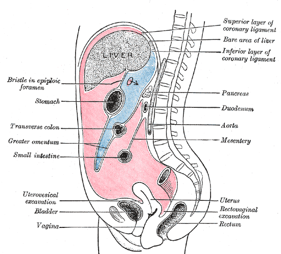
More on the folds, because tests name them:
- The greater omentum hangs like a fatty apron from the greater curvature of the stomach over the small intestine, then folds back up to attach to the transverse colon. It stores fat, carries immune cells ("milky spots") and can wall off an inflamed organ, which is why it is nicknamed the "policeman of the abdomen".
- The lesser omentum runs from the lesser curvature of the stomach and the first part of the duodenum to the liver. Its thick free edge (the hepatoduodenal ligament) carries the portal triad: portal vein, hepatic artery proper and common bile duct.
- The mesentery proper is a fan-shaped double fold that suspends the jejunum and ileum from the posterior wall. It carries their arteries, veins, lymphatics and nerves and holds the coiled intestine in place so it does not twist, while still letting it move during peristalsis.
- The transverse mesocolon and sigmoid mesocolon support those parts of the large intestine; the mesoappendix carries the appendicular artery.
- Behind the stomach is a pocket of the peritoneal cavity, the lesser sac (omental bursa), which opens to the main cavity through the epiploic foramen (of Winslow).
The peritoneum is a serous membrane: a single layer of simple squamous epithelium (mesothelium) on loose (areolar) connective tissue. It secretes a small volume of slippery serous fluid. Peritonitis, inflammation of the peritoneum, follows when a perforated appendix, ulcer or diverticulum spills gut contents into the cavity; the abdomen becomes rigid and tender. It is treated with IV antibiotics and surgery to repair the leak and wash out (drain) the cavity. Spontaneous bacterial peritonitis, infection of ascites fluid in cirrhosis without a perforation, is treated with antibiotics alone. Ascites is excess fluid in the peritoneal cavity, typical of cirrhosis. A large volume pushes up on the diaphragm and makes breathing hard; draining it with a needle (paracentesis) relieves the breathlessness and lets the fluid be tested for infection.
Spatial questions use two grids. The four quadrants are made by vertical and horizontal lines through the navel. The right upper quadrant (RUQ) holds most of the liver, the gallbladder, much of the duodenum, the head of the pancreas and the hepatic flexure. The left upper quadrant (LUQ) holds the stomach, the spleen, the body and tail of the pancreas and the splenic flexure. The right lower quadrant (RLQ) holds the cecum, appendix and terminal ileum, and the left lower quadrant (LLQ) holds the descending and sigmoid colon. Pain location in a vignette usually points to one of these groups; the spleen is the classic "not on the right side" answer.

The finer grid has nine regions, made by two vertical (midclavicular) lines and two horizontal lines (below the ribs and between the hip bones):
| Region | Main digestive contents |
|---|---|
| Right hypochondriac | Right lobe of the liver, gallbladder, hepatic flexure of the colon |
| Epigastric | Part of the stomach including the pylorus, left lobe of the liver, duodenum, pancreas |
| Left hypochondriac | Body and fundus of the stomach, spleen, tail of the pancreas, splenic flexure |
| Right lumbar | Ascending colon, some small intestine |
| Umbilical | Jejunum and ileum, transverse colon, part of the duodenum |
| Left lumbar | Descending colon, jejunum |
| Right iliac (inguinal) | Cecum, appendix, terminal ileum |
| Hypogastric (pubic) | Sigmoid colon, small intestine loops (with the bladder) |
| Left iliac (inguinal) | Sigmoid and lower descending colon |
So the stomach sits mainly in the epigastric and left hypochondriac regions, and the liver lies superior to the stomach, under the right dome of the diaphragm.
A hernia is an organ or fat pushing through a weak spot in the wall that should contain it. An indirect inguinal hernia (the most common type, mostly in males) passes through the deep inguinal ring along the spermatic cord, lateral to the inferior epigastric vessels. A direct inguinal hernia pushes straight through a weak posterior wall (Hesselbach's triangle), medial to those vessels, usually in older men. Femoral hernias are more common in women and more likely to strangle. Umbilical and incisional hernias come through the navel or an old scar. A hiatal hernia is part of the stomach sliding up through the esophageal hiatus of the diaphragm (the "sliding" type is the common one); it weakens the reflux barrier and is linked to GERD.
The wall and its linings
From the esophagus to the anus the wall has the same four layers, from the lumen outward:
| Layer | What it contains | Why it matters |
|---|---|---|
| Mucosa | Epithelium, lamina propria (loose connective tissue with capillaries, lymphatics and immune cells), muscularis mucosae | Secretion and absorption; most ulcers and cancers start here |
| Submucosa | Dense connective tissue, larger vessels, submucosal (Meissner) plexus, glands in some regions | Nerve control of secretion and local blood flow |
| Muscularis externa | Inner circular and outer longitudinal smooth muscle, myenteric (Auerbach) plexus between them | Peristalsis and segmentation |
| Serosa or adventitia | Visceral peritoneum (serosa) on intraperitoneal organs; fibrous connective tissue (adventitia) on the esophagus and retroperitoneal parts | Anchors the organ |

The lining changes with the job of each region, and comparing them is a favorite question:
| Region | Epithelium | Special features |
|---|---|---|
| Esophagus | Non-keratinized stratified squamous (resists abrasion) | Mucous glands in the submucosa; muscle is skeletal in the upper third, mixed in the middle, smooth below; adventitia instead of serosa for most of its length |
| Stomach | Simple columnar, every surface cell secretes mucus | Gastric pits and glands; rugae; an extra inner oblique muscle layer for churning |
| Small intestine | Simple columnar absorptive cells (enterocytes) with microvilli, plus goblet cells | Circular folds, villi and crypts; Brunner's glands in the duodenal submucosa make alkaline mucus; Peyer's patches in the ileum |
| Large intestine | Simple columnar with many goblet cells | No villi; straight deep crypts; taeniae coli; the anal canal changes to stratified squamous |
The sharp change from stratified squamous to simple columnar at the gastroesophageal junction explains why reflux hurts the esophagus: its lining is built for friction, not acid.
Why the two ends are different. The mouth, oropharynx, laryngopharynx, esophagus and lower anal canal all carry stratified squamous epithelium (non-keratinized, except keratinized patches such as the gums, hard palate and filiform papillae), because rough food and hard feces scrape them; many layers can be lost without exposing the tissue beneath. From the stomach to the rectum the lining is simple columnar, one cell thick, because that stretch secretes and absorbs, and a thin barrier lets molecules cross. (The nasopharynx, which carries only air, has the respiratory pseudostratified ciliated lining, whose job is to secrete and move mucus.)
Details of each layer that tests ask about:
- Mucosa (innermost): epithelium + lamina propria + muscularis mucosae, so it is the layer with the epithelium, its mucus-secreting exocrine cells (goblet cells, a one-celled gland), capillaries for absorbed nutrients, and MALT/GALT (lymphoid nodules in the lamina propria). The muscularis mucosae is a thin smooth-muscle sheet that throws the mucosa into small folds and moves the villi.
- Submucosa: dense irregular connective tissue (some books call it areolar) that binds the mucosa to the muscularis externa. It carries the larger blood and lymph vessels, the submucosal (Meissner) plexus, lymphoid tissue, elastic fibers, and glands in the esophagus and duodenum. It gives the tube its strength and stretch. Permanent folds such as the plicae circulares and the stomach's rugae are folds of mucosa and submucosa.
- Muscularis externa: inner circular and outer longitudinal smooth muscle, with the myenteric (Auerbach) plexus between them. It mixes and propels. The exceptions: the stomach adds an inner oblique layer (so the stomach has three layers: outer longitudinal, middle circular, inner oblique), the upper esophagus and the external anal sphincter are skeletal muscle, and in the colon the longitudinal layer is gathered into the three taeniae coli. Smooth-muscle sphincters (LES, pyloric, ileocecal, internal anal) are thickenings of the circular layer.
- Serosa (outermost): simple squamous mesothelium on areolar connective tissue, the visceral peritoneum. Its fluid reduces friction between the stomach or intestine and neighboring organs. Where there is no peritoneum (most of the esophagus, the retroperitoneal organs) a fibrous adventitia takes its place.
Mouth, salivary glands and esophagus
The mouth (oral or buccal cavity) is the first place where food is broken down, and both mechanical and chemical digestion happen there (as they also do in the stomach and small intestine). Chewing (mastication) tears food apart and raises the surface area enzymes can reach; the tongue mixes the food with saliva and shapes it into a bolus. Starch is the first macromolecule an enzyme acts on, so carbohydrate digestion is the first chemical digestion.
The oral cavity

- The oral vestibule is the slit between the lips and cheeks on the outside and the teeth and gums on the inside; the oral cavity proper lies inside the teeth. The lips are moved by the orbicularis oris muscle and the cheeks by the buccinator, which keeps food between the teeth.
- The labial frenula tie the lips to the gums, and the lingual frenulum ties the underside of the tongue to the floor of the mouth (cutting it treats "tongue-tie"). The floor of the mouth is supported by the mandible and the mylohyoid muscle.
- The hard palate (front of the roof) is bone: the palatine processes of the maxillae and the horizontal plates of the palatine bones. It separates the oral cavity from the nasal cavity and gives the tongue something to press food against.
- The soft palate (back of the roof) is muscle. It ends in the dangling uvula, and during swallowing both rise to close off the nasopharynx, so food does not enter the nose. The soft palate separates the oropharynx from the nasopharynx.
- The fauces are the opening from the mouth into the oropharynx, framed by the palatoglossal arch (in front) and the palatopharyngeal arch (behind, running down from the side of the soft palate to the pharynx). The palatine tonsils sit between them.
- The tonsils form Waldeyer's ring, a ring of MALT around the entrance to the pharynx: the pharyngeal tonsil (adenoids), tubal tonsils, palatine tonsils and lingual tonsil.
- Nerves: the trigeminal nerve (CN V, mandibular branch) drives the muscles of mastication (masseter, temporalis, the pterygoids), so trigeminal damage impairs chewing. The facial nerve (VII) carries taste from the front two-thirds of the tongue and drives the submandibular and sublingual glands; the glossopharyngeal (IX) carries taste from the back third and drives the parotid; the hypoglossal (XII) moves the tongue.
Tongue and taste
The tongue is skeletal muscle covered by stratified squamous epithelium. Intrinsic muscles change its shape; extrinsic muscles (genioglossus, hyoglossus, styloglossus, palatoglossus) move it. It mixes food, forms the bolus, pushes it back to start a swallow, and carries taste buds. The V-shaped groove at the back, the sulcus terminalis, divides the front two-thirds from the back third, where the lingual tonsil lies. Serous glands near the back (von Ebner glands) secrete lingual lipase.

| Papilla | Where | Taste buds? |
|---|---|---|
| Filiform | Whole front surface; the most numerous | None; rough, keratinized, grip food |
| Fungiform (mushroom-shaped) | Tip and sides, among the filiform | Yes, a few each |
| Foliate | Folds on the back sides | Yes (most prominent in children) |
| Circumvallate (vallate) | 8 to 12 large ones in a V before the sulcus terminalis | Yes, the most per papilla |
All five basic tastes (sweet, salty, sour, bitter, umami) can be detected on every part of the tongue that has taste buds. The old "tongue map" with one taste per zone is a myth. Chewing bread for several minutes makes it taste sweet anywhere on the tongue, because salivary amylase releases maltose.
Teeth

Human teeth are heterodont (different shapes for different jobs) and come in two sets (diphyodont). Teeth sit in sockets (alveoli) of two dental arches: the maxillary (upper) arch and the mandibular (lower) arch. Humans have two sets:
| Set | Number | Per quadrant (incisors-canine-premolars-molars) | When |
|---|---|---|---|
| Deciduous (primary, milk, baby) teeth | 20: 8 incisors, 4 canines, 8 molars, no premolars | 2-1-0-2 | Erupt from about 6 months; complete by about 2 to 3 years |
| Permanent (secondary) teeth | 32: 8 incisors, 4 canines (cuspids), 8 premolars (bicuspids), 12 molars | 2-1-2-3 | First permanent molar at about 6 years; all but the third molars by about 12 to 13; third molars (wisdom teeth) 17 to 25, or never |
Without the four wisdom teeth an adult has 28. The deciduous molars are replaced by the premolars (bicuspids); the permanent molars have no deciduous predecessors.
| Tooth | Shape | Job |
|---|---|---|
| Incisors | Chisel-shaped | Cut and bite off |
| Canines (cuspids) | One pointed cusp | Tear and pierce |
| Premolars (bicuspids) | Two cusps | Crush and grind |
| Molars | Broad, four or five cusps | Grind |

A tooth has a crown above the gum, a neck at the gumline and a root in the socket.
- Enamel covers the crown. It is about 96% mineral (hydroxyapatite), the hardest substance in the body, and has no cells, vessels or nerves, so it cannot repair itself once the enamel-forming cells (ameloblasts) are lost at eruption.
- Dentin forms the bulk of the tooth under the enamel and cementum. It is bone-like, harder than bone but softer than enamel, and is made throughout life by odontoblasts whose processes run in dentinal tubules. Like enamel, it has no blood vessels, which is the key difference between these calcified tissues and bone.
- Cementum is a thin, bone-like calcified layer covering the root. It is softer than dentin and is the anchor for the periodontal ligament.
- The pulp cavity in the center holds the pulp: connective tissue with blood vessels, nerves and lymphatics. Its extension through the root is the root canal, which opens at the root tip through the apical foramen. Losing the blood supply to a tooth means damage to the pulp.
- The periodontal ligament holds the root in its socket (a gomphosis joint). The gingiva (gum) covers the bone around the neck. These, with the cementum and alveolar bone, are the periodontal tissues.
Tooth decay (dental caries) happens when plaque bacteria, mainly Streptococcus mutans, ferment sugars into acid that dissolves enamel. Gingivitis is inflammation of the gums; untreated, it can progress to periodontitis, which destroys the periodontal ligament and bone and loosens teeth.
Salivary glands and saliva

| Gland | Location | Duct opening | Secretion | Share of daily (resting) saliva |
|---|---|---|---|---|
| Parotid (largest) | In front of and below the ear, over the masseter | Parotid (Stensen's) duct, inside the cheek opposite the upper second molar | Serous (watery, rich in amylase) | About 20 to 25%; becomes the main source during strong stimulation |
| Submandibular | Under the body of the mandible | Submandibular (Wharton's) duct, beside the lingual frenulum | Mixed, mostly serous with some mucus | About 60 to 70%; makes the most saliva |
| Sublingual (smallest) | Floor of the mouth, under the tongue | Many small ducts in the floor of the mouth | Mostly mucous (thick) | About 5 to 8% |
Many small buccal, labial and lingual glands add a little more. Mumps is a viral infection that swells the parotids.
Saliva is about 99.5% water; an adult makes roughly 1 to 1.5 L a day. It contains salivary amylase (ptyalin), a little lingual lipase, mucin (which becomes mucus), lysozyme and lactoferrin (antibacterial), IgA, bicarbonate and phosphate buffers, and electrolytes. It does not contain a protein-digesting enzyme (no digestive protease) or IgE. Its functions: moisten and lubricate food so it can be swallowed, dissolve food so it can be tasted, begin starch digestion, cleanse the mouth and control bacteria, and buffer acids that would dissolve enamel. Xerostomia is dry mouth.
Saliva is made in two steps. Acinar cells secrete a fluid much like plasma; the ducts then reabsorb Na⁺ and Cl⁻ and secrete K⁺ and HCO₃⁻, but their walls let little water through. The result is hypotonic saliva in which K⁺ and HCO₃⁻ are higher than in plasma and Na⁺ and Cl⁻ are lower. At high flow rates the fluid spends less time in the ducts, so less Na⁺ and Cl⁻ is removed and saliva becomes more like plasma (higher Na⁺ and Cl⁻), though still hypotonic.
Salivation is controlled almost entirely by the autonomic nervous system, through reflexes centered in the salivatory nuclei of the brainstem (pons and medulla). The sight, smell or thought of food and the touch of food in the mouth trigger it. Parasympathetic stimulation (facial and glossopharyngeal nerves) increases secretion from all the glands and produces large volumes of watery saliva. Sympathetic stimulation gives a small amount of thick, protein-rich saliva, which is why the mouth feels dry when you are frightened.
Salivary amylase cleaves internal α-1,4 bonds of starch, producing maltose, maltotriose and short branched fragments (α-limit dextrins). It works best near neutral pH (about 6.7 to 7) and is activated by chloride ions. It keeps working inside the bolus in the upper stomach until acid penetrates and denatures it; it is then digested like any other protein, and its amino acids are absorbed in the small intestine. Saliva has no digestive protease, so the mouth does not start protein digestion. Pancreatic amylase finishes the job; neither amylase can break the β-1,4 bonds of cellulose.
Pharynx, swallowing and esophagus
The pharynx (throat) is shared by air and food. The nasopharynx (behind the nose) carries only air and is lined with pseudostratified ciliated epithelium. The oropharynx (behind the mouth) and laryngopharynx (behind the larynx, down to the esophagus) carry both food and air and are lined with non-keratinized stratified squamous epithelium. Three pharyngeal constrictor muscles (skeletal) squeeze food downward.
Swallowing (deglutition) has three phases:
- Oral (buccal) phase, voluntary. The tongue presses against the hard palate and pushes the bolus back into the oropharynx.
- Pharyngeal phase, involuntary. Touch receptors in the pharynx signal the swallowing center in the medulla oblongata and lower pons (not the thalamus). The tongue blocks the mouth, the soft palate and uvula rise to close the nasopharynx, the larynx rises so the epiglottis (a flap of elastic cartilage) folds over the glottis (the vocal folds close too), breathing pauses, and the upper esophageal sphincter relaxes. The epiglottis keeps food out of the trachea; it does not push food anywhere.
- Esophageal phase, involuntary. A peristaltic wave carries the bolus to the stomach (about 8 s for a solid bolus, 1 to 2 s for liquid). If food sticks, its stretch starts a secondary peristalsis wave. Swallowing works against gravity because it is muscular, which is why you can swallow upside down.
The esophagus is a muscular tube about 25 cm (10 in) long. It runs from the pharynx (at about C6) behind the trachea and in front of the vertebral column, through the esophageal hiatus of the diaphragm (at about T10), to the cardia of the stomach. Its lining is non-keratinized stratified squamous epithelium, its submucosa has mucous glands that lubricate the bolus, and it has an adventitia rather than a serosa for most of its length. Its muscularis is skeletal in the upper third, mixed in the middle third and smooth in the lower third, always arranged inner circular, outer longitudinal. It secretes only mucus and digests and absorbs nothing. The upper esophageal sphincter keeps air out of the esophagus, and the lower esophageal sphincter (LES, or cardiac sphincter) keeps stomach contents out, which protects the esophagus from acid and prevents regurgitation; it relaxes as each swallow arrives. The LES is a physiological sphincter: a zone of thickened smooth muscle helped by the diaphragm around it, not a distinct anatomical ring.
The stomach
The stomach is a J-shaped sac in the left upper quadrant (epigastric and left hypochondriac regions), just below the diaphragm. It stores food temporarily, churns it into chyme, starts protein digestion, kills most microbes and releases chyme slowly into the duodenum. An empty stomach holds about 50 mL; it can stretch to hold about 1 to 1.5 L comfortably and up to about 4 L.
It has four regions: the cardia (around the opening from the esophagus), the fundus (the dome above and to the left of the cardia, where swallowed air collects), the body (the main part) and the pyloric part (the funnel-shaped pyloric antrum, then the narrow pyloric canal, ending at the pylorus and pyloric sphincter, which controls emptying into the duodenum). The concave lesser curvature faces the liver and is attached to it by the lesser omentum; the convex greater curvature gives rise to the greater omentum. When empty, the mucosa and submucosa fold into rugae, which flatten as the stomach fills; they are what lets it distend.

The surface is dotted with millions of gastric pits, each leading into tubular glands. Cardiac glands (near the cardia) and pyloric glands (in the pyloric region) secrete mostly mucus; the pyloric glands also hold most of the G cells. The gastric (oxyntic or fundic) glands of the fundus and body make most of the gastric juice. Within a gastric gland, parietal cells cluster in the upper part, chief cells toward the base, and enteroendocrine cells are scattered low down.

The cell types:
| Cell | Secretes | Function |
|---|---|---|
| Surface mucous (foveolar) cells | Thick alkaline mucus with bicarbonate | Main barrier against acid and pepsin; line the surface and the pits |
| Mucous neck cells | Thin, acidic mucus | Protects the gland neck |
| Parietal (oxyntic) cells | HCl and intrinsic factor | Acid lowers pH to about 2, denatures proteins, activates pepsinogen, kills microbes, and keeps dietary iron soluble so it can be reduced to the absorbable ferrous form (Fe²⁺); intrinsic factor is needed for vitamin B₁₂ absorption |
| Chief (zymogenic) cells | Pepsinogen and gastric lipase | Acid converts pepsinogen to pepsin |
| G cells (antrum and pylorus; also duodenum) | Gastrin | Stimulates acid secretion and motility |
| D cells | Somatostatin | Inhibits gastrin and acid |
| ECL (enterochromaffin-like) cells | Histamine | Stimulates parietal cells through H₂ receptors |
| X/A-like cells (fundus) | Ghrelin | "Hunger hormone": rises before meals |
| Stem cells (gland neck) | none | Replace the surface cells every few days |
G, D, ECL and ghrelin cells are enteroendocrine cells: they release hormones into the blood (or, for histamine and somatostatin, locally), so they are endocrine, not exocrine. "Ulcerin" does not exist; gastrin is the hormone that drives acid.
How a parietal cell makes HCl. Inside the cell, carbonic anhydrase combines CO₂ and H₂O into carbonic acid, which splits into H⁺ and HCO₃⁻. The H⁺/K⁺-ATPase (the proton pump) pumps H⁺ into the lumen against a million-fold gradient (primary active transport) in exchange for K⁺. Cl⁻ enters the cell from the blood in exchange for HCO₃⁻ and then diffuses into the lumen through channels, so H⁺ and Cl⁻ meet in the lumen as HCl. The HCO₃⁻ that goes into the blood makes the venous blood leaving the stomach alkaline after a meal, the alkaline tide. Three stimulants act on the parietal cell, and they amplify each other: acetylcholine (vagus), gastrin and histamine. Proton-pump inhibitors such as omeprazole block the pump itself and so raise stomach pH; H₂ blockers (famotidine) remove the histamine stimulus; antacids neutralize acid already secreted, raising gastric pH toward about 4, where pepsin is much less active.
Pepsin. Chief cells release inactive pepsinogen so they do not digest themselves. Below about pH 5, HCl unfolds pepsinogen and a piece is cut off, forming pepsin; pepsin then activates more pepsinogen (autocatalysis, a positive-feedback loop). Pepsin works best at about pH 1.5 to 3 and stops working above about pH 5, so it is inactive once it reaches the duodenum. Pepsin cuts proteins into large polypeptides, digesting perhaps 10 to 20% of dietary protein; most protein digestion happens in the small intestine, so losing pepsin causes discomfort rather than failure to digest protein. Pepsinogen placed on meat at pH 7 does nothing, because it is the inactive form and nothing activates it. Rennin (chymosin), which curdles milk protein, is made by the stomachs of young ruminants such as calves; it is not a significant enzyme in adult humans. The stomach makes about 2 L of gastric juice a day.
What the stomach absorbs. Almost no nutrients. Only a few lipid-soluble substances cross its lining: alcohol (about a fifth of it), aspirin and some other drugs, and a little water. Fatty food slows gastric emptying, which keeps alcohol in the stomach longer and slows its arrival in the small intestine, where most is absorbed; that is why eating before drinking lowers peak blood alcohol.
Acid secretion has three phases. The cephalic phase (about 30% of acid, before food arrives) starts at the sight, smell, taste or thought of food: signals pass from the cortex and hypothalamus to the vagal nuclei in the medulla, down the vagus nerve to enteric neurons and on to the glands. So gastric secretion can start before food enters the mouth. The gastric phase (about 60%) responds to stretch and to peptides and amino acids in the stomach, through local reflexes, vagovagal reflexes and gastrin. As the pH in the antrum falls below about 3, D cells release somatostatin and gastrin release is shut off, so very low pH inhibits further secretion (negative feedback). The intestinal phase starts as chyme enters the duodenum. A small excitatory part comes from intestinal gastrin, but mostly it inhibits the stomach through the enterogastric reflex and hormones (secretin, CCK, GIP), so the duodenum is not flooded with acid.
The stomach avoids digesting itself through overlapping defenses: a mucus-bicarbonate gel that keeps the surface near neutral, tight junctions that block back-leak of H⁺, rapid epithelial renewal, and prostaglandins (especially PGE₂) that boost mucus, bicarbonate and mucosal blood flow. When these fail, an ulcer forms (see Disorders).
Gastric motility has three parts. Receptive relaxation lets the fundus and body stretch to hold a meal without a pressure rise. Contractions then sweep toward the pylorus, and most chyme is pushed back (retropulsion) against a nearly closed sphincter, grinding food to a slurry. Peristaltic waves are strongest in the antrum. Small squirts (a few mL at a time) pass into the duodenum. Emptying is controlled mostly from the duodenum: when fat, acid, hypertonic chyme or stretch reaches it, the enterogastric reflex and the hormones CCK, secretin and GIP slow the stomach. A carbohydrate-rich meal leaves within a few hours; a fatty meal stays longest. A narrowed (stenosed) pylorus blocks passage of chyme into the duodenum.
Vomiting (emesis) is a reflex coordinated by the vomiting center in the medulla, triggered by stretch, irritation, toxins acting on the chemoreceptor trigger zone, motion or pain. The diaphragm and abdominal muscles squeeze the stomach while the LES and upper sphincter relax. Repeated vomiting loses HCl and causes metabolic alkalosis with low chloride and potassium.
Small intestine
The small intestine is where most digestion and almost all nutrient absorption happen, which is why it is the most important digestive organ. It is about 3 to 5 m long in a living adult and about 6 m (20 ft) in a cadaver, where muscle tone is lost, but only about 2.5 cm wide; "small" refers to the diameter. It is longer than the large intestine. Its three parts, in order: duodenum → jejunum → ileum.

The duodenum (about 25 cm, the shortest and widest part) receives chyme, bile and pancreatic juice; it is mostly retroperitoneal and curves in a C around the head of the pancreas. It has four parts. The first (superior) part begins with the duodenal bulb (cap), the site of most duodenal ulcers. The second (descending) part has the major duodenal papilla, where bile and pancreatic juice enter together through the hepatopancreatic ampulla (of Vater), guarded by the sphincter of Oddi; a minor papilla above it receives the accessory pancreatic duct. The third (horizontal) part crosses in front of the aorta under the superior mesenteric vessels. The fourth (ascending) part ends at the duodenojejunal flexure, held up by the suspensory ligament of the duodenum (ligament of Treitz), the landmark that divides the upper GI tract from the lower GI tract (bleeding above it is "upper GI bleeding"). Duodenal Brunner's glands in the submucosa pour alkaline mucus into the bottoms of the crypts to protect the duodenum from acid chyme and give intestinal enzymes the pH they need.
The jejunum (the middle part, about two-fifths of the length beyond the duodenum, roughly 2.5 m in a cadaver) is the main site of digestion and absorption; it has the tallest villi, the most prominent circular folds and the richest blood supply. The ileum (the last three-fifths, roughly 3.5 m in a cadaver, so the longest part) absorbs bile salts and vitamin B₁₂, has the most Peyer's patches, and ends at the ileocecal valve, where it joins the cecum. There is no sharp border between jejunum and ileum; the change is gradual.
| Feature | Jejunum | Ileum |
|---|---|---|
| Position | Mostly upper left of the abdomen | Mostly lower right |
| Wall and diameter | Thicker, wider, redder (more blood) | Thinner, narrower, paler |
| Circular folds and villi | Tall, numerous, close together | Fewer and lower; absent near the end |
| Mesenteric blood vessels | Long straight vessels, few arcades, little fat | Short straight vessels, many arcades, more fat |
| Lymphoid tissue | Little | Peyer's patches (large lymphoid nodules) |
| Special absorption | Most nutrients, water-soluble vitamins, folate | Vitamin B₁₂ and bile salts (terminal ileum) |
Surface area is multiplied at three scales: circular folds (plicae circulares), permanent folds of mucosa and submucosa that also slow chyme and make it spiral (about a 3-fold gain), villi, finger-like folds of the mucosa about 0.5 to 1 mm tall (about 10-fold), and microvilli (the brush border), folds of the cell membrane coated with a carbohydrate-rich glycocalyx that holds the brush-border enzymes (about 20-fold). Together they enlarge the absorptive area roughly 600-fold. Circular folds and villi become fewer and smaller toward the end of the ileum, and the large intestine has neither; rugae belong to the stomach, not the intestine.
Each villus has a capillary network that takes sugars and amino acids to the hepatic portal vein, a central lacteal (a blunt-ended lymphatic capillary) that takes up fat, and a strand of smooth muscle that makes it pump. Absorbed nutrients cross the epithelium and are picked up by vessels in the lamina propria. The crypts (of Lieberkühn) between villi are where new cells are made.
| Cell (small intestine epithelium) | Where | Job |
|---|---|---|
| Absorptive cells (enterocytes) | Villi, the most numerous | Absorb nutrients; carry brush-border enzymes |
| Goblet cells | Villi and crypts (more toward the ileum and colon) | Secrete mucus |
| Enteroendocrine cells (S, I, K, L, M and others) | Crypts and villi | Secrete secretin, CCK, GIP, GLP-1, motilin, serotonin |
| Paneth cells | Base of the crypts (most in the ileum) | Secrete lysozyme and defensins (antimicrobial); found in the small intestine, normally not in the colon |
| Stem cells | Crypt base | Divide to replace the lining every 3 to 5 days |
| M (microfold) cells | Over Peyer's patches | Pass antigens from the lumen to immune cells |
Mucus from goblet cells (and from Brunner's glands and the stomach's mucous cells) does three jobs along the whole tract: it lubricates contents so they slide without abrading the lining, it forms a gel that shields the epithelium from acid and digestive enzymes, and it traps microbes and holds antimicrobial molecules (IgA, defensins) as a barrier. In the colon it also binds feces together.
At the base of the microvilli the enterocyte membrane forms small pits and vesicles (pinocytosis and receptor-mediated endocytosis); this is how the B₁₂-intrinsic factor complex and small amounts of intact protein enter the cell.
Intestinal glands add about 1 to 2 L a day of watery, slightly alkaline intestinal juice that carries the chyme and enzymes; the enzymes that finish digestion are attached to the brush border rather than dissolved in this juice.
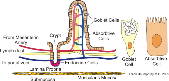
The brush border carries enzymes that finish digestion: maltase, sucrase-isomaltase (sucrose, and the branched α-limit dextrins amylase leaves behind), lactase, and peptidases. Enterokinase (enteropeptidase), made by the duodenal lining, converts trypsinogen to trypsin, which then activates the other pancreatic proteases.
Large intestine
The large intestine is about 1.5 m (5 ft) long and about 6 to 7 cm wide: wider but much shorter than the small intestine. It frames the small intestine on three sides. In order, it comprises the cecum (the first part, a blind pouch in the right iliac region, below the ileocecal valve) with the vermiform appendix hanging from it, the ascending colon (up the right side), the right colic (hepatic) flexure under the liver, the transverse colon (across the abdomen, hanging on its mesocolon), the left colic (splenic) flexure near the spleen (higher than the right), the descending colon (down the left side), the S-shaped sigmoid colon, the rectum and the anal canal. So the major regions are cecum, colon, rectum and anal canal; the jejunum and ileum are small intestine.

Three features recognize the colon from outside: the taeniae coli, three ribbons formed by gathering the outer longitudinal muscle; the haustra, pouches made because the taeniae are shorter than the wall and pucker it, which give the colon its segmented look; and the epiploic appendages (appendices epiploicae), small fat-filled tags of visceral peritoneum hanging from the surface. Inside, the lining is simple columnar epithelium with very many goblet cells in deep straight crypts. There are no villi, no circular folds and normally no Paneth cells; the mucus lubricates the drying feces. GALT is well developed in the lamina propria because so many microbes live in the lumen.
The colon's jobs: absorb water, Na⁺ and Cl⁻ from what is left (and secrete K⁺ and HCO₃⁻), house bacteria that ferment fiber and make vitamin K and some B vitamins, and form, store and eliminate feces. Little chemical digestion happens here except bacterial fermentation, and no mechanical digestion in the usual sense. Without the colon a person has watery stool but can survive; the gallbladder and the greater omentum can also be removed.
Feces are about 75% water and 25% solids. The solids are mostly bacteria (about a third), indigestible fiber, sloughed epithelial cells, fats, inorganic salts and some protein, colored brown by stercobilin from bilirubin.
The appendix (vermiform appendix, about 8 cm) is a narrow, worm-shaped blind tube rich in lymphoid tissue. It has no digestive function and has long been called vestigial; current evidence suggests it serves the immune system and acts as a reservoir ("safe house") that can reseed the colon with normal bacteria after diarrhea. Its narrow lumen is easily obstructed, causing appendicitis.
The rectum (about 12 to 15 cm) has three transverse folds (rectal valves) that hold feces while gas passes. The anal canal (about 3 to 4 cm) has vertical anal columns, and the pectinate (dentate) line at their base marks where the gut lining (simple columnar, painless visceral nerves) meets skin-like stratified squamous epithelium supplied by painful somatic nerves. That is why internal hemorrhoids (above the line) bleed painlessly and external hemorrhoids (below it) hurt. Two sphincters close the canal: the internal anal sphincter is a thickening of the circular smooth muscle, involuntary; the external anal sphincter is skeletal muscle, voluntary (pudendal nerve).
Slow haustral contractions (haustral churning) every half hour or so move contents from one haustrum to the next and mix them; the large intestine does not use segmentation, and the small intestine does not use haustral contractions. Three or four times a day a mass movement, a strong peristaltic wave, sweeps contents from the transverse colon toward the rectum, often after a meal (the gastrocolic reflex). When feces stretch the rectum, the defecation reflex (a spinal reflex through parasympathetic pelvic splanchnic nerves, S2 to S4) contracts the sigmoid colon and rectum and relaxes the internal sphincter. Defecation then happens only when the external sphincter is voluntarily relaxed. The Valsalva maneuver (closing the glottis and contracting the diaphragm and abdominal muscles) raises abdominal pressure and helps expel feces. Fast movement through the colon leaves too little time to absorb water (diarrhea); slow movement allows too much (constipation).
Blood and nerve supply
The gut tube develops as a foregut, midgut and hindgut, and each has its own unpaired artery from the abdominal aorta. Knowing the boundaries answers most blood-supply questions.
| Artery (level) | Region (embryonic gut) | Main branches |
|---|---|---|
| Celiac trunk (T12) | Foregut: lower esophagus, stomach, first half of the duodenum, liver, gallbladder, pancreas (part); also the spleen | Left gastric; splenic (short gastrics, left gastro-omental, pancreatic branches); common hepatic (proper hepatic to the liver, right gastric, gastroduodenal → right gastro-omental and superior pancreaticoduodenal) |
| Superior mesenteric artery (L1) | Midgut: second half of the duodenum to the proximal two-thirds of the transverse colon (jejunum, ileum, cecum, appendix, ascending colon) | Inferior pancreaticoduodenal, jejunal and ileal arteries, ileocolic (with the appendicular artery), right colic, middle colic |
| Inferior mesenteric artery (L3) | Hindgut: distal third of the transverse colon, descending and sigmoid colon, upper rectum | Left colic, sigmoid arteries, superior rectal |

Neighboring territories join through a chain of arcades along the colon (the marginal artery). The splenic (left colic) flexure sits at the border between the SMA and IMA territories, a "watershed" with the thinnest overlap, so it is the part of the colon most prone to ischemia when blood pressure falls.
Venous drainage is the key to the liver's role. Blood from the stomach, intestines, pancreas and spleen does not go straight to the heart. The splenic vein (usually joined by the inferior mesenteric vein) and the superior mesenteric vein unite behind the neck of the pancreas to form the hepatic portal vein, which carries absorbed nutrients (and absorbed toxins and bacteria) to the liver sinusoids for processing, storage and detoxification first. Blood then leaves through the hepatic veins into the inferior vena cava. When scarring blocks portal flow (portal hypertension in cirrhosis), blood detours through connections with systemic veins, which swell into esophageal varices, rectal varices and dilated veins around the navel (caput medusae).
Nerves. The gut has its own enteric nervous system (ENS) in the wall (below). The parasympathetic supply, mainly the vagus nerve (as far as about two-thirds of the way along the transverse colon) plus the pelvic splanchnic nerves (S2 to S4) for the distal colon and rectum, increases motility and secretion ("rest and digest"). The sympathetic supply (thoracic and lumbar splanchnic nerves relaying in the celiac, superior mesenteric and inferior mesenteric ganglia) decreases motility and secretion, contracts sphincters and constricts gut blood vessels. The small intestine's sympathetic fibers reach it through the superior mesenteric plexus.
Motility and its control
Motility is the general ability of the gut wall to move its contents: it covers mixing (segmentation, churning) as well as propulsion (moving contents forward: swallowing, peristalsis, mass movements). Some books list the digestive functions as motility, secretion, digestion and absorption.
Two patterns move the contents. Peristalsis is a wave in which the muscle contracts behind a bolus and relaxes in front of it, pushing it forward. Segmentation is a set of stationary ring contractions that chop and mix chyme against the mucosa with little net movement.

Peristalsis in the esophagus, stomach and small intestine is propulsion: circular muscle contracts behind the bolus while it relaxes ahead of it, and longitudinal muscle shortens the segment ahead. Segmentation is the major mixing movement of the small intestine and counts as mechanical digestion; it dominates after a meal, while the migrating motor complex takes over between meals. Churning in the stomach mixes and grinds. In the colon, haustral contractions and mass movements take over (see above).
Interstitial cells of Cajal act as pacemakers, producing slow waves (the basal electrical rhythm) that set the maximum contraction frequency. A slow wave on its own usually does not make the muscle contract; contraction happens when the wave's peak crosses threshold and fires spike potentials (action potentials carried mostly by calcium entry). Anything that depolarizes the muscle (stretch, acetylcholine, parasympathetic stimulation, some gut hormones) adds spikes and strengthens contraction. Sympathetic stimulation (norepinephrine) hyperpolarizes the muscle and decreases the number of spike potentials. The stomach runs at about 3 per minute; the duodenum is the fastest (about 11 to 12 per minute), with the rate falling toward the ileum (about 8 to 9). Textbooks differ by a cycle or two for the intestine, but the stomach is consistently the slowest. Between meals, the migrating motor complex sweeps residue and bacteria from the stomach through the ileum roughly every 90 to 120 minutes; it is triggered by the hormone motilin and stopped by eating. These "housekeeper" waves are the growling (borborygmi) of the stomach and small intestine you hear when hungry. Waves of peristalsis travel a short way and die out, and each new wave starts a little further along, so the complex creeps down to the ileum before a new one starts at the stomach. The antibiotic erythromycin also stimulates motilin receptors, so it speeds chyme through the gut and is used to treat slow gastric emptying.
The enteric nervous system (ENS) is a network of hundreds of millions of neurons in the gut wall that can run reflexes with no input from the brain. The myenteric plexus mainly controls motility and the submucosal plexus mainly controls secretion and local blood flow. In the peristaltic reflex, stretch causes contraction above the bolus (acetylcholine, substance P) and relaxation below it (nitric oxide, VIP). The autonomic system adjusts the ENS: parasympathetic (mostly vagal) input increases motility and secretion, and sympathetic input reduces them and constricts gut blood vessels, which is why stress and hard exercise slow digestion.
Gut reflexes come in two kinds. Short (local) reflexes run entirely inside the ENS: stretch or chemical receptors in the wall signal enteric interneurons and motor neurons, which adjust local motility and secretion (the peristaltic reflex is one). Long reflexes send sensory signals out of the gut to the CNS, often along vagal afferents, and back through autonomic efferents (vagovagal reflexes). They link distant parts of the tract and respond to outside cues such as the sight or smell of food (the cephalic phase), stress or emotion. The ENS can work if the vagus is cut; long reflexes cannot.
Hormones from enteroendocrine cells coordinate the organs:
| Hormone | Made by | Stimulus | Main effects |
|---|---|---|---|
| Gastrin | G cells (antrum) | Peptides, stretch, vagal input | ↑ acid; ↑ gastric motility; ↑ ECL histamine |
| Secretin | S cells (duodenum) | Acid in the duodenum | ↑ bicarbonate from pancreatic and bile ducts; ↓ acid |
| CCK | I cells (duodenum, jejunum) | Fatty acids, peptides | ↑ pancreatic enzymes; gallbladder contracts; sphincter of Oddi relaxes; ↓ gastric emptying; satiety |
| GIP | K cells (duodenum, jejunum) | Glucose, fatty acids | ↑ insulin; ↓ gastric acid |
| GLP-1 | L cells (ileum, colon) | Nutrients reaching the distal gut | ↑ insulin; ↓ glucagon; slows gastric emptying; satiety |
| Motilin | M cells (duodenum) | Fasting | Starts the migrating motor complex |
| Somatostatin | D cells (stomach, duodenum, pancreatic islets) | Acid | ↓ gastrin, acid and most GI secretion; ↓ insulin and glucagon |
| Ghrelin | X/A-like cells (gastric fundus) | Empty stomach, fasting | Hunger; ↑ growth hormone release; ↑ gastric motility |
| Peptide YY (PYY) | L cells (ileum, colon) | Nutrients, especially fat, in the distal gut | ↓ appetite after a meal; slows gastric emptying (the "ileal brake") |
| VIP (vasoactive intestinal peptide) | Enteric neurons | Distension, nerve activity | Relaxes smooth muscle and sphincters; ↑ water and electrolyte secretion by the intestine; ↑ pancreatic bicarbonate; ↓ acid |
| Serotonin (5-HT) | Enterochromaffin cells (most of the body's serotonin) | Stretch, food in the lumen | Starts the peristaltic reflex and secretion |
| Histamine | ECL cells (local, paracrine) | Gastrin, vagus | ↑ HCl through H₂ receptors |
Leptin is not a gut hormone but is part of the same appetite system: fat cells release it in proportion to how much fat is stored, and it signals the hypothalamus to reduce intake over the long term.
More details tests use:
- Gastrin is made in the stomach (G cells) and stimulates the stomach: acid (directly and by releasing histamine), motility, and growth of the gastric lining. Acid below about pH 3 shuts it off through somatostatin. A gastrinoma (Zollinger-Ellison syndrome) makes so much gastrin that severe, multiple ulcers form.
- Secretin (the first hormone ever discovered, 1902) is made in the small intestine (S cells) when acid chyme arrives. It increases bicarbonate from the pancreatic ducts and the bile ducts (so bile volume and bicarbonate rise, not bile salts), and it decreases gastrin release, acid and gastric emptying. It is the hormone that counteracts gastric acid in the small intestine.
- CCK is made in the small intestine (I cells), mainly in response to fats (fatty acids, monoglycerides) and amino acids. Its targets: pancreatic acinar cells (enzyme-rich juice), the gallbladder (contracts), the sphincter of Oddi (relaxes, so bile and juice can enter), and the stomach (slows emptying and secretion). It also contracts the pyloric sphincter (one way it slows emptying), stimulates insulin release a little, and signals fullness. Secretin and CCK both inhibit the stomach; secretin is the classic answer for "inhibits gastric acid secretion" (it is triggered by acid), while CCK is the classic answer for slowing gastric emptying after a fatty meal. Lacking CCK would mimic lacking a gallbladder, because bile would not be released in a timed squirt. CCK is used in imaging to test whether the gallbladder can contract.
- Enterogastrones are intestinal hormones that inhibit gastric secretion and motility: secretin, CCK and GIP, and also VIP, somatostatin and PYY. Gastrin, histamine and serotonin are not enterogastrones.
- Stretch of the stomach wall stimulates gastrin release; secretin, CCK and GIP respond mainly to chemicals in the duodenum (acid, fat, protein, glucose). Nothing causes "gastrin reuptake".
- Appetite. The hypothalamus integrates signals. Short-term, ghrelin (hunger) rises before meals and falls after eating, while CCK, GLP-1, PYY and stomach stretch signal fullness after a meal. Long-term, leptin (from fat) and insulin signal how much energy is stored. People with obesity usually have high leptin but respond poorly to it (leptin resistance).
Chemical digestion of proteins, fats and carbohydrates
Chemical digestion is hydrolysis: an enzyme adds water across the bond that joins two building blocks. Each macromolecule has its own monomers, and only monomers (plus a few di- and tripeptides) can be absorbed, so this is the step that makes absorption possible.
| Macromolecule | Built from | Bond cut |
|---|---|---|
| Carbohydrates (polysaccharides, disaccharides) | Monosaccharides (glucose, fructose, galactose) | Glycosidic bonds |
| Proteins (polypeptides) | Amino acids | Peptide bonds |
| Triglycerides (fats) | Glycerol (or monoglyceride) + fatty acids | Ester bonds |
| Nucleic acids (DNA, RNA) | Nucleotides → pentose sugars, phosphate and nitrogenous bases | Phosphodiester bonds |
Lipids are the odd one out. They do not dissolve in water, so they need bile to emulsify them and micelles to carry them, and they are absorbed into lymph rather than blood. Starch has two forms: amylose (straight chains of glucose linked α-1,4) and amylopectin (branched, with α-1,6 links at the branch points). Glycogen, the animal storage form, is even more branched. Cellulose is glucose linked β-1,4; humans have no enzyme for that bond, so it passes through as fiber and only colon bacteria ferment some of it.
Carbohydrates. Dietary carbohydrate is mostly starch plus sucrose, lactose and maltose. Salivary and pancreatic amylase cut starch into maltose, maltotriose and α-limit dextrins. Brush-border enzymes finish the job: maltase gives glucose, sucrase gives glucose + fructose, lactase gives glucose + galactose. Glucose and galactose enter enterocytes with sodium through SGLT1 (secondary active transport); fructose enters by facilitated diffusion through GLUT5; all three leave through GLUT2 into portal blood. Because each disaccharide has its own enzyme, a missing enzyme blocks only its substrate, and the undigested sugar draws water into the gut and is fermented by colon bacteria.
Proteins. Pepsin starts protein digestion in the stomach. In the duodenum, trypsin, chymotrypsin, elastase (endopeptidases that cut inside chains) and carboxypeptidase (an exopeptidase that trims from the end) make short peptides, and brush-border peptidases finish them. Amino acids enter by sodium-linked carriers (secondary active transport); di- and tripeptides enter by PEPT1, coupled to H⁺, and are split inside the cell. Peptides longer than about four amino acids are essentially not absorbed. Absorbed amino acids travel to the liver in the portal vein. Nine amino acids are essential (the body cannot make them, so they must be eaten): histidine, isoleucine, leucine, lysine, methionine, phenylalanine, threonine, tryptophan and valine. Tyrosine, alanine, glutamine and the rest are nonessential.
Fats. Bile salts emulsify triglyceride droplets. Pancreatic lipase, anchored by colipase, cuts triglycerides into 2-monoglyceride and free fatty acids, which pack into bile-salt micelles that ferry them to the brush border. Inside the enterocyte they are rebuilt into triglycerides and packaged with apolipoproteins as chylomicrons, which enter the lacteals, travel up the thoracic duct and join the blood at the left subclavian vein, bypassing the liver at first. Short- and medium-chain fatty acids (fewer than about 12 carbons) are water-soluble enough to skip micelles and chylomicrons and go straight into portal blood; lingual and gastric lipase handle much of their digestion. Bile salts are not the only emulsifiers: phospholipids (lecithin) in food and bile help too. Monoglycerides and fatty acids leave the micelle and cross the enterocyte membrane mostly by simple diffusion. Triglycerides are rebuilt in the smooth ER and packaged into chylomicrons in the Golgi. Rebuilding them at once keeps free fatty acids inside the cell low, which preserves the gradient that drives more diffusion in. Blocking triglyceride re-synthesis lets fatty acids and monoglycerides build up inside the cell, slows their uptake and leaves more of them in the micelles in the lumen. If the Golgi cannot release chylomicrons, fat and the fat-soluble vitamins A, D, E and K are malabsorbed, while water-soluble vitamins (B, C) are absorbed normally.
| Nutrient | Enzymes | Where | Absorbed as |
|---|---|---|---|
| Starch | Salivary and pancreatic amylase; brush-border maltase and isomaltase | Mouth, duodenum, jejunum | Glucose (SGLT1) |
| Protein | Pepsin; trypsin, chymotrypsin, elastase, carboxypeptidase; brush-border peptidases | Stomach, duodenum, jejunum | Amino acids, di- and tripeptides |
| Triglyceride | Lingual and gastric lipase (minor); pancreatic lipase + colipase | Stomach (minor), duodenum | Fatty acids and monoglycerides in micelles → chylomicrons |
| Nucleic acids | Pancreatic ribonuclease and deoxyribonuclease; brush-border nucleosidases and phosphatases | Duodenum, jejunum | Pentoses, phosphate, nitrogenous bases (active transport) |
The full list of enzymes, with what turns each one on, is the table tests are built from:
ReferenceDigestive enzymes· 20 rows
Source = where it is made; Acts in = where it works. "Zymogen" means it is secreted inactive.
No rows match.
| Enzyme | Source | Acts in (best pH) | Substrate → products | Notes |
|---|---|---|---|---|
| Salivary amylase (ptyalin) | Salivary glands (mostly parotid and submandibular) | Mouth, upper stomach until acid reaches the bolus (about 7) | Starch α-1,4 bonds → maltose, maltotriose, α-limit dextrins | Needs Cl⁻; the first enzyme food meets |
| Lingual lipase | Glands at the back of the tongue | Stomach (acidic) | Triglycerides (especially short- and medium-chain) → fatty acids + diglycerides | More important in infants |
| Pepsin | Chief cells, as pepsinogen | Stomach (1.5 to 3) | Proteins → large polypeptides (cuts next to aromatic and large hydrophobic amino acids) | Activated by HCl and by pepsin itself |
| Gastric lipase | Chief cells | Stomach | Triglycerides → fatty acids + diglycerides | Minor in adults |
| Pancreatic amylase | Acinar cells | Duodenum, jejunum (about 7) | Starch → maltose, maltotriose, α-limit dextrins | Cannot cut cellulose |
| Trypsin | Acinar cells, as trypsinogen | Small intestine | Proteins and polypeptides → shorter peptides (cuts after lysine and arginine) | Activated by enteropeptidase; then activates all the other pancreatic zymogens |
| Chymotrypsin | Acinar cells, as chymotrypsinogen | Small intestine | Peptides → shorter peptides (cuts after aromatic amino acids) | Activated by trypsin |
| Elastase | Acinar cells, as proelastase | Small intestine | Peptides, including elastin (cuts after small neutral amino acids) | Activated by trypsin |
| Carboxypeptidase A and B | Acinar cells, as procarboxypeptidases | Small intestine | Removes one amino acid at a time from the carboxyl end | An exopeptidase containing zinc; activated by trypsin |
| Pancreatic lipase + colipase | Acinar cells (colipase as procolipase) | Small intestine | Triglyceride → 2-monoglyceride + 2 free fatty acids | Colipase anchors lipase to bile-coated droplets; procolipase is activated by trypsin |
| Phospholipase A₂ | Acinar cells, as a zymogen | Small intestine | Phospholipids → lysophospholipid + fatty acid | Activated by trypsin |
| Cholesterol esterase | Acinar cells | Small intestine | Cholesterol esters → cholesterol + fatty acid | Needs bile salts |
| Ribonuclease, deoxyribonuclease | Acinar cells | Small intestine | RNA, DNA → nucleotides | |
| Enteropeptidase (enterokinase) | Duodenal brush border | Duodenum | Trypsinogen → trypsin | The trigger for the whole protease cascade |
| Aminopeptidase | Brush border | Small intestine | Removes amino acids from the amino end | Exopeptidase |
| Dipeptidase, tripeptidase | Brush border | Small intestine | Di- and tripeptides → amino acids | |
| Maltase (maltase-glucoamylase) | Brush border | Small intestine | Maltose and maltotriose → glucose | |
| Sucrase-isomaltase | Brush border | Small intestine | Sucrose → glucose + fructose; isomaltase (α-dextrinase) cuts the α-1,6 bonds of α-limit dextrins | |
| Lactase | Brush border | Small intestine | Lactose → glucose + galactose | Declines after weaning in most people |
| Nucleosidases, phosphatases | Brush border | Small intestine | Nucleotides → pentose + phosphate + base |
How to read it: the stomach contributes one real enzyme (pepsin) and a little lipase; the pancreas contributes enzymes for all four macromolecules, which is why it is the accessory organ whose loss most damages digestion; the brush border finishes carbohydrates, proteins and nucleic acids at the surface of the absorbing cell. The large intestine and esophagus make no digestive enzymes, and bile is not an enzyme. Endopeptidases (pepsin, trypsin, chymotrypsin, elastase) cut inside a chain; exopeptidases (carboxypeptidase, aminopeptidase) trim from an end. Digestive enzymes are hydrolases; oxidases, ligases and peroxidases are not digestive enzymes. Pepsin acts at low pH; the pancreatic proteases and amylases need near-neutral to slightly alkaline conditions.
Absorption of water, minerals and vitamins
About 9 L of fluid reaches the small intestine each day: 1 to 2 L from drinking and the rest from saliva, gastric juice, bile, pancreatic juice and intestinal secretions. The small intestine reabsorbs most of it, the colon takes up most of what is left (it can absorb several liters a day if pushed), and only about 0.1 to 0.2 L leaves in feces. Water is never pumped. It follows solutes osmotically, so absorbing water means absorbing sodium: through Na⁺-glucose cotransport (SGLT1), Na⁺/H⁺ exchange, and, in the distal colon, aldosterone-regulated ENaC channels. Oral rehydration solution works because glucose drives sodium uptake through SGLT1 even when a toxin is making the gut secrete.
| Substance | Main site | How |
|---|---|---|
| Iron | Duodenum | Fe³⁺ reduced to Fe²⁺, taken up by DMT1; exported by ferroportin; hepcidin from the liver blocks export when iron is high |
| Calcium | Duodenum (active), rest of small intestine (passive) | Active uptake needs vitamin D (calcitriol) |
| Water-soluble vitamins (B, C) | Jejunum mostly | Carrier-mediated |
| Vitamin B₁₂ | Terminal ileum | Bound to intrinsic factor; receptor-mediated endocytosis |
| Fat-soluble vitamins (A, D, E, K) | Jejunum and ileum | Carried in micelles, so they are lost when fat absorption fails |
| Bile salts | Terminal ileum | Active transport back to the liver |
| Sodium, chloride, water | Whole small intestine; colon finishes | Na⁺ transport, water follows |
Two water facts that look contradictory are both true: most water is absorbed in the small intestine (about 80% of the daily load), while the large intestine absorbs most of the water left in the undigested residue and so is where feces are formed. Without the colon's final absorption, stool would be watery.
How things cross the epithelium. Absorption uses every transport mechanism, and the type matters on tests:
| Mechanism | Examples |
|---|---|
| Simple diffusion | Fatty acids and monoglycerides (from micelles), fat-soluble vitamins, alcohol, water (osmosis) |
| Facilitated diffusion | Fructose in through GLUT5; glucose, galactose and fructose out of the cell into the villus core through GLUT2 |
| Secondary active transport (cotransport) | Glucose and galactose with Na⁺ (SGLT1); amino acids with Na⁺; di- and tripeptides with H⁺ (PEPT1); bile salts with Na⁺ in the ileum |
| Primary active transport | Na⁺/K⁺-ATPase on the basolateral side, which powers the cotransporters; Ca²⁺ pumped out of the cell |
| Endocytosis | B₁₂ bound to intrinsic factor; small amounts of intact protein (in human newborns this is minor; it matters more in calves and rodents) |
So the nutrients that ride with Na⁺ are glucose, galactose and amino acids; fructose is the monosaccharide that does not. Most nutrients are absorbed by active (carrier-mediated) processes, not passively. Almost everything is absorbed as monomers into the blood capillaries of the villi and goes to the liver first in the portal vein; fats are the exception, entering the lacteals and the lymph as chylomicrons.
Vitamins. Fat-soluble vitamins (A, D, E, K) travel with fat in micelles and chylomicrons, are stored in the liver and fat, and are lost whenever fat absorption fails (bile shortage, pancreatic failure, celiac disease, cholestasis). Water-soluble vitamins (the B group and C) are absorbed by carriers, mostly in the jejunum (B₁₂ in the ileum), and are mostly not stored, except B₁₂, which the liver holds for years. Because fat-soluble vitamins are stored, megadoses (especially of A and D) can build up to toxic levels; excess water-soluble vitamins such as C are mostly excreted in urine, so an overdose is usually harmless.
| Vitamin | Main role | Deficiency |
|---|---|---|
| A (retinol) | Vision pigment, epithelial health, immunity | Night blindness, dry eyes |
| D (calciferol) | Ca²⁺ and phosphate absorption; made in skin, activated by liver then kidney | Rickets (children), osteomalacia (adults) |
| E (tocopherol) | Antioxidant in membranes | Nerve damage, red-cell fragility |
| K | Liver makes clotting factors II, VII, IX and X with it; colon bacteria make some of it | Bleeding; newborns get a vitamin K shot because their gut is not yet colonized |
| B₁ (thiamine) | Carbohydrate metabolism | Beriberi; Wernicke-Korsakoff in alcohol use disorder |
| B₃ (niacin) | NAD⁺ | Pellagra: dermatitis, diarrhea, dementia |
| B₉ (folate) | DNA synthesis | Megaloblastic anemia; neural tube defects |
| B₁₂ (cobalamin) | DNA synthesis, myelin | Pernicious (megaloblastic) anemia plus nerve damage |
| C (ascorbic acid) | Collagen synthesis; helps iron absorption | Scurvy: bleeding gums, poor wound healing |
B₁₂ step by step. Acid and pepsin free B₁₂ from food proteins; a salivary binding protein (haptocorrin) holds it through the stomach; pancreatic proteases release it in the duodenum; it binds intrinsic factor; the complex is taken up by receptors only in the terminal ileum. Each step is a separate way to become deficient: atrophic gastritis, pernicious anemia, gastrectomy, pancreatic failure, Crohn's disease of the ileum, or ileal resection.
Minerals. Iron is absorbed mainly in the duodenum: stomach acid keeps iron soluble, and vitamin C and a brush-border reductase reduce Fe³⁺ to Fe²⁺, and heme iron is taken up separately. Calcium is actively absorbed in the duodenum and passively further down. Active uptake needs calcitriol (active vitamin D), whose production the kidneys raise under parathyroid hormone; calcitriol makes enterocytes build calcium-binding proteins (calbindin). So the duodenum is the absorption site for iron and calcium, and duodenal disease (celiac disease) causes iron-deficiency anemia. Sodium, chloride and potassium are absorbed along the whole small intestine and colon.
Fiber is plant carbohydrate that human enzymes cannot digest. Soluble fiber (oats, beans, fruit pectin) forms a gel that slows gastric emptying and glucose absorption, binds bile acids so the liver must use cholesterol to replace them (lowering LDL), and is fermented by colon bacteria into short-chain fatty acids. Insoluble fiber (wheat bran, vegetable skins) holds water and adds bulk, speeding transit and softening stool, which lowers colonic pressure and is linked to less diverticular disease.
Liver and gallbladder
The liver, the largest gland and largest internal organ (about 1.5 kg), sits under the right dome of the diaphragm in the right hypochondriac and epigastric regions, superior to the stomach. About three-quarters of its blood (roughly 75%) comes from the hepatic portal vein, nutrient-rich but oxygen-poor blood from the gut and spleen, and about a quarter from the hepatic artery, oxygen-rich blood for the liver's own metabolism. The portal circulation exists so that absorbed nutrients reach the liver for processing and storage, and absorbed toxins for removal, before they enter the general circulation.
Lobes and ligaments. Seen from the front, the falciform ligament (a peritoneal fold attaching the liver to the diaphragm and anterior abdominal wall) divides a large right lobe from a smaller left lobe; that is why the liver is sometimes said to have two lobes. On the underside two small lobes appear: the quadrate lobe, in front, between the gallbladder and the round ligament, and the caudate lobe, behind, between the inferior vena cava and the ligamentum venosum. Between them lies the porta hepatis, where the portal vein and hepatic artery enter and the hepatic ducts leave. So: four lobes by surface anatomy (right, left, quadrate, caudate), and functionally eight surgical segments, each with its own blood supply and duct. Under the microscope the liver is built from tens of thousands of tiny lobules (roughly 50,000 to 100,000).
| Ligament or structure | What it is |
|---|---|
| Falciform ligament | Attaches the liver to the diaphragm and anterior abdominal wall; marks the right/left lobe border |
| Round ligament (ligamentum teres hepatis) | In the free edge of the falciform ligament; the remnant of the fetal umbilical vein |
| Ligamentum venosum | Remnant of the fetal ductus venosus |
| Coronary and triangular ligaments | Attach the top of the liver to the diaphragm, around the bare area (no peritoneum) |
| Lesser omentum | Liver to the stomach and duodenum; carries the portal triad in its free edge |
| Glisson's capsule | The fibrous capsule around the liver |
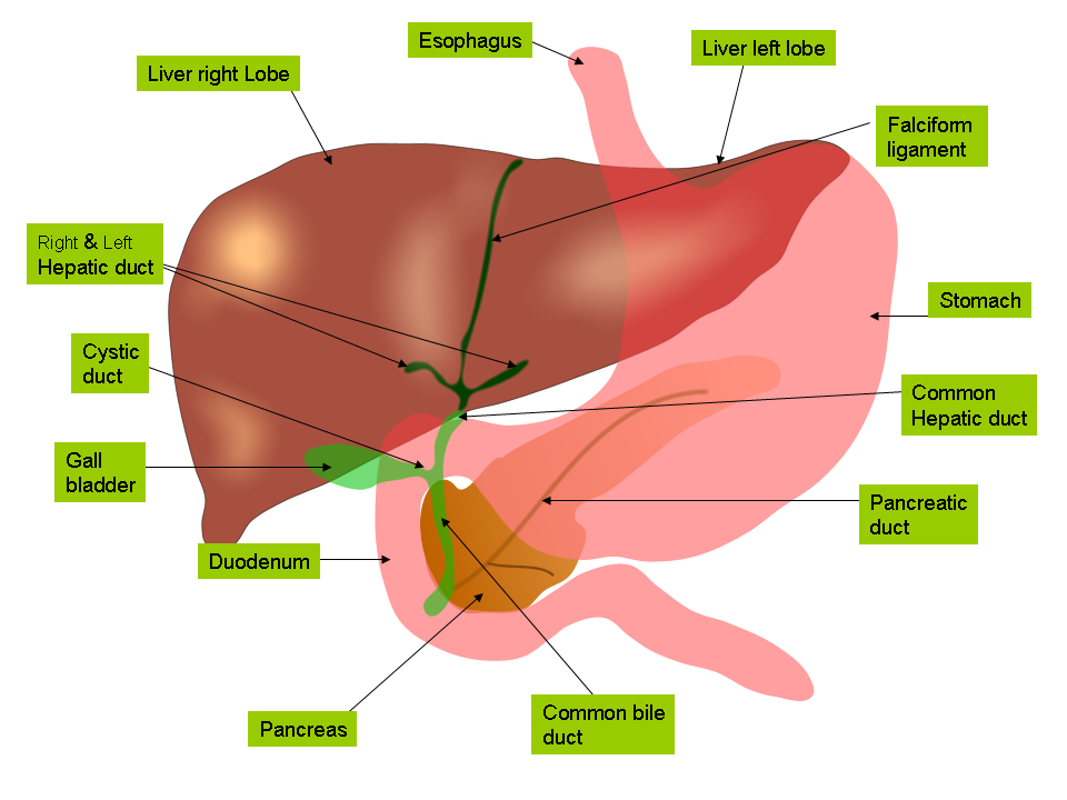
The biliary tree. Bile canaliculi → bile ductules → right and left hepatic ducts → common hepatic duct, which joins the cystic duct from the gallbladder to form the common bile duct. The common bile duct passes behind the duodenum and through the head of the pancreas and joins the main pancreatic duct at the hepatopancreatic ampulla (of Vater), which opens into the second part of the duodenum through the major papilla, guarded by the hepatopancreatic sphincter (of Oddi). Between meals the sphincter is closed, so bile backs up the cystic duct into the gallbladder for storage. When the gallbladder contracts, bile leaves first through the cystic duct.
What the liver does. It is the body's main metabolic and chemical-processing organ:
- Bile production: its main digestive job (bile salts emulsify fat). It makes no digestive enzymes.
- Carbohydrate metabolism: stores glucose as glycogen, releases glucose, makes new glucose (below).
- Lipid metabolism: makes cholesterol, lipoproteins and bile acids; burns fatty acids; makes ketone bodies in fasting.
- Protein metabolism: removes amino groups from amino acids and converts the toxic ammonia into urea (the main nitrogen waste, excreted by the kidneys; urea is not turned back into ammonia); makes plasma proteins such as albumin (which holds water in the blood by oncotic pressure), most clotting factors (fibrinogen, prothrombin and others) and transport proteins.
- Storage: glycogen, iron (as ferritin), copper, and vitamins A, D, B₁₂ (and K and E). The spleen does not store these.
- Detoxification and excretion: metabolizes drugs, alcohol and hormones, and excretes bilirubin and cholesterol in bile.
- Activation: converts vitamin D to its storage form (the first of two activation steps).
- Defense: Kupffer cells filter bacteria arriving from the gut.
When the liver fails, each job shows up as a sign: low albumin → fluid leaks out of capillaries (by Starling forces, lower plasma oncotic pressure raises net filtration) → edema and ascites; low clotting factors → bleeding; poor bilirubin handling → jaundice; high blood ammonia → confusion (hepatic encephalopathy); poor glucose release → low blood sugar; poor bile output → poor fat digestion. Of the macronutrients, lipid digestion suffers most when the liver or gallbladder is damaged.
Its anatomical unit is the hexagonal lobule: plates of hepatocytes radiating from a central vein, with a portal triad (hepatic artery branch, portal vein branch, bile duct) at the corners. Blood runs through the sinusoids toward the central vein; bile runs the other way in bile canaliculi toward the bile ducts.

Liver cells (State & National)
| Cell | Location | Job |
|---|---|---|
| Hepatocytes | Plates between sinusoids | Metabolism, bile secretion, plasma proteins (albumin, clotting factors), detoxification |
| Sinusoidal endothelial cells | Line sinusoids; fenestrated, no basement membrane | Let plasma reach hepatocytes through the space of Disse |
| Kupffer cells (stellate macrophages) | Inside sinusoids | Resident macrophages, the largest group in the body: engulf bacteria and endotoxin arriving from the gut, old red cells and debris, protecting the liver from infection. In chronic alcohol use, gut endotoxin activates them and they release TNF-α and other cytokines that drive alcoholic liver inflammation. They are not involved in protein breakdown |
| Stellate (Ito) cells | Space of Disse | Store vitamin A; when activated by injury they become collagen-making myofibroblasts, causing fibrosis and cirrhosis |
| Cholangiocytes | Bile ducts | Modify bile, adding water and bicarbonate (secretin-driven) |
Do not confuse the two "stellate" names: Kupffer cells are star-shaped macrophages inside the sinusoids, while hepatic stellate (Ito) cells are fat- and vitamin-A-storing cells in the space of Disse that make scar collagen. Hepatocytes that die by apoptosis in viral hepatitis appear on slides as pink Councilman (acidophil) bodies.
The lobule also has zones. Zone 1, near the portal triad, gets the most oxygen and does most gluconeogenesis and fatty-acid oxidation. Zone 3, around the central vein, gets the least oxygen and has the most cytochrome P450 drug-metabolizing enzymes, so it is injured first by low blood flow and by acetaminophen overdose.
Energy metabolism and glucose homeostasis
The liver buffers blood glucose. After a meal, insulin (from pancreatic beta cells) tells hepatocytes to store glucose as glycogen (glycogenesis) and convert the excess into fatty acids, exported as VLDL. Between meals, glucagon (alpha cells) drives glycogenolysis, which covers the first day or so of fasting, and then gluconeogenesis from lactate, amino acids (especially alanine) and glycerol. The liver can release free glucose because it has glucose-6-phosphatase; skeletal muscle lacks it, so muscle glycogen fuels only the muscle itself. In fasting, blood glucose comes first from liver glycogen, then from gluconeogenesis, while fat supplies more and more of the energy; in exercise, working muscle burns mainly its own glycogen, topped up by glucose released from liver glycogen, with fat contributing more as exercise lasts longer. In prolonged fasting the liver also makes ketone bodies from fatty acids for the brain and heart. The liver also makes urea from ammonia, which is why liver failure raises blood ammonia.
Cholesterol balance
Cholesterol is not burned for energy; the body uses it to build cell membranes and as the starting material for steroid hormones, vitamin D and bile acids. The liver makes cholesterol (rate-limited by HMG-CoA reductase, the target of statins), takes it up from LDL and chylomicron remnants, and gets rid of it by converting it to bile acids or secreting it into bile. Lipoproteins carry it in blood:
| Lipoprotein | Made by | Carries | Clinical meaning |
|---|---|---|---|
| Chylomicron | Enterocytes | Dietary triglyceride to tissues | Remnants go to the liver |
| VLDL | Liver | Triglyceride made by the liver | Becomes IDL, then LDL, as triglyceride is removed |
| LDL | From VLDL in blood | Cholesterol to tissues; cleared by liver LDL receptors | High LDL deposits in artery walls ("bad") |
| HDL | Liver and intestine | Collects cholesterol from tissues back to the liver (reverse cholesterol transport) | Higher HDL is protective ("good") |
Fewer hepatic LDL receptors means higher blood LDL. Anything that forces the liver to make more bile acid (soluble fiber, bile-acid sequestrant drugs) pulls cholesterol out of the blood.
Bile, bilirubin and the gallbladder
Hepatocytes make about 0.5 to 1 L of bile a day, containing bile salts (amphipathic cholesterol derivatives), phospholipid, cholesterol, bicarbonate and bilirubin. Bile salts emulsify fat and form micelles; about 95% are reabsorbed in the terminal ileum and return in portal blood, the enterohepatic circulation.

Bilirubin comes from heme. Macrophages convert heme to unconjugated (indirect) bilirubin, which travels bound to albumin. Hepatocytes conjugate it with glucuronic acid using the enzyme UGT1A1, making water-soluble conjugated (direct) bilirubin for bile. Colon bacteria turn it into urobilinogen and stercobilin (the brown of feces). Jaundice has three sources: too much hemolysis (unconjugated bilirubin rises), liver-cell disease or a conjugation defect, or blocked bile flow (conjugated bilirubin rises, with pale stools and dark urine).
The gallbladder is a pear-shaped sac (fundus, body, neck) in a fossa on the underside of the liver's right lobe. It holds only about 30 to 50 mL, so it concentrates bile roughly 5- to 10-fold by absorbing water and electrolytes. CCK contracts it and relaxes the sphincter of Oddi, so bile arrives in a squirt timed to a fatty meal. Bile is a yellow-green, slightly alkaline fluid: water, bile salts, phospholipid (lecithin), cholesterol, bilirubin, bicarbonate and electrolytes. It has no enzymes. Its bile salts and phospholipid are the emulsifiers. Only the bile salts are recycled efficiently by the enterohepatic circulation; bilirubin and much of the cholesterol leave in feces, which makes bile the body's main route for getting rid of cholesterol.
Gallstone vocabulary. Cholelithiasis = stones in the gallbladder. Choledocholithiasis = a stone in the common bile duct. Cholecystitis = inflammation of the gallbladder, usually from a stone blocking the cystic duct. Cholangitis = infection of the bile ducts. Cholecystectomy = removal of the gallbladder; biliary colic = the pain of a stone blocking a duct.
| Stone type | Share | How it forms | Risk factors |
|---|---|---|---|
| Cholesterol (yellow) | About 75 to 80% in the US | Bile too concentrated in cholesterol, too little bile salt or lecithin, or the gallbladder empties poorly | The classic "female, fertile, forty, fat (obese)": estrogen (pregnancy, oral contraceptives) raises cholesterol output into bile; obesity; rapid weight loss or fasting (bile sits unused); diseased or resected ileum (bile salts lost); diabetes; some ethnic groups |
| Pigment (black or brown) | The rest | Excess unconjugated bilirubin precipitates with calcium | Hemolytic anemias (black), cirrhosis, bile duct infection (brown) |
Ultrasound is the first test for stones. Cholecystitis gives right upper quadrant pain after fatty meals that may spread to the right shoulder, fever, nausea, vomiting and loss of appetite, and Murphy's sign (a catch in breath when the gallbladder is pressed during inspiration). A low-fat diet helps because fat triggers CCK, the gallbladder contracts against the stone, and that causes the pain.
Living without a gallbladder. People live normally after cholecystectomy: the liver still makes bile, but it now trickles continuously into the duodenum instead of being stored and released in a squirt. A large fatty meal can then meet too little bile, so some people get fatty, loose stools for a while; smaller, lower-fat meals help, and absorption of fat and fat-soluble vitamins can be somewhat less efficient. The gallbladder never made bile or trypsin, and the duodenal pH is unchanged.
Pancreas
The pancreas is a soft gland about 12 to 15 cm long lying behind the stomach across the posterior abdominal wall; it is retroperitoneal except for its tail. Its head sits in the C-shaped curve of the duodenum (with a hook, the uncinate process, tucked behind the superior mesenteric vessels), its neck lies in front of the portal vein, and its body and tail extend left to the spleen. It is a mixed gland, both exocrine and endocrine. The exocrine pancreas, about 98 to 99% of its mass (acinar cells in grape-like clusters plus their ducts), secretes digestive juice into ducts. The endocrine islets of Langerhans, about 1 to 2%, release hormones into the blood. The ducts join into the main pancreatic duct (of Wirsung), which meets the common bile duct at the hepatopancreatic ampulla; a smaller accessory duct (of Santorini) often opens separately at the minor papilla. Juice goes into the duodenum.
Pancreatic juice amounts to about 1.2 to 1.5 L a day with a pH of about 8. It is the only secretion with enzymes for all four classes of food (amylase, proteases, lipases, nucleases; see the enzyme table), and its bicarbonate neutralizes acid chyme so those enzymes can work. It contains no pepsin and no bile.
Its secretion has the same three phases as the stomach. The cephalic phase (vagus nerve; about a quarter of enzyme output comes from sight, smell and taste) and the gastric phase (stretch of the stomach, vagovagal reflexes) are small. The intestinal phase is the largest: acid in the duodenum releases secretin, which makes the duct cells pour out a bicarbonate-rich, enzyme-poor juice, and fat and protein release CCK, which makes the acinar cells release an enzyme-rich juice. Each hormone strengthens the other's effect. The parasympathetic (vagal) supply drives secretion; sympathetic fibers mainly control the gland's blood vessels.
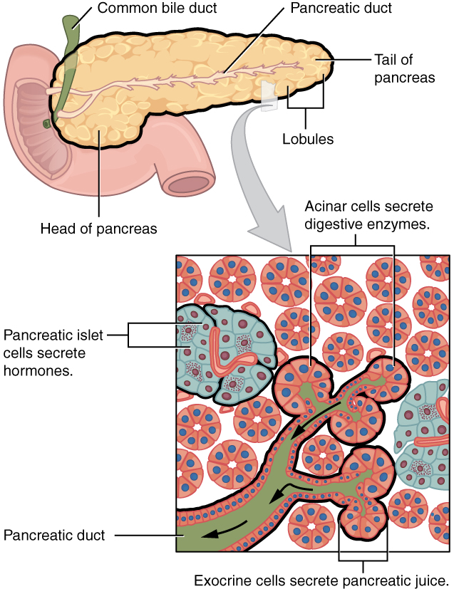
At the cellular level, acinar cells store enzymes in apical zymogen granules and release them under CCK and vagal stimulation. Duct cells secrete bicarbonate under secretin: CFTR channels let chloride out, and a Cl⁻/HCO₃⁻ exchanger swaps it for bicarbonate, which is why cystic fibrosis thickens pancreatic secretions. The juice contains amylase, lipase with colipase, nucleases, and the inactive zymogens trypsinogen, chymotrypsinogen, proelastase and procarboxypeptidase. Making proteases inactive, plus a trypsin inhibitor in the juice, protects the gland; pancreatitis is what happens when trypsin is activated inside it.
| Islet cell | Hormone | Main effect |
|---|---|---|
| Beta (most numerous, about 60 to 80%; central in rodent islets, mixed with other cells in human islets) | Insulin (and amylin) | Lowers glucose: uptake into muscle and fat, storage as glycogen and fat |
| Alpha (about 20 to 30%) | Glucagon | Raises glucose: glycogenolysis and gluconeogenesis in the liver |
| Delta (about 4 to 10%) | Somatostatin | Inhibits insulin and glucagon locally |
| PP (F or gamma) cells (a few %) | Pancreatic polypeptide | Inhibits pancreatic exocrine secretion and gallbladder contraction |
| Epsilon (under 1%) | Ghrelin | Small local role |
Insulin is the chief hormone for handling sugar. It moves glucose into muscle and fat cells (GLUT4), makes the liver and muscle build glycogen, promotes fat and protein synthesis, and stops gluconeogenesis and fat breakdown. Glucagon acts mainly on the liver: glycogen breakdown, gluconeogenesis and ketone production. Type 1 diabetes is autoimmune destruction of beta cells (no insulin). Type 2 diabetes begins with insulin resistance, usually with obesity, and later beta-cell failure. Fasting plasma glucose is normal below 100 mg/dL, prediabetic at 100 to 125, and diabetic at 126 or more. Long-term high glucose damages small vessels, so diabetes can cause kidney failure, blindness and nerve damage; diabetic damage to the vagus can also slow gastric emptying (gastroparesis).
Islet-cell tumors (pancreatic neuroendocrine tumors) are much rarer than ductal adenocarcinoma, which arises from the exocrine ducts. They often overproduce one hormone: insulinoma (low blood sugar), gastrinoma (Zollinger-Ellison syndrome with severe ulcers), VIPoma (large-volume watery diarrhea with low potassium, because VIP drives intestinal secretion) and glucagonoma.
Exocrine pancreatic insufficiency (chronic pancreatitis, cystic fibrosis, pancreatic cancer, removal of the pancreas) cuts off lipase first in practice. Fat goes undigested and shows up as steatorrhea: pale, greasy, foul-smelling, floating stools, with weight loss and deficiency of vitamins A, D, E and K. It is treated with pancreatic enzyme capsules taken with meals. In cystic fibrosis, a defective CFTR chloride channel makes thick secretions that plug the pancreatic ducts (and the airways); most patients have pancreatic insufficiency, and newborns may have meconium ileus. Pale ("clay-colored") stool with jaundice instead points to blocked bile flow.
A beta cell senses glucose by metabolizing it: rising ATP closes ATP-sensitive K⁺ channels, the membrane depolarizes, Ca²⁺ enters and insulin is released. Gut hormones (GIP and GLP-1) add to this response, which is why oral glucose triggers more insulin than the same glucose given intravenously (the incretin effect).
Intestinal microbes (Division C, State & National)
The colon holds the densest microbial community in the body, mostly anaerobic bacteria. The stomach and upper small intestine have far fewer because acid, bile and the migrating motor complex keep numbers down. The microbes help digestion by fermenting fiber and resistant starch into short-chain fatty acids: butyrate is the main fuel of colon cells, and acetate and propionate travel to the liver. They also make vitamin K and some B vitamins and convert primary bile acids into secondary bile acids.
They help defense in three ways. Colonization resistance: resident bacteria occupy niches and nutrients, so pathogens such as Clostridioides difficile cannot take hold. Barrier support: butyrate strengthens tight junctions and mucus production. Immune training: microbial signals sampled through M cells and dendritic cells drive IgA production and regulatory T cells. Broad-spectrum antibiotics disturb this community (dysbiosis), which is why they can cause diarrhea, C. difficile colitis or lower vitamin K. An altered microbiome is also linked to inflammatory bowel disease, obesity and MASLD.
Numbers and names that tests use. The community is called the gut microbiota (older terms: gut flora, intestinal flora, normal flora); its genes are the microbiome. The gut holds roughly as many bacterial cells as the body has human cells (about 40 trillion, the large majority in the colon) belonging to hundreds of species. Numbers rise from about 10³ per mL in the stomach to about 10¹¹ to 10¹² per gram in the colon. Two phyla dominate, Bacillota (Firmicutes) and Bacteroidota (Bacteroidetes), with Actinobacteria (Bifidobacterium), Proteobacteria (Escherichia coli) and Verrucomicrobia (Akkermansia). Named examples: Bacteroides, Bifidobacterium, Lactobacillus, E. coli (makes vitamin K), and Faecalibacterium prausnitzii, a major butyrate producer that is anti-inflammatory and depleted in Crohn's disease.
More on the jobs:
- Digestion. Colon bacteria break down carbohydrates humans cannot: fiber, resistant starch, some cellulose and undigested sugars such as lactose in lactase deficiency. Fermentation also makes gas (H₂, CO₂, methane), the basis of breath tests.
- Vitamins. Bacteria make vitamin K (K₂), biotin (B₇), folate (B₉), riboflavin (B₂), B₁₂ and other B vitamins. Bacterial B₁₂ made in the colon is mostly not absorbed, because B₁₂ uptake happens upstream in the ileum with intrinsic factor. Some of the body's vitamin K comes from them, though diet (K₁ from leafy greens) is the main source and how much bacterial K₂ is absorbed is debated.
- Immune balance. Butyrate encourages naive T cells to become regulatory T cells (Tregs), which damp inflammation. Losing butyrate producers lets pro-inflammatory cells and cytokines (TNF-α, IL-6) dominate. Commensals also drive secretory IgA, the main antibody of the gut.
- Bile acids. Bacteria deconjugate bile salts and convert primary to secondary bile acids.
Probiotics are live beneficial microbes (yogurt cultures, Lactobacillus, Bifidobacterium); prebiotics are fibers that feed them (inulin, oligosaccharides). Fecal microbiota transplantation (stool from a healthy donor) restores a normal community and is used for recurrent C. difficile infection, which thrives after antibiotics wipe out competitors. Regular exercise and a high-fiber diet are associated with a more diverse microbiome and more butyrate producers. At birth the gut is nearly sterile; vaginal birth and breastfeeding (milk oligosaccharides feed Bifidobacterium) shape the first community.
Effects of exercise and obesity
During hard exercise, sympathetic activity diverts blood from the gut to working muscle, and splanchnic blood flow can fall by more than half. Gastric emptying and absorption slow, and endurance athletes commonly get nausea, cramps or "runner's diarrhea"; very long efforts can injure the gut lining from low blood flow. Eating a large or fatty meal just before exercise worsens this.
Regular moderate exercise helps the system: it shortens colonic transit time (less constipation), and active people have lower rates of colon cancer, diverticular disease, gallstones and MASLD. Exercise also improves insulin sensitivity, so the liver and pancreas handle glucose more easily.
Obesity affects almost every organ in the tract:
| Organ | Effect of obesity |
|---|---|
| Esophagus | Higher abdominal pressure pushes acid upward: GERD, hiatal hernia, Barrett's esophagus and esophageal adenocarcinoma are more common |
| Gallbladder | Cholesterol-supersaturated bile → gallstones and cholecystitis |
| Liver | Fat builds up in hepatocytes → MASLD, which can progress to cirrhosis |
| Pancreas | Fat infiltration; more severe pancreatitis; insulin resistance strains beta cells |
| Colon | Higher risk of colorectal cancer and diverticulitis |
Obesity is also tied to high triglycerides and low HDL (the lipid pattern of metabolic syndrome), often with high LDL. Outside the gut it raises the risk of hypertension, type 2 diabetes, heart disease, stroke, sleep apnea and osteoarthritis, and an enlarged fatty liver (hepatomegaly) that can progress to cirrhosis. Hormonally, people with obesity usually have high leptin (with leptin resistance) and high insulin (insulin resistance).
Aging also changes the tract: less saliva and worn or missing teeth make chewing and swallowing harder, the LES weakens (more reflux), stomach acid and intrinsic factor often fall (atrophic gastritis, so B₁₂, iron and calcium absorption decline), colonic motility slows (constipation), diverticula become common, liver blood flow and drug clearance fall, and cancer risk rises.
Measuring it. Obesity is excess body fat, usually screened with the body mass index (BMI, below). Adult categories: under 18.5 underweight, 18.5 to 24.9 healthy, 25 to 29.9 overweight, 30 or more obese (class 3, "severe", at 40 or more). BMI does not distinguish fat from muscle, so waist circumference is added: above about 102 cm (40 in) in men or 88 cm (35 in) in women marks the visceral (abdominal) fat that drives insulin resistance.
Energy and nutrients. A calorie (small calorie) is the heat needed to raise 1 g of water by 1 °C; a food Calorie is a kilocalorie (1,000 calories). Carbohydrate and protein each give about 4 kcal per gram, fat 9 and alcohol 7. Food labels use a 2,000 kcal reference diet. U.S. guidelines suggest 45 to 65% of energy from carbohydrate (mostly complex carbohydrates rather than sugars), 20 to 35% from fat and 10 to 35% from protein, plus about 25 to 38 g of fiber a day. A pound of body fat stores roughly 3,500 kcal, so a lasting surplus of 500 kcal a day adds about a pound a week (a rule of thumb; real change slows over time). Essential nutrients are those the body cannot make and must eat: nine amino acids, two fatty acids (linoleic and α-linolenic), vitamins (including vitamin C, which humans cannot make; its lack causes scurvy) and minerals.
Exercise and appetite. Hard aerobic exercise briefly suppresses appetite: it lowers ghrelin and raises PYY, GLP-1 and CCK. Exercise intensity is often rated on the Borg RPE (rating of perceived exertion) scale. Even light exercise right after eating diverts some blood from the gut; a gentle walk is fine, but hard training on a full stomach causes cramps and nausea.
Bariatric surgery is considered when diet, exercise and drugs fail, traditionally at a BMI of 40 or more, or 35 or more with obesity-related disease (newer guidance lowers these thresholds). Sleeve gastrectomy removes most of the stomach along the greater curvature, leaving a narrow tube; it restricts intake and removes much of the ghrelin-producing fundus. Roux-en-Y gastric bypass staples off a small upper pouch and connects it directly to the jejunum, so food bypasses most of the stomach and the duodenum. It restricts intake and reduces absorption, so patients need lifelong iron, calcium, vitamin D and B₁₂ supplements.
Dumping syndrome follows bypass or gastrectomy. Without a pylorus to meter it, concentrated (hyperosmolar) chyme rushes into the small intestine. Early dumping (within about 30 minutes): water is pulled into the gut, causing cramps, diarrhea, nausea, dizziness and palpitations. Late dumping (1 to 3 hours): a rapid sugar load triggers an insulin surge and then low blood sugar. Prevention: eat smaller meals more often, avoid simple sugars, favor protein, fiber and complex carbohydrates, and drink liquids between meals rather than with them.
Metabolic syndrome and toxins (Division C, State & National)
Metabolic syndrome is a cluster of risk factors that share a root in insulin resistance and excess visceral fat. Under the widely used NCEP ATP III definition, a person has it with any three of five: waist circumference above 102 cm (40 in) in men or 88 cm (35 in) in women; triglycerides of 150 mg/dL or more; HDL below 40 mg/dL in men or 50 mg/dL in women; blood pressure of 130/85 mmHg or more; and fasting glucose of 100 mg/dL or more (treatment for any of these counts as meeting it). In the liver, insulin resistance keeps fat flowing from adipose tissue to hepatocytes and keeps the liver making new fat, causing steatosis (MASLD). In the pancreas, beta cells must secrete more insulin to compensate until they fail (type 2 diabetes), and very high triglycerides can trigger pancreatitis.
The 2027 Division C list names six toxins:
| Toxin | How it injures the liver or pancreas |
|---|---|
| Acetaminophen (paracetamol) | At normal doses most is made water-soluble by conjugation (glucuronide and sulfate) and a small fraction is converted by cytochrome P450 (CYP2E1) to the toxic NAPQI, which glutathione neutralizes at once. In an overdose conjugation saturates, more NAPQI forms, glutathione runs out, and NAPQI binds cell proteins and kills zone 3 hepatocytes. Regular heavy drinking raises the risk (alcohol induces CYP2E1 and depletes glutathione). It is the leading cause of acute liver failure in the US. The antidote N-acetylcysteine restores glutathione and works best within about 8 hours |
| Anabolic steroids | Impair bile flow (cholestatic jaundice) and are linked to liver adenomas, blood-filled cysts (peliosis hepatis) and liver cancer |
| Alcohol | Alcohol dehydrogenase converts ethanol to toxic acetaldehyde, then aldehyde dehydrogenase to acetate. The extra NADH this makes pushes the liver to make fat and stop burning it, so fat builds up. Gut endotoxin leaking through the barrier activates Kupffer cells, whose cytokines inflame the liver. The sequence is fatty liver (reversible) → alcoholic hepatitis → cirrhosis; AST is typically more than twice ALT. Alcohol is also a leading cause of acute and chronic pancreatitis |
| Heavy metals (lead, mercury, cadmium, arsenic) | Bind protein sulfur groups and cause oxidative stress; the liver concentrates many of them; arsenic exposure is linked to liver fibrosis and cancer |
| Bisphenols (such as BPA) | Endocrine disruptors; animal and population studies link exposure to fatty liver and insulin resistance |
| Polychlorinated biphenyls (PCBs) | Persistent, fat-stored industrial chemicals that induce liver enzymes; exposure is associated with fatty liver disease and diabetes |
Formulas and worked calculations
Disorders
Rows follow the order of the 2027 lists: the shared Regional list first, then the State and National additions. The level column gives B / C. In Division B, treatment and prevention are tested at State and National; in Division C, prevention, diagnosis and treatment are National only.
| Disorder | Level (B / C) | Cause / mechanism | Signs and symptoms | Diagnosis | Treatment / prevention |
|---|---|---|---|---|---|
| Stomach (gastric) and duodenal ulcers; peptic ulcer disease | Regional (as ulcers) and S&N (as PUD) / Regional | H. pylori (urease makes ammonia; inflammation weakens the barrier) or NSAIDs (block COX → less prostaglandin) | Burning epigastric pain: gastric ulcer pain worse with food, duodenal often eased by food; bleeding (black stool) | Urea breath test or stool antigen for H. pylori; endoscopy with biopsy | Antibiotics plus a proton-pump inhibitor; stop NSAIDs; surgery for perforation |
| Cancers of the digestive system | Regional (any) / see below | Accumulated mutations; risk factors vary by organ | Weight loss, bleeding, anemia, obstruction | Endoscopy or imaging with biopsy | Surgery, chemotherapy, radiation; screening |
| Diarrhea | Regional / not separately listed | Osmotic (unabsorbed solute, e.g. lactose), secretory (toxins raise cAMP and open CFTR chloride channels, as in cholera), inflammatory (damaged mucosa) or fast transit | Frequent loose stool, dehydration, low potassium | History; stool tests; secretory diarrhea continues when fasting | Oral rehydration solution; treat the cause; sanitation and handwashing prevent infectious causes |
| Lactose intolerance | Regional / Regional | Low lactase; lactose reaches the colon and is fermented | Bloating, gas, osmotic diarrhea after dairy | Hydrogen breath test | Less lactose, lactase tablets, lactose-free products |
| Hepatitis | Regional / S&N | Inflammation of the liver: viruses (A and E fecal-oral; B, C and D by blood and body fluids), alcohol, drugs, autoimmunity | Jaundice, fatigue, dark urine, right upper quadrant pain | Raised ALT and AST; viral antigens and antibodies | Vaccines for A and B; antivirals for B; curative antivirals for C; avoid alcohol |
| Appendicitis | Regional / Regional | Lumen blocked (fecalith in adults; lymphoid hyperplasia in children) → bacterial overgrowth, swelling, ischemia | Pain near the navel moving to McBurney's point (right lower quadrant), fever, vomiting | Exam, raised white count, ultrasound or CT | Appendectomy; antibiotics; perforation causes peritonitis |
| Diverticular disease | S&N / Regional | Pouches of mucosa push through weak points in the colon wall where vessels enter; linked to low fiber, high pressure, obesity | Ranges from none to left lower quadrant pain and fever | CT for diverticulitis; colonoscopy after recovery | Fiber; antibiotics or rest for uncomplicated disease; drainage or surgery for complications |
| GERD | S&N / Regional | LES weak or relaxing too often; hiatal hernia and obesity contribute | Heartburn, regurgitation, cough; complications include esophagitis, stricture and Barrett's esophagus | Symptoms and response to PPIs; endoscopy; pH monitoring | Lifestyle changes; antacids, H₂ blockers, PPIs; fundoplication |
| Ulcerative colitis | S&N / Regional | Autoimmune inflammation of the mucosa, starting at the rectum and spreading continuously | Bloody diarrhea, urgency, cramping | Colonoscopy and biopsy (crypt abscesses, no granulomas) | 5-ASA drugs, steroids, immunosuppressants, biologics; colectomy is curative |
| Crohn's disease | S&N / Regional | Autoimmune transmural inflammation anywhere from mouth to anus, most often the terminal ileum, with skip lesions | Pain, diarrhea, weight loss, fistulas, strictures, B₁₂ deficiency | Colonoscopy and biopsy (granulomas, cobblestoning); imaging | Steroids, immunosuppressants, biologics; surgery for complications; stop smoking |
| Celiac disease | S&N / Regional | Gluten peptides trigger a T-cell attack in people with HLA-DQ2 or DQ8 → villous atrophy | Diarrhea, bloating, weight loss, iron-deficiency anemia, itchy rash (dermatitis herpetiformis) | Anti-tissue transglutaminase IgA; duodenal biopsy | Lifelong gluten-free diet |
| Pancreatitis | not named / S&N | Trypsin activated inside the gland; gallstones and alcohol are the top causes, then very high triglycerides | Severe epigastric pain radiating to the back, vomiting | Lipase at least 3 times normal; CT | Fluids, pain control, early feeding; remove gallstones; stop alcohol |
| MASLD | not named / S&N | Fat in over 5% of hepatocytes plus a cardiometabolic risk factor (formerly NAFLD); can progress to inflammation (MASH) and fibrosis | Usually silent; fatigue; enlarged liver | Ultrasound, liver enzymes, elastography | Weight loss of 7 to 10%, exercise; resmetirom for MASH with fibrosis |
| Cirrhosis | not named / S&N | Chronic injury → stellate-cell fibrosis and regenerating nodules | Jaundice, ascites, varices, bleeding, confusion (high ammonia) | Imaging, elastography, low albumin, prolonged clotting time | Treat the cause; manage complications (diuretics, lactulose); transplant |
| Colorectal, gastric and hepatic cancer | covered by "cancers" / S&N | Colorectal: polyp-to-cancer sequence; gastric: H. pylori, salted foods; hepatic: cirrhosis, hepatitis B or C | Blood in stool, anemia; early satiety; liver mass | Colonoscopy, endoscopy, imaging, biopsy | Surgery, chemotherapy; colonoscopy screening; H. pylori treatment; hepatitis B vaccine |
The diverticular forms named in the Division C list form a spectrum:
| Form | What it means |
|---|---|
| Diverticulosis | Pouches present, no inflammation; often found by chance |
| Symptomatic uncomplicated diverticular disease | Ongoing pain or bowel changes with diverticula but no clear inflammation |
| Uncomplicated diverticulitis | Inflamed diverticula without abscess, perforation, fistula or obstruction |
| Complicated diverticulitis | Diverticulitis with an abscess, perforation, fistula, obstruction or peritonitis |
| Segmental colitis associated with diverticulosis | Chronic mucosal inflammation limited to the diverticula-bearing segment, usually the sigmoid; the rectum is spared |
| Smoldering diverticulitis | Persistent low-grade inflammation and symptoms for weeks to months after an attack that never fully settles |
| Disorder | Cause / mechanism | Key finding |
|---|---|---|
| Achalasia | Loss of inhibitory neurons in the esophageal myenteric plexus: LES cannot relax and peristalsis is lost | Difficulty swallowing solids and liquids; "bird's beak" on barium swallow; treated by dilation or myotomy |
| Hirschsprung disease | Neural crest cells fail to reach the distal colon, so it has no ganglion cells and stays contracted | Newborn does not pass meconium; dilated colon above the narrow segment; rectal biopsy shows no ganglia; surgical removal |
| Cholecystitis | A stone blocks the cystic duct → gallbladder inflammation | Right upper quadrant pain after fatty meals, fever, Murphy's sign; ultrasound; cholecystectomy |
| Choledocholithiasis | Stone in the common bile duct | Obstructive jaundice (conjugated bilirubin, raised ALP); removed by ERCP |
| Ascending cholangitis | Infection above a blocked bile duct | Fever, jaundice and right upper quadrant pain (Charcot's triad); antibiotics and urgent drainage |
| Mirizzi syndrome | Stone stuck in the cystic duct or gallbladder neck compresses the common hepatic duct | Obstructive jaundice without a stone in the duct itself; surgery |
| Primary biliary cholangitis | Autoimmune destruction of small bile ducts inside the liver, mostly middle-aged women | Itching, fatigue, raised ALP, anti-mitochondrial antibodies; ursodeoxycholic acid |
| Primary sclerosing cholangitis | Inflammation and scarring of bile ducts inside and outside the liver; strongly linked to ulcerative colitis | "Beaded" ducts on MRCP; raises cholangiocarcinoma risk; transplant |
| Gilbert syndrome | Reduced UGT1A1 activity | Mild unconjugated jaundice during fasting, illness or stress; harmless |
| Crigler-Najjar syndrome | Absent (type I) or severely reduced (type II) UGT1A1 | Severe unconjugated jaundice in infancy with risk of brain damage (kernicterus); phototherapy, transplant |
| Esophageal cancer | Squamous cell (smoking, alcohol) or adenocarcinoma (GERD, Barrett's, obesity) | Progressive difficulty swallowing, weight loss |
| Pancreatic cancer | Usually ductal adenocarcinoma; smoking, diabetes, chronic pancreatitis | Painless jaundice when in the head; often found late |
| Neuroendocrine tumors | Tumors of enteroendocrine (hormone-making) cells | May secrete hormones; liver spread can cause carcinoid syndrome (flushing, diarrhea) |
The listed disorders in more depth follow, in the order tests most often ask about them.
Peptic ulcers in detail
An ulcer is a break in the lining that goes through the muscularis mucosae into the submucosa or deeper; a shallower break that stays within the mucosa is an erosion. Gastritis is inflammation of the gastric mucosa, with the same main causes. Peptic ulcers form where acid and pepsin overwhelm the defenses: the stomach (gastric ulcer) or the first part of the duodenum (duodenal ulcer, the more common kind).
- Causes and risk factors. Helicobacter pylori infection and NSAIDs, including aspirin. H. pylori is a bacterium (not a virus or fungus), traditionally named the most common cause of both kinds, and it is the reason ulcers are treated with antibiotics; in the US, NSAIDs now rival it as a cause of gastric ulcers. Smoking, heavy alcohol use, corticosteroids taken with NSAIDs, severe physical stress (burns, ICU illness) and a gastrin-secreting tumor (Zollinger-Ellison) add risk. Emotional stress and spicy food do not cause ulcers, though they can worsen symptoms. Pancreatitis is not a risk factor.
- How H. pylori survives. It burrows into the mucus layer and makes urease, which splits urea into ammonia and CO₂; the ammonia neutralizes acid around it and also injures cells.
- Symptoms. Burning or gnawing epigastric pain ("pit of the stomach"). A duodenal ulcer typically hurts 2 to 5 hours after a meal and at night, and food or antacids relieve it (food buffers acid). A gastric ulcer is typically made worse by eating. Heartburn from GERD, by contrast, is a chest burning made worse by large meals and lying down. The pain of an ulcer comes and goes over weeks; pain that is sudden, unrelenting and worsening suggests a complication.
- Complications. Bleeding: vomiting blood (bright red or "coffee-ground") and melena, black tarry stool from digested blood; chronic bleeding causes pale skin and anemia. Perforation: sudden severe pain, a rigid abdomen and peritonitis, with free air under the diaphragm on X-ray. Obstruction of the pyloric outlet from scarring.
- Diagnosis. Endoscopy with biopsy (older tests used a barium X-ray). For H. pylori: the urea breath test (the patient swallows urea labeled with carbon-13 or carbon-14; if urease is present, it releases labeled CO₂, which is absorbed and breathed out, so a positive test is labeled CO₂ in the second breath sample), the stool antigen test, or biopsy.
- Treatment. A proton-pump inhibitor (omeprazole; blocks the H⁺/K⁺-ATPase, raising pH) plus antibiotics to eradicate H. pylori (for example clarithromycin with amoxicillin, or a bismuth-based four-drug regimen); stop NSAIDs; H₂ blockers, antacids or protective drugs such as sucralfate and misoprostol (a prostaglandin analog) help. Before acid-blocking drugs, severe ulcers were treated by cutting the vagus nerve (vagotomy) or removing part of the stomach.
Gastric ulcers are sometimes sorted by the modified Johnson classification: type I on the lesser curvature of the body (the most common; normal or low acid), type II gastric plus duodenal ulcer, type III prepyloric, type IV high on the lesser curvature near the gastroesophageal junction, and type V anywhere, caused by NSAIDs. Types II and III behave like duodenal ulcers and are linked to acid hypersecretion.
Diagnostic tests in one place
| Test | What it shows |
|---|---|
| Upper endoscopy (EGD) | Looks at esophagus, stomach and duodenum; biopsies (ulcers, Barrett's, celiac, cancer) |
| Colonoscopy, sigmoidoscopy | Colon and rectum; removes polyps; IBD and cancer biopsies |
| Barium swallow or meal (X-ray) | Outline of esophagus and stomach (achalasia "bird's beak", strictures, hiatal hernia) |
| Esophageal manometry | Pressures along the esophagus and LES: the test that confirms achalasia and other motility disorders; done before reflux surgery |
| 24-hour pH (impedance) monitoring | How often acid refluxes into the esophagus: confirms GERD |
| Urea breath test, stool antigen | H. pylori |
| Hydrogen breath test | Lactose intolerance, bacterial overgrowth |
| Ultrasound; HIDA scan; MRCP or ERCP | Gallstones; gallbladder function (with CCK); bile-duct images (ERCP can also remove stones) |
| CT | Appendicitis, diverticulitis, pancreatitis, tumors |
| Blood tests | ALT and AST (liver injury), ALP and GGT (blocked bile flow), bilirubin, albumin and clotting time (liver function), lipase (pancreatitis), tTG-IgA (celiac), hepatitis serology |
| Stool tests | Hidden blood (FIT), fecal elastase (pancreatic function), fecal fat, infection |
Hepatitis viruses and hepatitis B tests
Hepatitis means inflammation of the liver. Viruses are the commonest cause; alcohol, drugs (acetaminophen), toxins and autoimmunity also cause it. All forms can cause jaundice (the inflamed liver cannot take up, conjugate and excrete bilirubin), fatigue, nausea, dark urine, pale stool and right upper quadrant pain, with raised ALT and AST. Many infections, especially hepatitis A in children, are mild or silent, which is why hepatitis A is underreported.
| Virus | Genome | Spread | Chronic? | Prevention and treatment |
|---|---|---|---|---|
| Hepatitis A | RNA | Fecal-oral: contaminated food and water, raw shellfish; the type most often caught from food | Never; acute only | Vaccine; supportive care. Contagious from about 2 weeks before symptoms until about a week after jaundice |
| Hepatitis B | DNA (the only DNA one) | Blood, sexual contact, mother to baby at birth | Yes: about 90% of infected newborns but only about 5% of infected adults | Vaccine (made from surface antigen); antiviral drugs control but rarely cure; raises liver cancer risk |
| Hepatitis C | RNA | Blood (shared needles; transfusions before screening) | Yes, in most (roughly 55 to 85%) | No vaccine; direct-acting antivirals cure more than 95%; a leading cause of cirrhosis and liver cancer |
| Hepatitis D | RNA, incomplete | Blood; can only infect someone who also has hepatitis B | Yes, worsens B | The hepatitis B vaccine prevents it |
| Hepatitis E | RNA | Fecal-oral, contaminated water | Usually not | Dangerous in pregnancy; vaccine only in a few countries |
Hepatitis B tests read like a story:
| Marker | Meaning |
|---|---|
| HBsAg (surface antigen) | Virus present now: acute or chronic infection |
| Anti-HBs (surface antibody) | Immunity, from vaccination or recovery |
| IgM anti-HBc (core antibody, IgM) | Recent or acute infection; the first antibody to appear |
| IgG anti-HBc | Past or chronic infection; never produced by the vaccine |
| HBeAg | Virus actively multiplying; highly infectious |
Acute viral hepatitis runs through phases: incubation (no symptoms; weeks for A, up to months for B and C), a prodromal phase of flu-like symptoms, an icteric phase with jaundice, dark urine and pale stool (flu-like symptoms often ease as jaundice appears), and recovery (convalescence). Acute hepatitis A is confirmed by IgM anti-HAV. Interferon was the older treatment for B, C and D (with ribavirin for C); oral antivirals have replaced it for C and largely for B, but pegylated interferon is still used for D.
So anti-HBs alone (HBsAg negative, anti-HBc negative) means immune from vaccination; anti-HBs plus anti-HBc means recovered from a past infection; HBsAg for more than six months means chronic infection. The core antigen itself is not measured in blood.
Appendicitis signs
Appendicitis classically starts as vague pain around the navel (the appendix shares visceral nerve supply with the midgut) that moves within hours to the right lower quadrant at McBurney's point, one-third of the way from the right anterior superior iliac spine to the navel. Loss of appetite, nausea and vomiting, low fever and a high white blood cell count follow. The obstruction comes from a hardened bit of stool (fecalith) in adults, swollen lymphoid tissue in children, and occasionally worms or a tumor.
| Exam sign | What it means |
|---|---|
| McBurney's point tenderness | Inflamed appendix under the spot |
| Rebound tenderness | Pain when pressure is released quickly: the parietal peritoneum is inflamed |
| Guarding and rigidity | Reflex tightening of the abdominal muscles |
| Rovsing's sign | Pressing the left lower quadrant causes pain in the right lower quadrant |
| Psoas sign | Pain when the right hip is extended: appendix behind the cecum |
| Obturator sign | Pain when the flexed right hip is rotated inward: appendix low in the pelvis |
Rectal bleeding is not a sign of appendicitis. Diagnosis: exam plus raised white count, then ultrasound (children, pregnancy) or CT. Treatment: appendectomy, usually laparoscopic, with antibiotics given first; some uncomplicated cases are treated with antibiotics alone. Untreated, it can perforate, causing peritonitis, an abscess and sepsis.
Lactose intolerance types
Lactose intolerance means too little lactase, so lactose reaches the colon, where it draws water in by osmosis (osmotic diarrhea) and bacteria ferment it into gas and acids (bloating, cramps, flatulence, sometimes nausea and vomiting), usually 30 minutes to 2 hours after dairy. Lactase is made by the small intestine and acts at the brush border; its activity is highest in the jejunum (lower in the duodenum and ileum).
| Type | Cause |
|---|---|
| Primary (lactase non-persistence), the most common | Lactase production falls after weaning; about two-thirds of the world's adults; less common in people of northern European descent, who usually carry a lactase-persistence gene variant |
| Secondary | Damage to the small intestine lining: celiac disease, Crohn's disease, infection (gastroenteritis), bowel surgery, chemotherapy; often improves when the gut heals |
| Congenital | Rare inherited absence of lactase from birth |
| Developmental | Premature babies, whose lactase has not yet developed |
Diagnosis: the hydrogen breath test (breath hydrogen rises after a lactose drink because colon bacteria ferment it) or a lactose tolerance test (blood glucose fails to rise). Management: limit lactose, use lactose-free products or lactase tablets; hard cheeses and yogurt are often tolerated. A deficiency of any brush-border disaccharidase, such as sucrase-isomaltase, produces the same kind of osmotic symptoms with its own sugar.
Diarrhea, vomiting and acid-base balance
Diarrhea is frequent, loose, watery stool. It has four mechanisms:
| Type | Mechanism | Clues | Examples |
|---|---|---|---|
| Osmotic | Unabsorbed solute holds water in the lumen | Stops when the person fasts | Lactose intolerance, sorbitol, magnesium laxatives |
| Secretory | Toxins or hormones make the gut secrete Cl⁻ and water | Large volumes; continues during fasting | Cholera: cholera toxin keeps cAMP high, which holds CFTR chloride channels open, so Cl⁻ pours into the lumen and water follows ("rice-water" stool, up to about 1 L an hour); VIPoma |
| Inflammatory (invasive, exudative) | Damaged mucosa leaks and absorbs poorly | Fever; blood, mucus and white blood cells (pus) in stool | Shigella, Salmonella, Campylobacter, E. coli O157, C. difficile, ulcerative colitis |
| Motility | Contents move too fast to be absorbed | Irritable bowel syndrome, hyperthyroidism |
Common infectious causes: norovirus (all ages) and rotavirus (young children; vaccine available), traveler's diarrhea (enterotoxigenic E. coli), and Clostridioides difficile after antibiotics.
Diarrhea loses water, Na⁺, K⁺ and especially bicarbonate (intestinal and pancreatic fluid are alkaline), causing dehydration, low potassium and metabolic acidosis. The body compensates by hyperventilating to blow off CO₂ and raise pH. Vomiting does the opposite: losing HCl causes metabolic alkalosis (with low chloride and potassium). An infant with projectile, non-bile-stained vomiting after feeds and a palpable "olive" in the upper abdomen has hypertrophic pyloric stenosis, a thickened pyloric sphincter, and becomes alkalotic; it is treated by cutting the muscle (pyloromyotomy).
Bismuth subsalicylate (Pepto-Bismol) reduces secretion and binds bacterial toxins. Treatment of diarrhea is replacing water and electrolytes, ideally with oral rehydration solution (glucose plus salt; glucose drives Na⁺ uptake through SGLT1 and water follows), then treating the cause. Sanitation, safe water and handwashing prevent infectious diarrhea.
Constipation is infrequent (fewer than three a week) or hard, difficult stools, from slow colonic transit allowing too much water absorption. Causes: low fiber, low fluid, inactivity, opioids, hypothyroidism and ignoring the urge. Treat with fiber, fluids, exercise and, if needed, laxatives.
Inflammatory bowel disease compared
| Feature | Ulcerative colitis | Crohn's disease |
|---|---|---|
| Where | Colon only: starts in the rectum and spreads continuously upward | Anywhere from mouth to anus; most often the terminal ileum; the rectum is often spared |
| Pattern | Continuous | Skip lesions (patches of normal bowel between) |
| Depth | Mucosa and submucosa | Transmural (the whole wall thickness) |
| Look on colonoscopy | Red, friable mucosa that bleeds easily, shallow ulcers, pseudopolyps; colon loses haustra ("lead pipe") | Cobblestone mucosa, deep linear ulcers, narrowed segments ("string sign" on X-ray) |
| Biopsy | Crypt abscesses, no granulomas | Non-caseating granulomas |
| Main symptoms | Bloody diarrhea, urgency | Abdominal pain, (usually non-bloody) diarrhea, weight loss |
| Complications | Toxic megacolon and fulminant colitis; colon cancer; primary sclerosing cholangitis | Fistulas, strictures, abscesses, perianal disease; B₁₂ and bile-salt loss, gallstones and kidney stones from ileal disease |
| Smoking | Smokers have lower risk | Smoking raises risk and worsens it (the main controllable risk factor) |
| Antibody blood test | p-ANCA positive | ASCA positive |
| Surgery | Removing the colon cures it | Not curative; disease recurs |
Both can cause problems outside the gut (extraintestinal manifestations): aphthous mouth ulcers (especially Crohn's, along with cracked mouth corners from nutrient loss), arthritis, eye inflammation (uveitis), skin lesions (erythema nodosum) and, mainly with UC, primary sclerosing cholangitis. Both are chronic, relapsing autoimmune (immune-mediated) diseases with genetic and microbial triggers, and both raise colon cancer risk. Both are treated with anti-inflammatory 5-ASA drugs (mainly UC), corticosteroids for flares, immunosuppressants and biologics (anti-TNF antibodies such as infliximab). Irritable bowel syndrome is different: recurrent pain with altered bowel habits but no inflammation or visible damage.
Celiac disease details
In celiac disease, gluten (the gliadin fraction of wheat, barley and rye) is modified by the enzyme tissue transglutaminase and presented to T cells in people with HLA-DQ2 or HLA-DQ8. The immune attack flattens the villi of the small intestine, mainly the duodenum and proximal jejunum, so the absorptive surface shrinks. Results: diarrhea (often watery or fatty), bloating, weight loss, iron-deficiency anemia (iron is absorbed in the duodenum), osteoporosis (calcium and vitamin D), secondary lactose intolerance, and in children poor growth. The itchy blistering rash is dermatitis herpetiformis.
Diagnosis: blood test for IgA anti-tissue transglutaminase antibodies while the person is still eating gluten, then duodenal biopsy. Biopsy changes are graded by the Marsh classification: Marsh 1, more lymphocytes in the epithelium; Marsh 2, plus enlarged (hyperplastic) crypts; Marsh 3, plus villous atrophy (3a partial, 3b subtotal, 3c total). Treatment is a lifelong gluten-free diet; the villi recover. Untreated disease raises the risk of intestinal lymphoma.
Pancreatitis details
In acute pancreatitis digestive enzymes, especially trypsin, are activated inside the gland and digest it ("autodigestion"). Enzymes leak into the surrounding tissue (causing severe pain and inflammation), and fewer reach the duodenum (disturbing digestion, with nausea and vomiting). Causes are remembered as "I GET SMASHED": idiopathic, gallstones, ethanol, trauma, steroids, mumps, autoimmune, scorpion sting, high triglycerides or calcium, ERCP, drugs. Gallstones and alcohol cause most cases.
- Symptoms: severe, steady epigastric pain radiating to the back, often eased by leaning forward; nausea and vomiting; fever and a fast pulse.
- Diagnosis: two of three of: typical pain, serum lipase (or amylase) at least three times the upper limit of normal (lipase is the more specific), and CT or ultrasound findings. Bruising around the navel (Cullen sign) or flanks (Grey Turner sign) means bleeding in severe cases.
- Treatment: generous IV fluids, pain control, early feeding as tolerated; remove gallstones and later the gallbladder; stop alcohol. Complications include fluid-filled pseudocysts, necrosis, low calcium and lung injury.
Chronic pancreatitis (mostly alcohol and smoking, also cystic fibrosis and genetic causes) slowly replaces the gland with scar and calcifications, causing chronic pain, steatorrhea from lipase loss and eventually diabetes from islet loss.
Cirrhosis and MASLD details
Cirrhosis is the end stage of chronic liver injury: activated stellate cells lay down fibrous scar, normal lobules are replaced by nodules of regenerating cells, and the liver can no longer repair itself. Causes: alcohol, chronic hepatitis B and C, MASLD, autoimmune and biliary diseases, and inherited iron or copper overload (hemochromatosis, Wilson disease). Its consequences follow from the lost functions: portal hypertension (ascites, enlarged spleen, esophageal varices that can bleed), low albumin (edema), clotting problems, jaundice, high ammonia (confusion, hepatic encephalopathy; treated with lactulose, a sugar humans cannot digest that colon bacteria ferment into acids, which trap ammonia as ammonium so it is excreted in stool), and a high risk of liver cancer. The only cure for end-stage disease is a liver transplant.
MASLD (metabolic dysfunction-associated steatotic liver disease, formerly non-alcoholic fatty liver disease, NAFLD) is the most common liver disease in the US, affecting roughly a third of adults. It is fat in more than 5% of hepatocytes with at least one cardiometabolic risk factor (obesity, type 2 diabetes, high triglycerides, low HDL, high blood pressure) and little or no alcohol. Its inflammatory form is MASH (formerly NASH), now one of the fastest-growing reasons for liver transplants in the US. Diagnosis: ultrasound, liver enzymes, elastography (liver stiffness) and, as the reference standard, biopsy showing fat, inflammation and fibrosis. Treatment: weight loss (about 5% reduces liver fat; 7 to 10% can reverse inflammation), exercise and control of diabetes and lipids; drugs (resmetirom, and the GLP-1 drug semaglutide) are approved for MASH with moderate to advanced fibrosis but not cirrhosis.
Cancers of the digestive system
A neoplasm is any abnormal new growth of cells (a tumor). Benign neoplasms stay local; malignant ones (cancers) invade and spread (metastasize), often first to the liver through the portal vein. Most digestive cancers are carcinomas (from epithelium): adenocarcinoma from glandular columnar epithelium, squamous cell carcinoma from stratified squamous epithelium. Others: lymphomas (including gastric MALT lymphoma linked to H. pylori), gastrointestinal stromal tumors and neuroendocrine tumors.
| Cancer | Type and risk factors | Typical findings | Detection and treatment |
|---|---|---|---|
| Colorectal | Adenocarcinoma from adenomatous polyps (the adenoma-carcinoma sequence); risks: age, family history and inherited syndromes (familial adenomatous polyposis, Lynch syndrome), IBD, low fiber and red or processed meat, obesity, inactivity, smoking, alcohol, diabetes | Change in bowel habits, blood in stool, iron-deficiency anemia, narrowing stool or obstruction | Screening from age 45: colonoscopy every 10 years (removes polyps), or stool tests for hidden blood (FIT/FOBT) yearly; FOBT is a screening test, not a treatment. Surgery, chemotherapy, radiation (rectal) |
| Gastric | Adenocarcinoma; H. pylori, smoked, salted and pickled foods (nitrosamines), smoking, alcohol, atrophic gastritis, pernicious anemia; a high-sugar diet is not a recognized risk | Often silent until late; early satiety, weight loss, upper abdominal pain, anemia; a hard left supraclavicular lymph node (Virchow's node) | Endoscopy with biopsy; gastrectomy, chemotherapy; treating H. pylori lowers risk |
| Hepatic (hepatocellular carcinoma) | Cirrhosis from any cause, hepatitis B and C, alcohol, MASLD, aflatoxin from moldy grain | Liver mass, weight loss, worsening liver function; raised alpha-fetoprotein | Ultrasound every 6 months in people with cirrhosis; resection, ablation, transplant; hepatitis B vaccine prevents many cases |
| Esophageal (National) | Squamous cell (upper and middle esophagus; smoking, alcohol, very hot drinks) or adenocarcinoma (lower esophagus; GERD → Barrett's esophagus, obesity) | Progressive dysphagia, solids first then liquids; weight loss | Endoscopy; surgery, chemoradiation |
| Pancreatic (National) | Ductal adenocarcinoma (about 90%), most in the head; smoking (the biggest modifiable risk), obesity, diabetes, chronic pancreatitis; slightly more common in men | Painless jaundice with a palpable non-tender gallbladder (Courvoisier sign), weight loss, new diabetes; usually found late and nearly always fatal (5-year survival about 13%) | CT; CA 19-9 marker; Whipple operation (pancreaticoduodenectomy) when resectable |
| Neuroendocrine tumors (National) | From hormone-making cells, most often in the small intestine, appendix, rectum and pancreas | Slow-growing; may secrete hormones; liver spread of serotonin-secreting (carcinoid) tumors causes carcinoid syndrome (flushing, diarrhea, wheezing) | Urine 5-HIAA test; surgery; somatostatin analogs (octreotide) |
Oral cancer is usually squamous cell carcinoma linked to tobacco, alcohol and HPV. Leukoplakia, a white patch in the mouth that cannot be scraped off, is a precancerous lesion common in smokers; a white coating that does scrape off is usually thrush (Candida).
Other conditions tests use
ReferenceOther digestive conditions· 26 rows
These are not all on the rules list, but they appear as distractors, complications or context in tournament questions.
No rows match.
| Condition | What it is | Key clue |
|---|---|---|
| Barrett's esophagus | Chronic acid reflux turns the lower esophageal lining from stratified squamous into columnar epithelium with goblet cells (intestinal metaplasia) | Precursor of esophageal adenocarcinoma; caused by GERD, not nausea |
| Hiatal hernia | Stomach slides up through the esophageal hiatus | Reflux, heartburn |
| Achalasia (National) | LES fails to relax; myenteric neurons lost | "Bird's beak" barium swallow |
| Zenker's diverticulum | Pouch of pharyngeal mucosa pushing out just above the upper esophageal sphincter | Bad breath, regurgitating old food |
| Mallory-Weiss tear | Tear in the mucosa at the gastroesophageal junction from forceful vomiting | Vomiting blood after retching or binge drinking |
| Esophageal varices | Swollen veins from portal hypertension | Cirrhosis; can bleed massively |
| Dysphagia | Difficulty swallowing (odynophagia = painful swallowing) | Neurologic, stricture, cancer, achalasia; nasal regurgitation and aspiration when the pharyngeal phase fails |
| Gastroparesis | Delayed gastric emptying without blockage | Diabetes damaging the vagus; nausea, early fullness |
| Zollinger-Ellison syndrome | Gastrin-secreting tumor | Multiple, severe ulcers |
| Pernicious anemia | Autoimmune destruction of parietal cells or intrinsic factor | B₁₂ deficiency; treat with B₁₂ injections |
| Dumping syndrome | Rapid gastric emptying after gastric surgery | Cramps, diarrhea, palpitations after sugary meals |
| Peritonitis | Inflammation of the peritoneum | Rigid abdomen; follows perforation of an ulcer, appendix or diverticulum |
| Intussusception | One segment of intestine telescopes into the next (often ileum into colon) | Child under about 3 with episodes of severe crying pain, vomiting and "currant jelly" stool; sausage-shaped mass; ultrasound; treated with an air or contrast enema |
| Volvulus | Twisting of a bowel loop on its mesentery | Obstruction and ischemia (sigmoid in the elderly; midgut in infants) |
| Meckel's diverticulum | Remnant of the embryonic yolk (vitelline) duct on the ileum; a true diverticulum | "Rule of 2s": about 2% of people, about 2 feet from the ileocecal valve, may contain stomach tissue and bleed painlessly in young children |
| Bowel obstruction | Blockage by adhesions, hernia, tumor or twisting | Cramping pain, distension, vomiting, no gas passed |
| Irritable bowel syndrome (IBS) | Functional disorder of gut-brain signaling | Pain related to defecation, altered habits, no structural damage |
| Hemorrhoids | Swollen anal cushions of veins | Internal: painless bright-red bleeding; external: painful |
| Anal fissure | Tear in the anal canal lining | Pain with defecation |
| Cystic fibrosis | Defective CFTR chloride channel | Thick pancreatic secretions, malabsorption, meconium ileus in newborns |
| Gallstone ileus | A large stone erodes into the bowel and blocks it | Obstruction in an older woman |
| Mumps | Viral parotitis | Swollen parotid glands; can cause pancreatitis |
| Xerostomia | Dry mouth | Drugs, Sjögren syndrome, radiation |
| Dental caries | Acid from bacterial fermentation dissolves enamel | Streptococcus mutans |
| Hemochromatosis | Excess iron absorption in the duodenum stored in liver and pancreas | Cirrhosis, diabetes, bronze skin |
| Wilson disease | Copper cannot be excreted into bile | Liver disease, neurologic signs, Kayser-Fleischer eye rings |
Common pitfalls
Stomach acid does not digest carbohydrate
Pepsin acts on protein at low pH. Carbohydrate digestion starts in the mouth and mostly happens in the small intestine.
Enterokinase is made by the duodenum, not the stomach or pancreas
Enterokinase is a brush-border enzyme that activates trypsinogen. Some archive keys place it in the stomach; that is wrong.
Sucrase-isomaltase deficiency is not a lactose problem
Sucrase-isomaltase handles sucrose and the limit dextrins. A question about sucrose intolerance points to that complex, not lactase.
The stomach is the slowest pacemaker
Among stomach, duodenum and ileum, the stomach has the slowest basal electrical rhythm (about 3 per minute).
Bile is made in the liver and stored in the gallbladder
Removing the gallbladder does not stop bile production; bile just arrives as a steady trickle.
Kupffer cells are not the scarring cells
Kupffer cells are macrophages. Stellate (Ito) cells make the collagen of fibrosis.
Retroperitoneal is not the same as "behind the abdomen"
The transverse and sigmoid colon lie toward the back in places but have mesocolons and are intraperitoneal. The duodenum is retroperitoneal except for the short duodenal cap at the start of its first part.
Obesity is linked with low HDL
Obesity raises LDL and triglycerides and lowers HDL. A question asking for the exception usually hinges on this.
The ileum is the longest part of the small intestine
The jejunum does the most absorbing and has the richest blood supply, but the ileum is longer. Some bank keys say "jejunum" for longest; that is wrong.
Fructose does not ride with sodium
Glucose, galactose and amino acids are cotransported with Na⁺. Fructose enters by facilitated diffusion (GLUT5). An answer pairing "amino acids and fructose" with Na⁺ is wrong.
Most water is absorbed in the small intestine
The colon finishes the job and forms feces, but the small intestine absorbs most of the roughly 9 L a day.
Deciduous molars become premolars
Children have no premolars. The permanent premolars replace the deciduous molars, and the permanent molars come in behind.
Check the level for your division
Hepatitis is Regional in B but State in C; GERD and the inflammatory bowel diseases are the reverse. Treatment questions appear at State in B but only at Nationals in C.
How tournaments test this
The Scio.ly archive holds 1,852 digestive questions from two past seasons: 1,065 from 2017-18 and 787 from 2022-23. Division C accounts for 1,235, Division B for 567, and 50 are tagged for both. The practice bank holds 2,964 digestive questions: about 1,250 match questions in these archive tests, and about 1,600 come from other tournament tests, mostly 2017-18 and 2022-23 invitationals, that the archive does not have.
- Format. Free response is the largest type at 884 questions (47.7%), then multiple choice at 775 (41.8%), multiple-select at 105 (5.7%) and true/false at 87 (4.7%).
- Task profile. The most common tasks are "which of the following" (9.8%), naming a structure in a figure (4.6%), explain or why (4.6%) and patient scenarios (3.8%). Calculation is rare (0.5%). Figure identification is common, so practice unlabeled diagrams of the tract, the gut wall, the villus and the liver lobule.
- Topics. Intestines are tagged on 831 archived questions, stomach on 521, liver on 299, mouth on 281 and pancreas on 196.
- Recurring terms. "Small intestine" appears in 375 prompts, "large intestine" in 154, "chemical digestion" in 83, "lactose intolerance" in 59, "mechanical digestion" in 44, "celiac disease" in 39, "parietal cells" in 36, "chief cells" in 31, "lamina propria" in 31 and "intrinsic factor" in 27.
- Answers that recur. As the correct option, "small intestine" appears 34 times, "stomach" 34, "liver" 32, "mouth" 31 and "duodenum" 30; secretin (15), CCK (10) and gastrin (9) are frequent hormone answers, and submucosa (16) and mucosa (16) frequent layer answers.
- Where the bank's questions fall. Sorting the roughly 2,960 currently visible, de-duplicated practice-bank digestive questions by main topic gives approximate shares: absorption and nutrients about 12%, chemical digestion and enzymes 11%, pancreas 10%, wall layers and cell types 8%, liver and gallbladder structure 7% (plus 2% liver metabolism), disorders together about 25% (lactose intolerance, appendicitis and diarrhea 6%; ulcers and GERD 5%; biliary and National disorders 4%; hepatitis, cirrhosis and MASLD 3%; cancers 3%; IBD and celiac 2%), mouth and esophagus 5%, stomach 4%, motility 4%, hormones and nerves 4%, and exercise, obesity, toxins and microbes about 4% between them. Most questions test one specific fact (a cell, enzyme, site, hormone or sign), so the reference tables above repay memorizing.
- What the archive does not show. Past tests predate several 2027 additions: abdominal wall layers, intraperitoneal versus retroperitoneal organs, gut microbes, metabolic syndrome, the six toxins, MASLD and the National-only biliary list. Expect new questions on these, likely as labeled diagrams and patient cases.
Test yourself
These are real questions from public tournament tests in the Scio.ly archive. Click an answer to check it; each question links to its full test.
- 1
The Lamina Propria is a layer of the
Practice more Anatomy and Physiology questions →
- 1
Through which of the following pancreatic ducts does both bile and pancreatic secretions empty into the duodenum?
From: Columbia University (CUSO) Invitational, 2022-23, Division C
1 question from this set is no longer available.
Practice more Anatomy and Physiology questions →
- 1
Which of the following statements is true concerning the role of NSAIDs in causing gastric ulcers? I. They inhibit the enzyme cyclooxygenase II. They decrease the synthesis of inflammatory prostaglandins III. They artificially increase secretion of gastrin by G cells in the antrum IV. They contain proteases that directly damage the mucosa and mucosal cells
Practice more Anatomy and Physiology questions →
- 1
Which of the following statements are true regarding cementum? SELECT ALL THAT APPLY.
From: Camas Invitational 2022-23, Division B
1 question from this set is no longer available.
Practice more Anatomy and Physiology questions →
Condensed facts
| Wall layer (lumen outward) | Contents |
|---|---|
| Mucosa | Epithelium, lamina propria (capillaries, lacteals, MALT), muscularis mucosae |
| Submucosa | CT, big vessels, Meissner plexus (secretion), Brunner (duodenum) and esophageal glands |
| Muscularis externa | Inner circular, outer longitudinal (+ inner oblique in stomach); Auerbach plexus (motility) |
| Serosa / adventitia | Visceral peritoneum (simple squamous); adventitia on esophagus and retroperitoneal parts |
| Region | Epithelium | Hallmarks |
|---|---|---|
| Mouth, oropharynx, esophagus, lower anal canal | Stratified squamous, mostly non-keratinized (keratinized on gums, hard palate and anal skin) | Abrasion; esophagus skeletal → mixed → smooth |
| Stomach | Simple columnar, all mucus cells | Rugae, gastric pits and glands, 3 muscle layers |
| Small intestine | Simple columnar + microvilli, goblet | Plicae, villi, crypts, Paneth; Brunner (duodenum), Peyer's patches (ileum) |
| Large intestine | Simple columnar, many goblet | No villi; taeniae coli, haustra, epiploic appendages |
| Gastric cell | Product |
|---|---|
| Surface (foveolar) mucous | Alkaline mucus + HCO₃⁻ |
| Parietal (oxyntic) | HCl (H⁺/K⁺-ATPase), intrinsic factor |
| Chief (zymogenic) | Pepsinogen, gastric lipase |
| G / D / ECL / X-A | Gastrin / somatostatin / histamine / ghrelin |
| Hormone | Cell, site | Trigger | Effect |
|---|---|---|---|
| Gastrin | G, antrum | Peptides, stretch, vagus | ↑ HCl, motility |
| Secretin | S, duodenum | Acid | ↑ HCO₃⁻ (pancreas, bile ducts); ↓ acid |
| CCK | I, duodenum | Fat, protein | ↑ enzymes, GB contracts, Oddi relaxes, ↓ emptying |
| GIP | K, duodenum | Glucose, fat | ↑ insulin, ↓ acid |
| GLP-1, PYY | L, ileum and colon | Nutrients | ↑ insulin (GLP-1); satiety; ↓ emptying |
| Motilin | M, duodenum | Fasting | MMC |
| Ghrelin | Fundus | Empty stomach | Hunger |
| Somatostatin | D | Acid | Inhibits nearly everything |
| Enzyme | From | Substrate → product |
|---|---|---|
| Salivary amylase | Salivary glands | Starch → maltose, dextrins |
| Pepsin(ogen) | Chief cells | Protein → polypeptides (pH 2) |
| Trypsin(ogen) | Pancreas; activated by enteropeptidase | Proteins → peptides; activates other zymogens |
| Chymotrypsin, elastase, carboxypeptidase | Pancreas; activated by trypsin | Peptides → smaller peptides, amino acids |
| Pancreatic amylase | Pancreas | Starch → maltose |
| Lipase + colipase | Pancreas | Triglyceride → 2-MG + 2 FFA |
| Nucleases | Pancreas | DNA, RNA → nucleotides |
| Maltase, sucrase-isomaltase, lactase | Brush border | Disaccharides, limit dextrins → monosaccharides |
| Aminopeptidase, dipeptidase | Brush border | Peptides → amino acids |
| Enteropeptidase | Duodenal brush border | Trypsinogen → trypsin |
| Absorbed | Where | How |
|---|---|---|
| Glucose, galactose | Jejunum | SGLT1 with Na⁺ in; GLUT2 out |
| Fructose | Jejunum | GLUT5 (facilitated) |
| Amino acids; di-, tripeptides | Jejunum | Na⁺ cotransport; PEPT1 with H⁺ |
| Fats, A D E K | Jejunum, ileum | Micelles → diffusion → chylomicrons → lacteals → thoracic duct |
| Iron, calcium | Duodenum | Fe²⁺ via DMT1; Ca²⁺ needs vitamin D |
| B₁₂, bile salts | Terminal ileum | Intrinsic factor; Na⁺ cotransport |
| Water | Small intestine (most), colon (rest) | Osmosis after Na⁺ |
| Disorder | One-line key |
|---|---|
| Peptic ulcer | H. pylori or NSAIDs; duodenal eased by food; urea breath test; PPI + antibiotics |
| GERD | Weak LES; heartburn; Barrett's → adenocarcinoma; PPIs; Nissen fundoplication |
| Lactose intolerance | Lactase low; osmotic diarrhea + gas; H₂ breath test |
| Celiac | Gluten, HLA-DQ2/8, tTG-IgA, villous atrophy, dermatitis herpetiformis; gluten-free diet |
| Crohn's | Mouth to anus, terminal ileum, skip, transmural, granulomas, cobblestone, fistulas; smoking worsens |
| UC | Rectum up, continuous, mucosal, bloody diarrhea, toxic megacolon, p-ANCA; colectomy cures |
| Appendicitis | Periumbilical → McBurney's; rebound, Rovsing, psoas, obturator; appendectomy |
| Diverticular | Low fiber, high pressure; sigmoid; LLQ pain in diverticulitis |
| Hepatitis | A, E fecal-oral; B (DNA), C, D blood; vaccines A, B; anti-HBs alone = vaccinated |
| Pancreatitis | Gallstones, alcohol; epigastric pain to back; lipase ≥3×; fluids |
| Cirrhosis | Stellate fibrosis + nodules; ascites, varices, jaundice, encephalopathy |
| MASLD | Fat >5% hepatocytes + metabolic risk; weight loss 7 to 10% |
| Gallstones | 80% cholesterol; cholecystitis RUQ pain after fat, Murphy's sign |
| Hirschsprung | No ganglion cells in distal colon; no meconium; megacolon |
| Achalasia | LES cannot relax; bird's beak |
References and further reading
- OpenStax, Anatomy and Physiology 2e, chapter 23 (The Digestive System): https://openstax.org/books/anatomy-and-physiology-2e/pages/23-introduction
- OpenStax, Anatomy and Physiology 2e, chapter 24 (Metabolism and Nutrition), for glucose homeostasis and lipoproteins: https://openstax.org/books/anatomy-and-physiology-2e/pages/24-introduction
- NIDDK, digestive diseases (GERD, peptic ulcers, celiac disease, diverticular disease, IBD, cirrhosis, MASLD, gallstones, pancreatitis): https://www.niddk.nih.gov/health-information/digestive-diseases
- NIDDK, Definition and facts for NAFLD and NASH (now MASLD and MASH): https://www.niddk.nih.gov/health-information/liver-disease/nafld-nash/definition-facts
- National Heart, Lung, and Blood Institute, Metabolic syndrome: https://www.nhlbi.nih.gov/health/metabolic-syndrome
- NIH LiverTox database, for acetaminophen, anabolic steroids and other liver toxins: https://www.ncbi.nlm.nih.gov/books/NBK547852/
- CDC, Viral hepatitis (hepatitis A, B, C, D and E; hepatitis B serology): https://www.cdc.gov/hepatitis/
- NCBI Bookshelf (StatPearls), reviews used for appendicitis signs, peptic ulcer disease, acute pancreatitis, celiac disease and inflammatory bowel disease: https://www.ncbi.nlm.nih.gov/books/NBK430685/
- American Cancer Society, cancer facts for colorectal, stomach, liver, esophageal and pancreatic cancer: https://www.cancer.org/cancer/types.html
- U.S. Preventive Services Task Force, Colorectal cancer screening (2021): https://www.uspreventiveservicestaskforce.org/uspstf/recommendation/colorectal-cancer-screening
- Dietary Guidelines for Americans 2020-2025 (macronutrient ranges and fiber): https://www.dietaryguidelines.gov/
- SciOly Wiki, Anatomy and Physiology (Digestive): https://scioly.org/wiki/index.php/Anatomy_and_Physiology/Digestive
- Practice more: the archived Anatomy & Physiology tests in the Scio.ly test archive.