Anatomy and Physiology - Respiratory System
On this page(18 sections)
The respiratory system moves air in and out of the lungs (ventilation), moves gases between that air and the blood (external respiration), and sets how much oxygen the blood can carry to the tissues. It is one of the three systems in the 2027 Anatomy & Physiology rotation, alongside the digestive and immune systems. Tests mix anatomy (label the airway, name the cartilage, match the epithelium) with physiology, and in the question bank physiology topics outnumber anatomy topics about two to one (see How tournaments test this). Almost every physiology question comes down to three ideas: airflow follows pressure differences created by the chest wall and diaphragm, gases move down partial-pressure gradients, and hemoglobin's grip on O₂ changes with CO₂, pH and temperature. Learn those three and the lung volumes, the breathing patterns, the altitude questions and the disorders become bookkeeping. The condensed facts at the end collects the numbers and lists in one place.
What the rules cover
The 2027 rules manual tests the anatomy and physiology of the respiratory, digestive and immune systems in both divisions. Teams have up to 2 members and about 50 minutes. Each team may bring writing utensils, up to two Class II calculators and one 8.5 × 11 inch sheet of paper with notes on both sides in any form; the sheet may be laminated or in a sheet protector, but affixed labels and extra sheets are not allowed. The event is either a written test or a rotation of lab-practical stations (models, specimens, illustrations, data to analyze, problems to solve). The high score wins, and preselected questions break ties.
The respiratory outline is similar in the two divisions, but Division C is more quantitative and has a National-only tier. "Regional" below means every level, including invitationals.
| Topic | Division B | Division C |
|---|---|---|
| Anatomy and function of the respiratory system (C adds the cellular level) | Regional | Regional |
| Mechanics of breathing: pressure gradients, compliance, elastic recoil, airway resistance | Regional ("mechanisms of pulmonary ventilation") | Regional, named explicitly |
| Comparing the walls of pharynx, larynx, trachea, bronchi, bronchioles and alveoli | Not listed separately | Regional |
| Measures and patterns of breathing; reading a spirogram | Regional | Regional, quantitative, plus flow–volume loops and obstructive vs restrictive patterns |
| Gas exchange, O₂ transport and the O₂–hemoglobin dissociation curve | Regional | Regional, with diffusion principles |
| Effects of exercise and high altitude | Regional | Regional |
| Asthma, COPD, emphysema, pneumonia, sleep apnea, cystic fibrosis | Regional | Regional |
| Blood chemistry, respiratory rhythm, regulation and control | State & National | State & National (respiratory centers, chemoreceptors) |
| Tuberculosis, pulmonary edema, pleurisy, pulmonary fibrosis, pneumoconiosis (silicosis, coal worker's lung, asbestosis) | State & National | State & National |
| Small cell and non-small cell lung cancer | State & National | National only |
| ARDS, infant respiratory distress syndrome, pulmonary hypoplasia | Not listed | National only |
| Treatment and prevention of the listed disorders | State & National | National only (prevention, diagnosis and treatment) |
Note the reversal: lung cancer and treatments are State-level content in Division B but National-only in Division C. At a Division C State tournament you still need to recognize and explain every State-listed disorder, just not its prevention, diagnosis or treatment, which Division C saves for Nationals.
Functions and stages of respiration
The respiratory system's main job is to bring O₂ into the blood and remove CO₂, but tests ask for the full list of functions, often as "which is NOT a function". Know these:
| Function | How the respiratory system does it |
|---|---|
| Gas exchange | O₂ diffuses from alveolar air into pulmonary capillary blood; CO₂ diffuses out |
| Regulation of blood pH | Breathing sets PCO₂, and CO₂ forms carbonic acid, so faster breathing raises pH and slower breathing lowers it (see Regulation and control of breathing) |
| Voice production (phonation) | Air pushed past the vocal folds in the larynx makes them vibrate |
| Olfaction (smell) | Odorants reach the olfactory epithelium in the roof of the nasal cavity |
| Protection | The nose filters, warms and humidifies air; mucus, cilia, macrophages and reflexes (cough, sneeze) remove particles and microbes |
| Non-respiratory air movements | Coughing, sneezing, laughing, crying, hiccuping, yawning and the Valsalva maneuver all use the respiratory apparatus |
| Helping venous and lymph return | Pressure swings during breathing (the respiratory pump) help push blood and lymph back to the heart |
| Chemical processing | Angiotensin-converting enzyme (ACE) on pulmonary capillary endothelium converts angiotensin I to angiotensin II; the pulmonary capillaries also trap small clots |
Things the respiratory system does not do: produce CO₂ (cells do that in cellular respiration), excrete nitrogenous waste (kidneys), digest nutrients, or "deliver CO₂ to the tissues".
"Respiration" names several processes, and tests use the terms precisely. In order:
- Pulmonary ventilation (breathing): moving air in (inspiration) and out (expiration) so alveolar gas is continuously refreshed. This is the first step.
- External respiration (pulmonary gas exchange, sometimes called hematosis, the oxygenation of venous blood in the lungs): O₂ and CO₂ diffuse between alveolar air and pulmonary capillary blood.
- Transport of respiratory gases: the blood carries O₂ and CO₂ between the lungs and the tissues (this needs the cardiovascular system).
- Internal respiration (systemic gas exchange): O₂ and CO₂ diffuse between systemic capillary blood and tissue cells. Here O₂ is unloaded and CO₂ is loaded.
- Cellular respiration: mitochondria use O₂ to make ATP and produce CO₂. It is the reason for the other four steps but is not itself a respiratory-system process.
Anatomy of the airway
Air takes a long, branching path, and many questions simply ask you to put structures in order or name the next one.

Upper versus lower respiratory tract. Textbooks draw the line in two places. In the convention most Anatomy & Physiology texts use, the upper tract is the nose, nasal cavity, paranasal sinuses and pharynx, and the lower tract starts at the larynx and continues through the trachea, bronchi and lungs. Many clinical sources instead put the larynx in the upper tract and start the lower tract at the trachea. If a question asks for "the first structure of the lower respiratory tract", answer the larynx; never place the bronchi in the upper tract or the pharynx in the lower one.
Nose and nasal cavity
The external nose is shaped by bone (the nasal bones and frontal processes of the maxillae) above and plates of hyaline cartilage below: the septal cartilage, the paired lateral (upper lateral) cartilages, and the major and minor alar cartilages, which shape the flared wings (alae) around the nostrils. You can feel the bridge and the alar cartilages of the nose. Air enters through the nostrils (external nares) into the nasal vestibule, the skin-lined space just inside the nostrils. The vestibule has stratified squamous epithelium, sebaceous and sweat glands, and coarse hairs called vibrissae that trap large particles.
Behind the vestibule, the nasal cavity is split into right and left halves by the nasal septum (septal cartilage in front, the vomer and the perpendicular plate of the ethmoid bone behind). The hard palate (maxillae and palatine bones) forms its floor and separates it from the oral cavity; the soft palate continues backward and ends in the uvula. The roof is the cribriform plate of the ethmoid bone, whose holes let the olfactory nerve fibers pass to the olfactory bulbs.
The lateral walls carry three scroll-shaped shelves, the superior, middle and inferior nasal conchae (turbinates). The groove under each concha is a meatus (superior, middle and inferior meatus). The conchae:
- greatly increase the mucosal surface area;
- make incoming air turbulent, so it tumbles against the moist, warm mucosa, where particles stick to mucus and the air is warmed and humidified;
- on expiration, reclaim some heat and moisture from outgoing air.
The respiratory mucosa lining most of the cavity is pseudostratified ciliated columnar epithelium with goblet cells over a lamina propria rich in seromucous glands and thin-walled veins. The blood warms the air; mucus (which contains lysozyme and defensins) traps dust and microbes; the cilia sweep the mucus backward to the pharynx, where it is swallowed. A small patch in the roof, over the superior concha and upper septum, is olfactory epithelium, which has olfactory receptor cells and olfactory (Bowman's) glands. The nasolacrimal duct drains tears into the inferior meatus, which is why crying gives you a runny nose.
The nasal cavity opens backward into the nasopharynx through the choanae (posterior or internal nares).
Paranasal sinuses
Four paired air-filled cavities in the skull bones surround the nasal cavity: frontal, ethmoid, sphenoid and maxillary (the maxillary are the largest). They are lined with the same ciliated respiratory mucosa and drain through small openings (ostia) into the nasal cavity. Their functions:
- lighten the skull;
- act as resonance chambers that shape the quality of the voice;
- produce mucus that drains into the nasal cavity;
- help warm and humidify inhaled air.
Pharynx
The pharynx (throat) is a funnel-shaped muscular tube about 13 cm long, running from the choanae down to the larynx and esophagus. Its wall has skeletal constrictor muscles. It has three regions:
| Region | Boundaries | Carries | Epithelium | Contents to know |
|---|---|---|---|---|
| Nasopharynx | Behind the nasal cavity, above the soft palate | Air only | Pseudostratified ciliated columnar | Pharyngeal tonsil (adenoids) on the posterior wall; openings of the auditory (Eustachian) tubes, ringed by the tubal tonsils. The soft palate and uvula rise to close it off during swallowing |
| Oropharynx | Behind the oral cavity, from the soft palate to the level of the hyoid bone (tip of the epiglottis) | Air and food | Nonkeratinized stratified squamous | Entered from the mouth through the fauces; palatine tonsils in its lateral walls (the tonsils people usually mean); lingual tonsil at the base of the tongue |
| Laryngopharynx | From the hyoid to where it splits into the larynx (front) and esophagus (back) | Air and food | Nonkeratinized stratified squamous | Continuous with the esophagus behind and opens into the larynx in front |
The change from ciliated to stratified squamous epithelium is about wear: the oropharynx and laryngopharynx are scraped by swallowed food, and stratified squamous epithelium withstands abrasion.
The pharyngeal, tubal, palatine and lingual tonsils together make Waldeyer's ring of lymphoid tissue guarding the entrance to both the airway and the gut. The auditory tube connects the nasopharynx to the middle ear and equalizes pressure (yawning or swallowing opens it), which is also how throat infections spread to the middle ear.

Larynx and voice production
The larynx (voice box) sits in the anterior neck at about the C3–C6 vertebral levels and is about 5 cm long. It has three jobs: it keeps an open airway into the trachea, it acts as a switching valve that sends food and drink into the esophagus instead of the airway, and it produces voice. Its framework is nine cartilages joined by membranes and ligaments:
| Cartilage | Number | Type | What to know |
|---|---|---|---|
| Thyroid | 1 (unpaired) | Hyaline | Largest; two plates fused in front into the laryngeal prominence ("Adam's apple"), larger in males after puberty |
| Cricoid | 1 (unpaired) | Hyaline | Signet-ring shape below the thyroid; the only complete ring of cartilage in the airway; sits on top of the trachea |
| Epiglottis | 1 (unpaired) | Elastic | Spoon-shaped flap anchored to the thyroid cartilage; tips down over the laryngeal inlet during swallowing |
| Arytenoid | 2 (paired) | Hyaline (mostly) | Pyramid-shaped, on top of the cricoid; anchor the back ends of the vocal folds and pivot to open and close them |
| Corniculate | 2 (paired) | Elastic | Small horns on top of the arytenoids; move with them |
| Cuneiform | 2 (paired) | Elastic | Small rods in the aryepiglottic folds; stiffen them |
The hyoid bone sits just above the thyroid cartilage and suspends the larynx by the thyrohyoid membrane, but it is a bone and is not one of the laryngeal cartilages. In the neck you can feel the hyoid bone, the thyroid cartilage (laryngeal prominence), the cricoid cartilage and the upper tracheal rings; the small arytenoid, corniculate and cuneiform cartilages lie inside the larynx and cannot be felt.

Inside, two pairs of mucosal folds run from front to back:
- The vestibular folds (false vocal cords) are the upper pair. They do not make sound; they help close the airway, for example when you hold your breath against a closed glottis.
- The vocal folds (true vocal cords) are the lower pair. Each contains a vocal ligament of elastic fibers, stretched from the inner (posterior) surface of the thyroid cartilage in front to the vocal process of an arytenoid cartilage behind (the vestibular ligaments of the false folds run between the same two cartilages a little higher), covered by nonkeratinized stratified squamous epithelium, which is why the folds look pearly white (they have almost no blood vessels).
The glottis is the vocal folds plus the opening between them, the rima glottidis. Stratified squamous epithelium covers the surfaces that take friction from food and vibration (the epiglottis and the vocal folds); the rest of the larynx, including everything below the vocal folds, is pseudostratified ciliated columnar.
Voice production. Air forced up from the lungs makes the adducted vocal folds vibrate.
- Pitch depends on how fast the folds vibrate, which depends on their tension (and length and thickness). Tighter folds vibrate faster and give a higher pitch. The cricothyroid muscle tenses them. Adult males have longer, thicker folds, so their voices are lower.
- Loudness depends on the force of the airflow across the folds (more forceful exhalation gives larger vibrations).
- Resonance comes from the pharynx, oral and nasal cavities and the paranasal sinuses; articulation into words comes from the tongue, lips, teeth and soft palate.
- The intrinsic laryngeal muscles move the arytenoids. The posterior cricoarytenoid is the only muscle that abducts (opens) the vocal folds; the lateral cricoarytenoids and the arytenoid muscles adduct them. All intrinsic muscles are supplied by branches of the vagus nerve (mainly the recurrent laryngeal nerve). During speech the folds are pulled together; during quiet breathing they are apart.
- Voice quality depends on how tightly the folds are pressed together, measured by the closed quotient (the fraction of each vibration cycle the glottis is closed). Pressed voice has strong adduction and a long closed phase; breathy voice has incomplete closure, a short closed phase and audible air leak; modal (normal) voice lies between; creaky voice (vocal fry) is a low-pitched, irregular vibration of short, slack, tightly adducted folds.
Swallowing. The larynx is pulled up and forward under the tongue, the epiglottis tips back over the laryngeal inlet, and the vestibular and vocal folds close, so the bolus slides over the epiglottis into the esophagus. If anything gets past, the cough reflex expels it. In infants the larynx sits higher in the neck, so the epiglottis nearly meets the soft palate; this lets a baby breathe while it swallows milk. The larynx descends during childhood. After surgical removal of the larynx (laryngectomy), a person can still breathe through a stoma but cannot speak normally.
Trachea and bronchial tree
The trachea (windpipe) runs from the larynx (about C6) to its split at about the T4–T5 level (the sternal angle). It is about 10–12 cm long and 2–2.5 cm wide, and it lies anterior to the esophagus. Its wall, from the lumen out, has a mucosa of pseudostratified ciliated columnar epithelium with goblet cells over a lamina propria, a submucosa with seromucous glands, 16–20 C-shaped rings of hyaline cartilage, and an outer adventitia.
- The rings hold the trachea open against the pressure changes of breathing; their open side faces the esophagus, so the esophagus can bulge forward when a bolus passes.
- The gap at the back is bridged by the trachealis smooth muscle and elastic connective tissue. During coughing the trachealis contracts, narrowing the lumen so expelled air moves faster and clears mucus more forcefully.
- The last tracheal cartilage forms a ridge, the carina, where the trachea splits into the right and left main bronchi. Its mucosa is the most sensitive cough-trigger site in the airway.
Artificial airways. A tracheostomy is a surgical opening through the front of the neck into the trachea, below the larynx; a tube placed through it bypasses a blocked upper airway (and is used for long-term ventilation). A cricothyrotomy is a quicker emergency cut through the cricothyroid membrane, between the thyroid and cricoid cartilages, when the upper airway is blocked and there is no time for a tracheostomy. Endotracheal intubation needs no incision: a tube passes through the mouth (or nose), between the vocal folds and into the trachea.
The airways then branch about 23 times. Anatomists number these branchings as "generations" (Weibel's classic model of the airway tree counts the trachea as generation 0): roughly the first 16 conduct air and the rest exchange gas.
- Primary (main) bronchi, one per lung, enter at the hilum. The right main bronchus is wider, shorter and more vertical than the left, so aspirated food or a swallowed object usually lodges on the right (most often in the right lower lobe).
- Secondary (lobar) bronchi: three on the right and two on the left, one per lobe.
- Tertiary (segmental) bronchi: one per bronchopulmonary segment.
- Smaller bronchi branch into bronchioles (under about 1 mm wide), which have no cartilage.
- Terminal bronchioles are the last purely conducting airways.
- Respiratory bronchioles have scattered alveoli in their walls and begin the respiratory zone.
- Alveolar ducts are lined with alveoli; they end in an atrium that opens into alveolar sacs, clusters of alveoli.
An acinus is everything served by one terminal bronchiole (its respiratory bronchioles, alveolar ducts, sacs and alveoli). The number of tubes rises at each step, so there is one trachea, two main bronchi, five lobar bronchi, hundreds of thousands of respiratory bronchioles and hundreds of millions of alveoli.

Conducting and respiratory zones
The conducting zone runs from the nose to the terminal bronchioles. It has no alveoli, so no gas exchange happens there; its jobs are to carry, warm, humidify and clean the air. The respiratory zone begins at the respiratory bronchioles, the first airways with alveoli budding from their walls, and includes the alveolar ducts, sacs and alveoli. The conducting zone is also the anatomic dead space (about 150 mL in an adult): air that fills it never reaches an exchange surface. The terminal bronchioles are a common trick: they are anatomically part of the lower tract and deep in the lung, but functionally they are still conducting zone.
| Conducting zone | Respiratory zone | |
|---|---|---|
| Structures | Nose, pharynx, larynx, trachea, bronchi, bronchioles, terminal bronchioles | Respiratory bronchioles, alveolar ducts, alveolar sacs, alveoli |
| Gas exchange | None | Yes |
| Functions | Conduct, filter, warm, humidify air; voice | Exchange O₂ and CO₂ |
| Cartilage | Down to the small bronchi | None |
Alveoli and the respiratory membrane
The adult lungs hold roughly 300–500 million alveoli, each about 0.2 mm across, giving a total exchange surface of about 70 m² (textbooks often compare it to a tennis court, though 70 m² is really only about a quarter to a third of one: a doubles court is about 260 m²). Each alveolus is a thin-walled cup wrapped in a dense net of capillaries and surrounded by elastic fibers, which store energy during inspiration and recoil during expiration. Small alveolar pores (pores of Kohn) connect neighboring alveoli, so air can reach an alveolus whose own duct is blocked. The number of alveoli is fixed after childhood; exercise does not add more.
Three cell types matter:
| Cell | Shape and share | Function |
|---|---|---|
| Type I alveolar cell (type I pneumocyte) | Simple squamous; covers about 95% of the alveolar surface | Forms the thin exchange surface |
| Type II alveolar cell (type II pneumocyte, septal cell) | Cuboidal; more numerous than type I but covers only about 5% of the surface | Secretes surfactant (mostly phospholipid) and antimicrobial proteins; divides to replace type I cells after injury |
| Alveolar macrophage ("dust cell") | Wanders over the alveolar surface and in the septa | Phagocytoses dust, microbes and debris; loaded macrophages ride the mucociliary escalator out or move into the lymphatics |

The respiratory membrane is the barrier gas crosses, about 0.5 µm thick. From the air side to the blood side it is:
- the thin film of alveolar fluid with surfactant;
- the type I alveolar cell;
- the fused basement membranes of the alveolar epithelium and the capillary endothelium (with a little interstitium where they are not fused);
- the capillary endothelial cell.
It is moist, very thin and very large in area, the three features that make diffusion fast (see Gas exchange). Anything that thickens it (fibrosis, edema fluid) or removes area (emphysema) slows exchange.
Comparing the airway walls
Division C lists this comparison explicitly; Division B does not, but its anatomy topic can still draw on the same micrographs. The trend from top to bottom is simple: epithelium gets thinner, cartilage disappears, goblet cells and glands vanish, cilia thin out, and smooth muscle becomes relatively more important, until the alveolar wall is just a single flat cell layer.
| Structure | Epithelium | Cartilage | Muscle | Glands and goblet cells |
|---|---|---|---|---|
| Nasal vestibule | Stratified squamous (keratinized near the nostril) with hairs | Hyaline (external nose) | None | Sebaceous and sweat glands |
| Nasal cavity (respiratory part) | Pseudostratified ciliated columnar; olfactory epithelium in the roof | None in the cavity wall (bone and septal cartilage around it) | None | Many goblet cells and seromucous glands; olfactory glands in the olfactory mucosa |
| Pharynx | Pseudostratified ciliated in the nasopharynx; nonkeratinized stratified squamous in the oro- and laryngopharynx | None | Skeletal (constrictor muscles) | Mucous glands |
| Larynx | Stratified squamous on the true vocal folds and epiglottis surface; pseudostratified ciliated elsewhere | Hyaline (thyroid, cricoid, most of the arytenoids) and elastic (epiglottis, corniculate, cuneiform) | Skeletal (intrinsic laryngeal muscles) | Present |
| Trachea | Pseudostratified ciliated columnar | 16–20 C-shaped hyaline rings, open side toward the esophagus | Trachealis smooth muscle closes the open side | Many goblet cells; seromucous glands in the submucosa |
| Bronchi | Pseudostratified ciliated columnar, shortening as they branch | Irregular hyaline plates | Continuous spiral smooth-muscle layer | Present, decreasing |
| Bronchioles | Simple ciliated columnar → simple cuboidal; club cells | None | Thick smooth muscle relative to wall size | None (few goblet cells only in larger ones) |
| Alveoli | Simple squamous type I cells plus cuboidal type II cells | None | None | None; alveolar macrophages patrol the surface |
Other cells you may be asked to name:
- Goblet cells secrete mucus in the conducting airways.
- Basal cells are the stem cells of the pseudostratified epithelium.
- Club cells (formerly Clara cells) in the bronchioles secrete protective proteins and a surfactant-like material, detoxify inhaled chemicals, and act as local stem cells. They play the role in the bronchioles that type II cells play in the alveoli.
- Neuroendocrine (Kulchitsky) cells are scattered hormone-secreting cells; small cell lung cancer arises from them.
Two consequences come up constantly. Because bronchioles have smooth muscle but no cartilage, they set airway diameter and are the airways that narrow in asthma (their autonomic control is in Airway resistance). Because the alveolar wall is simple squamous epithelium fused to the capillary endothelium, the respiratory membrane is extremely thin.

Airway defenses and non-respiratory air movements
The conducting airways defend the lungs in layers:
- Filtering in the nose: vibrissae catch large particles; turbulence from the conchae throws smaller ones onto the mucus.
- The mucociliary escalator: goblet cells and submucosal glands make a sticky mucus layer that traps particles and microbes; the cilia beneath it beat in a coordinated wave toward the pharynx (backward in the nasal cavity, upward from the trachea and bronchi), where the mucus is swallowed or coughed out. If a trachea were transplanted upside down, its cilia would push mucus the wrong way. Cold air slows the cilia (one reason for a runny nose in winter), and cigarette smoke paralyzes and then destroys them; thick, dehydrated mucus in cystic fibrosis cannot be moved at all.
- Chemical defenses in the mucus: lysozyme, defensins, lactoferrin and secretory IgA.
- Alveolar macrophages clean the respiratory zone, which has no cilia or mucus.
- Reflexes: coughing and sneezing.
Non-respiratory air movements use the breathing apparatus for something other than ventilation. Most are reflexes; some can also be voluntary.
| Movement | What happens | Purpose |
|---|---|---|
| Cough | Deep inspiration, glottis closes, abdominal and internal intercostal muscles contract to build pressure, glottis bursts open | Clears the lower airway (larynx, trachea, bronchi) |
| Sneeze | Like a cough, but the uvula and soft palate drop so air is forced out mainly through the nose | Clears the upper airway (nasal cavity) |
| Hiccup | Sudden involuntary spasm of the diaphragm; the glottis snaps shut, making the sound | No useful purpose; triggered by irritation of the diaphragm or phrenic nerve |
| Yawn | Very deep inspiration with the jaws wide open | May ventilate all alveoli; opens the auditory tubes |
| Sigh | Longer, deeper inspiration followed by a slow expiration | Re-expands collapsed alveoli |
| Laughing and crying | Inspiration followed by many short convulsive expirations | Emotional expression |
| Valsalva maneuver | Deep breath, glottis closed, abdominal muscles contract | Raises abdominal pressure for defecation, urination and childbirth |
Choking (foreign-body airway obstruction). If food or an object blocks the larynx or trachea and the person cannot cough, speak or breathe (often clutching the throat, the universal choking sign), give cycles of 5 back blows (heel of the hand between the shoulder blades) and 5 abdominal thrusts (the Heimlich maneuver), as both the American Red Cross and the 2025 AHA guidelines teach. For abdominal thrusts, stand behind, fist just above the navel, and thrust inward and upward. The sudden rise in abdominal pressure pushes the diaphragm up and forces a burst of residual air out of the lungs, like an artificial cough. Infants under 1 year get alternating back blows and chest thrusts instead; pregnant or very large people get chest thrusts; an unconscious victim gets CPR. If the airway cannot be cleared from above, an emergency cricothyrotomy or tracheostomy bypasses it.
The lungs and pleura
The two lungs fill the thoracic cavity on either side of the mediastinum, each in its own pleural cavity. Each lung has an apex that rises just above the clavicle, a concave base that rests on the diaphragm, a broad costal surface against the ribs, and a medial (mediastinal) surface with an indentation, the hilum, where the root of the lung enters: the primary bronchus, a pulmonary artery, the pulmonary veins, bronchial vessels, lymphatics and nerves. The functional tissue of the lung (its alveoli and respiratory passages) is called its parenchyma. Because they are mostly air, healthy lungs float in water.
Lobes, fissures and segments
The two lungs are not mirror images, and "which lung has…" questions are common. Compare them row by row:
| Right lung | Left lung | |
|---|---|---|
| Lobes | 3: superior (upper), middle, inferior (lower) | 2: superior (upper), inferior (lower) |
| Fissures | Oblique and horizontal | Oblique only |
| Special features | Shorter and wider, because the liver pushes the diaphragm higher on the right | Cardiac notch (impression) for the heart; lingula, a tongue-like flap of the superior lobe that corresponds to the right middle lobe |
| Size | Larger and heavier (about 55% of total lung volume) | Smaller, because the heart tilts to the left and takes up space |
| Bronchopulmonary segments | 10 | 8–10 |
Each lobe is divided into bronchopulmonary segments, each supplied by its own segmental (tertiary) bronchus and a branch of the pulmonary artery and separated from its neighbors by connective tissue. Because of this, a surgeon can remove one diseased segment without damaging the others. Segments are made of lobules, each served by a bronchiole. Surgeons can likewise remove a whole lobe (lobectomy) or an entire lung (pneumonectomy, usually for cancer); a person can live with one lung, because the remaining lung handles gas exchange at rest, though exercise capacity and reserve are reduced.

Pleura
Each lung sits in its own pleural sac, a double serous membrane (a single layer of squamous mesothelium on connective tissue).
- The visceral pleura is fused to the lung surface and dips into its fissures. It is the innermost layer; a surgeon cutting into the lung meets it right on the lung.
- The parietal pleura lines the chest wall, the top of the diaphragm and the side of the mediastinum. It has pain fibers, so pleural inflammation hurts sharply; the visceral pleura has no pain fibers.
- Between them is the pleural cavity, a potential space containing only a thin film of serous pleural fluid (only about 10–20 mL per side, roughly 0.1–0.3 mL/kg of body weight), not an open gap.
The pleural fluid has three jobs: it lubricates, so the layers slide without friction as the lungs move; it makes the layers adhere, because its surface tension holds them together like two wet glass slides, so the lung is pulled outward whenever the chest wall moves out (this sets up the negative intrapleural pressure, see Mechanics of breathing); and the separate sacs compartmentalize the chest, which keeps an infection or a collapsed lung on one side. Air entering the space (pneumothorax) or fluid collecting in it (pleural effusion) breaks the seal and the lung recoils; inflamed pleural layers rubbing together cause the sharp pain of pleurisy (see Disorders).

The mediastinum is the central compartment between the two pleural sacs. It holds the heart in its pericardium, the great vessels, the trachea and main bronchi, the esophagus, the thymus, the vagus and phrenic nerves, the thoracic duct and lymph nodes.
Blood supply and innervation of the lungs
The lungs have two blood supplies:
- Pulmonary circulation (for gas exchange). The right ventricle pumps deoxygenated blood into the pulmonary trunk and the pulmonary arteries, which branch alongside the bronchi down to the capillary networks around the alveoli. Oxygenated blood returns through the pulmonary veins (usually four) to the left atrium. These are the only arteries in the adult that carry deoxygenated blood and the only veins that carry oxygenated blood. The circuit is low pressure (about 25/8 mmHg), which keeps fluid from leaking into the alveoli.
- Bronchial circulation (to feed the lung tissue). Small bronchial arteries, mostly from the thoracic aorta, carry oxygenated blood to the bronchi, the connective tissue and the visceral pleura. Bronchial venous blood from the large airways near the hilum drains to the azygos system, but much of the rest drains into the pulmonary veins, a small normal shunt that makes arterial PO₂ slightly lower than alveolar PO₂.
Innervation. Nerves reach the lungs through the pulmonary plexus at the hilum. Parasympathetic fibers in the vagus nerve release acetylcholine, which constricts bronchiolar smooth muscle and increases mucus secretion. Sympathetic stimulation, mainly through circulating epinephrine acting on β₂ receptors, dilates the bronchioles. Visceral sensory fibers, also in the vagus, carry signals from stretch and irritant receptors to the brainstem. The diaphragm itself is skeletal muscle driven by the phrenic nerves (C3–C5).
The diaphragm has three main openings: the caval opening (T8; inferior vena cava), the esophageal hiatus (T10; esophagus and vagus nerves) and the aortic hiatus (T12; aorta, thoracic duct, azygos vein).
Lung development and aging
The lungs start as a lung bud off the foregut in about the fourth week of development and pass through five overlapping stages:
| Stage | Approximate timing | What forms |
|---|---|---|
| Embryonic | Weeks 4–7 | Lung bud, trachea, main and lobar bronchi |
| Pseudoglandular | Weeks 5–17 | Branching of the conducting airways down to the terminal bronchioles; no exchange possible |
| Canalicular | Weeks 16–26 | Respiratory bronchioles; capillaries grow close to the airways; type I and type II cells appear and type II cells begin secreting small amounts of surfactant (about weeks 20–24) |
| Saccular | Week 24 to about week 38 | Terminal sacs (primitive alveoli) and more surfactant; survival outside the womb becomes possible with intensive care |
| Alveolar | About week 36 to childhood (about age 8) | Mature alveoli multiply |
Surfactant and viability timeline (the one to memorize):
- Weeks 20–24: type II cells start making small amounts of surfactant.
- Weeks 22–24: the limit of viability; a baby born this early can survive only with intensive care, because the exchange surface and surfactant are just appearing.
- Third trimester: production rises steadily. Fetal cortisol, which rises late in pregnancy, speeds this maturation, which is why mothers in preterm labor are given corticosteroids. Thyroid hormones also stimulate surfactant production, while high fetal insulin (maternal diabetes) delays it.
- Weeks 34–36: surfactant is usually adequate to keep the alveoli open without help. A baby born before this is at risk of infant respiratory distress syndrome.
Before birth the lungs are filled with fluid and gas exchange happens across the placenta; fetal "breathing" movements move amniotic fluid and train the respiratory muscles. At birth the first breaths need a large pressure to open the fluid-filled alveoli, and surfactant keeps them from collapsing again. Too little surfactant in a premature baby causes infant respiratory distress syndrome (see Disorders).
With aging, costal cartilages stiffen and the chest wall becomes less compliant, elastic tissue in the lungs decreases, vital capacity falls and residual volume rises, and cilia and macrophages work less well, so older adults get more respiratory infections and have a weaker cough. Lung function peaks in the early to mid-20s and declines slowly after that.
Mechanics of breathing
Pulmonary ventilation is moving air into and out of the lungs. It has two phases, inspiration (inhalation) and expiration (exhalation). Air moves only because of a pressure difference between the atmosphere and the alveoli, and the lungs cannot create that difference themselves. Muscles change the volume of the thoracic cavity. The lungs follow because the pleural fluid holds the visceral pleura against the parietal pleura (see The lungs and pleura), and Boyle's law ( at constant temperature) means a larger volume holds the same air at a lower pressure.

Pressures in the thorax
Four pressures matter. By convention they are given relative to atmospheric pressure: "−4 mmHg" means 4 mmHg below 760, which is 756 mmHg absolute.
| Pressure | Where | Value | Notes |
|---|---|---|---|
| Atmospheric (P_atm) | Air around the body | 760 mmHg at sea level (written as 0) | 1 atm = 760 mmHg = 760 torr |
| Intrapulmonary, also called intra-alveolar (P_alv) | Inside the alveoli | 0 between breaths; about −1 in inspiration; about +1 in quiet expiration | Always moves back to equal P_atm. It rises far above P_atm in a forced cough or sneeze. |
| Intrapleural (P_ip) | Pleural cavity, between the pleural layers | About −4 at rest; about −6 at the end of quiet inspiration | Always below P_alv. Positive only during forced expiration, coughing, straining. |
| Transpulmonary | Across the lung wall: P_alv − P_ip | About +4 at rest | Holds the lungs open. A larger transpulmonary pressure means a larger lung. |
Why intrapleural pressure is negative. Two elastic structures are coupled by the pleural fluid. The lungs recoil inward (elastic fibers plus alveolar surface tension), and the chest wall springs outward. Pulled in opposite directions, the sealed pleural space is slightly stretched, so its pressure falls below atmospheric. The pleural fluid itself is drained by lymphatics, which also helps keep the space at negative pressure.
Pneumothorax. If air enters the pleural space, intrapleural pressure rises to atmospheric, transpulmonary pressure falls to zero, and the lung collapses while the chest wall on that side springs outward. Causes, types and treatment are in Pleurisy and other pleural disorders.

The breathing cycle step by step
Quiet inspiration
- The medulla fires the phrenic nerves (to the diaphragm) and the intercostal nerves.
- The diaphragm contracts, flattens and moves down (lengthening the thorax). It pushes the abdominal contents down, so abdominal pressure rises. The external intercostals lift the ribs up and out ("bucket-handle" motion widens the chest; the sternum moves forward in the "pump-handle" motion).
- Thoracic volume rises, the parietal pleura moves out, and intrapleural pressure falls from about −4 toward −6 mmHg.
- Transpulmonary pressure rises, so the lungs expand.
- Lung volume rises, so intrapulmonary pressure falls to about −1 mmHg.
- Air flows in down the pressure gradient. Flow is fastest early to midway through inspiration, when the gap between alveolar and atmospheric pressure is largest. It stops at the end of inspiration, when intrapulmonary pressure is back to 0.
Quiet expiration
- The inspiratory muscles relax. The diaphragm domes back up into the thorax and the ribs drop.
- Elastic recoil of the lungs and chest wall reduces thoracic and lung volume. No muscle contraction is needed, so quiet expiration is passive and costs no extra energy.
- Intrapulmonary pressure rises to about +1 mmHg, and air flows out until it is back to 0.
Forced expiration (blowing out candles, coughing, playing a wind instrument) is active. The internal intercostals pull the ribs down, and the abdominal muscles push the abdominal organs up against the diaphragm. Intrapleural pressure can become positive. A cough combines this with a closed glottis that then opens suddenly (non-respiratory air movements are covered in Anatomy of the airway).
Negative versus positive pressure breathing. Humans and other mammals use negative-pressure breathing: they lower the pressure in the lungs below atmospheric. Frogs gulp air and push it in (positive-pressure breathing), as does a mechanical ventilator.
Muscles of quiet and forced breathing
| Phase | Muscles that contract | Action |
|---|---|---|
| Quiet inspiration | Diaphragm (main muscle), external intercostals | Diaphragm flattens: thorax lengthens. Ribs rise: thorax widens front to back and side to side. |
| Forced (deep) inspiration, accessory muscles | Above, plus sternocleidomastoid, scalenes, pectoralis minor, serratus anterior (also serratus posterior superior; pectoralis major when the arms are braced) | Lift the sternum and upper ribs further |
| Quiet expiration | None | Passive recoil |
| Forced expiration | Internal intercostals (the interosseous part), rectus abdominis, external and internal obliques, transversus abdominis | Pull ribs down; raise abdominal pressure to push the diaphragm up |
The diaphragm is skeletal muscle, so it can be controlled voluntarily as well as automatically. Textbooks credit it with about two-thirds to three-quarters of the volume change in quiet breathing (about 67–75%). If it is paralyzed, the accessory inspiratory muscles take over ("costal" breathing), and the abdomen may move inward during inspiration (paradoxical breathing). Abdominal muscles cannot help with inspiration.
Compliance, elastic recoil and surfactant
Compliance is how easily the lungs and chest wall stretch: the change in volume for each unit change in pressure. Elastic recoil is the opposite tendency, the pull back toward a smaller volume. Three things set lung compliance:
- Elastic tissue (elastin) in the alveolar walls and around the airways. More or stiffer connective tissue (fibrosis) lowers compliance. Loss of elastin (emphysema, and to a lesser degree normal aging) raises compliance and lowers recoil: air goes in easily but is hard to push out. A healthy 90-year-old has more compliant lungs but a stiffer chest wall than a 20-year-old.
- Alveolar surface tension. The thin film of water lining each alveolus pulls inward. Surface tension accounts for a large share of the lung's recoil.
- The chest wall. Rib or spine deformity (scoliosis), obesity, or a stiff, scarred pleura makes the chest wall less compliant. Thoracic wall compliance is the chest wall's ability to stretch under pressure.
Functional residual capacity (FRC) is the resting volume where the inward recoil of the lungs exactly balances the outward recoil of the chest wall. Reaching it takes no muscle effort, so the body spends no metabolic energy to get there.
Surfactant. Water molecules cling to each other, so a water film tries to shrink each alveolus. By the Laplace relation, a small alveolus with the same surface tension needs more pressure to stay open than a large one, so small alveoli would empty into large ones and collapse. Type II alveolar cells (type II pneumocytes) secrete surfactant, a detergent-like mix of phospholipids (mainly dipalmitoylphosphatidylcholine) and proteins. It gets between water molecules and reduces their cohesion, lowering surface tension, and more so as an alveolus shrinks, which keeps small and large alveoli stable together. A deep breath or sigh stretches type II cells and stimulates surfactant release.
Lower surface tension means higher compliance and less work of breathing. Losing surfactant reverses all of that:
- Alveoli collapse at the end of expiration (atelectasis), and each breath must reopen them.
- Compliance falls, so the work of breathing rises.
- FRC falls, because more alveoli are closed at rest.
- Fluid is drawn into the alveoli (pulmonary edema), and gas exchange suffers.
Surfactant timeline. Surfactant appears in mid-gestation but becomes adequate only late in pregnancy; the full timeline (start, viability, adequate amounts) is in Lung development and aging, and infant respiratory distress syndrome in ARDS, infant RDS and pulmonary hypoplasia.
Airway resistance
Airflow follows , and resistance in a tube scales with (Poiseuille). Halving an airway's radius raises its resistance 16-fold, which is why bronchoconstriction, mucus and inflammation matter so much. In healthy lungs the medium-sized bronchi contribute most resistance; the many small airways are in parallel, so together they add little. Resistance also falls as lung volume rises, because the surrounding tissue pulls airways open, and forced expiration can compress floppy airways, which limits flow in emphysema.
Friction between the moving air and the airway walls is the main non-elastic source of resistance. Its size depends mostly on airway diameter, and the diameter that actually changes is that of the bronchioles, which have smooth muscle but no cartilage:
| Bronchoconstriction (↑ resistance, ↓ flow) | Bronchodilation (↓ resistance, ↑ flow) |
|---|---|
| Parasympathetic input (vagus nerve, acetylcholine on muscarinic receptors) | Sympathetic input and epinephrine on β₂ receptors (albuterol mimics this) |
| Histamine and leukotrienes from mast cells (asthma, allergy) | Rising local PCO₂ |
| Irritants: smoke, dust, cold air | |
| Falling local PCO₂ | |
| Mucus, edema, inflammation (narrow the lumen without muscle) |
Lung volumes, spirometry and patterns of breathing
A spirogram plots lung volume against time as a person breathes into a spirometer. Reading one is a Regional skill in both divisions.

There are four volumes that do not overlap, and four capacities, each a sum of two or more volumes. Learn the definitions in the exact words tests use; most volume questions are definition matching. The table gives typical young-adult values from two standard textbooks. Many tests use the Guyton column (VC 4,600 mL, TLC 5,800 mL), while others use 4,800 and 6,000 mL. Either is fine unless the question supplies its own numbers.
| Volume or capacity | Definition in test language | Typical male (mL) | Typical female (mL) | Guyton value (mL) |
|---|---|---|---|---|
| Tidal volume (TV) | Air moved into or out of the lungs in one quiet, resting breath | 500 | 500 | 500 |
| Inspiratory reserve volume (IRV) | Extra air that can be forcibly inhaled after a normal inspiration | 3,100 | 1,900 | 3,000 |
| Expiratory reserve volume (ERV) | Extra air that can be forcibly exhaled after a normal expiration | 1,200 | 700 | 1,100 |
| Residual volume (RV) | Air that remains after a maximal (forced) expiration and cannot be expelled | 1,200 | 1,100 | 1,200 |
| Inspiratory capacity (IC) = TV + IRV | Maximum air that can be inhaled from the resting end-expiratory position | 3,600 | 2,400 | 3,500 |
| Functional residual capacity (FRC) = ERV + RV | Air left in the lungs after a normal, quiet expiration | 2,400 | 1,800 | 2,300 |
| Vital capacity (VC) = IRV + TV + ERV | Maximum air that can be exhaled after the deepest possible inhalation: the total exchangeable air | 4,800 | 3,100 | 4,600 |
| Total lung capacity (TLC) = VC + RV | Maximum air the lungs can hold after a maximal inspiration | 6,000 | 4,200 | 5,800 |
Male and female values from Marieb's Human Anatomy & Physiology; the last column is Guyton and Hall.
How to read it: the volumes stack. Starting from the bottom of a spirogram: RV, then ERV, then TV, then IRV. FRC is the level the trace returns to after each quiet breath. Tidal volume is only about 10% of TLC. Among the four volumes, IRV is largest; among all eight, TLC is largest.
What spirometry cannot measure. A spirometer records air that moves, so it gives VC and its parts. It cannot give RV, FRC or TLC, because residual air never leaves; those need gas dilution (helium or nitrogen washout) or body plethysmography.
Helium dilution. The patient starts breathing, at the end of a quiet expiration, from a closed spirometer holding a known volume of gas with a known helium concentration. Helium is inert and barely dissolves in blood, so the amount of helium stays constant while it spreads into the air already in the lungs (the FRC). From then on, RV = FRC − ERV and TLC = FRC + IC.
Why the residual volume matters. RV keeps the alveoli from collapsing completely and lets gas exchange continue between breaths, so blood gases do not swing with each breath.
Static versus dynamic measures. Static volumes (TV, IRV, ERV, VC, IC; RV, FRC and TLC) describe how much air there is, with no time limit. Dynamic measures depend on both the volume moved and the speed of movement: forced vital capacity (FVC), FEV₁, peak expiratory flow, and maximum voluntary ventilation. Dynamic measures reveal obstruction, which slows air without necessarily shrinking the lungs.
What changes lung volumes.
- Sex and body size: males and taller people have larger volumes; height is the strongest single predictor.
- Age: elastic recoil falls and the chest wall stiffens, so VC falls and RV rises while TLC changes little. A healthy 90-year-old has a lower VC than a 20-year-old.
- Posture: lying flat lets the abdominal organs push the diaphragm up, so FRC and ERV fall. Obesity and pregnancy do the same.
- Training: endurance training strengthens respiratory muscles and raises maximum ventilation, but the number of alveoli does not change and adult lung size changes only a little. Even so, athletes in some sports (especially swimmers), singers and wind-instrument players often have a higher VC and FVC than untrained people of the same size, partly from stronger inspiratory and expiratory muscles and partly because people with big lungs are drawn to those activities. If a question asks who would have the larger FVC, the trained swimmer or musician is the expected answer.
- Disease: obstruction raises RV and FRC (trapped air) and lowers FEV₁; restriction lowers TLC, VC and FRC.
Minute ventilation, alveolar ventilation and dead space
Only air that reaches the alveoli exchanges gas. Anatomic dead space (V_D) is about 150 mL in an adult (roughly 1 mL per pound of body weight). Physiologic dead space adds alveoli that are ventilated but not perfused.
The anatomic dead space is the whole conducting zone: nose, pharynx, larynx, trachea, bronchi and bronchioles down to the terminal bronchioles. Alveoli are not part of it. Alveolar dead space is ventilated alveoli with no blood flow. It is near zero in health and rises with a pulmonary embolism or emphysema. Physiologic (total) dead space = anatomic + alveolar.
Typical resting values: TV 500 mL × 12 breaths/min = 6 L/min minute ventilation (also called pulmonary ventilation), of which about 4.2 L/min reaches the alveoli. Alveolar ventilation is the better measure of effective ventilation because it removes the wasted dead-space air. Rearranged, .
The lesson is general. Deeper breathing raises alveolar ventilation more than faster breathing, because the dead space is subtracted once per breath. Rapid, shallow breathing (pneumonia, pain, anxiety, or a pons that cuts each breath short) can leave alveolar ventilation low even when the rate looks impressive.
Snorkels add dead space. A snorkel is an extension of the conducting zone. Its volume (a cylinder: ) adds to the anatomic dead space, so a long snorkel can leave too little fresh air for the alveoli however hard the diver breathes.
Dead space can be measured from exhaled CO₂, because dead-space air contains none and dilutes the alveolar gas.
Flow–volume loops: obstructive versus restrictive
A flow–volume loop plots airflow against volume during a forced breath out and in. Division C names it and the obstructive/restrictive distinction at Regional level; Division B students who read spirograms will meet the same FEV₁ and FVC numbers.
Four forced-breathing measures do most of the work:
- FVC (forced vital capacity): the total volume forcibly exhaled after a maximal inhalation. It is close to VC in healthy lungs and smaller than VC when airways collapse during forced expiration.
- FEV₁ (forced expiratory volume in 1 second): the part of the FVC exhaled in the first second. FEV₁ is always less than FVC. Healthy young adults exhale about 75–85% of their FVC in the first second.
- FEV₁/FVC: the ratio that separates obstruction (low) from restriction (normal or high).
- Peak expiratory flow (PEF): the fastest flow at the start of a forced breath out, in L/min (some figures label it MEF, maximal expiratory flow). A handheld peak-flow meter tracks asthma at home.
The loop adds point flows. FEF₂₅, FEF₅₀ and FEF₇₅ are the forced expiratory flows when 25%, 50% and 75% of the FVC has been exhaled. FEF₂₅–₇₅ is the average flow over the middle half of the FVC; it reflects the small airways and falls early in obstruction. FIF values are the matching forced inspiratory flows, drawn below the volume axis.

Compared with that normal loop, an obstructive loop has a low peak and a scooped (concave) expiratory limb, because flow collapses as the airways narrow during the breath out; it is often shifted toward high lung volumes by air trapping. A restrictive loop keeps the normal shape but is small and narrow, because the total volume is small. The table sets out the full spirometry signature of each pattern:
| Feature | Obstructive (asthma, COPD, CF) | Restrictive (fibrosis, pneumoconiosis, neuromuscular) |
|---|---|---|
| FEV₁ | Low | Low |
| FVC | Normal or low | Low |
| FEV₁/FVC | Low (below 0.70) | Normal or high |
| TLC, RV | High (air trapping, hyperinflation) | Low |
| Flow–volume loop | Scooped, concave expiratory limb | Small, narrow loop with normal shape |
| Diffusing capacity for carbon monoxide (DLCO; see Factors that affect external respiration) | Low in emphysema, normal in asthma | Low in fibrosis |
In short, obstruction reduces flow (air gets out slowly) and restriction reduces volume (the lungs or chest cannot expand). Obstructed patients breathe slowly and deeply, with a long expiration. Restricted patients take rapid, shallow breaths, because each deep breath costs a lot of work against stiff lungs. Which disorders fall on each side, with a worked case, is in Obstructive versus restrictive disorders.
Patterns of breathing
Both divisions list breathing patterns. Know the vocabulary and the abnormal patterns that point to a cause. Normal adult resting rate is 12–20 breaths/min (OpenStax gives 12–18). Infants breathe 30–60 times a minute and 10-year-olds about 18–30, so the normal range for rate depends mostly on age. Infants and children breathe faster because they use more O₂ and make more CO₂ per kilogram (a higher metabolic rate), their tidal volume is small in absolute terms, their very compliant (soft, cartilaginous) chest wall and mostly diaphragmatic breathing make deep breaths inefficient, and their breathing control is still immature. At 12–20 breaths/min an adult takes roughly 17,000–29,000 breaths a day (12 × 1,440 minutes ≈ 17,300); about 20,000 a day is the figure usually quoted. Quiet breathing with only the diaphragm and external intercostals is quiet (tidal) breathing. Breathing that recruits accessory and expiratory muscles is forced breathing.
The table is long, so it lives in a filterable block. Read the "Clue" column as the phrase a question stem is likely to use.
ReferenceBreathing patterns and their causes· 17 rows
No rows match.
| Pattern | What it looks like | Clue in a question | Typical causes |
|---|---|---|---|
| Eupnea | Quiet, regular, unlabored breathing at a normal rate (12–20/min) | "Normal resting breathing", "healthy person asleep" | Normal rest |
| Apnea | Pause or cessation of breathing | "Skipped breaths", "stops breathing" | Sleep apnea, breath-holding, after hyperventilation (low CO₂), opioid overdose |
| Dyspnea | The feeling of difficult, labored breathing; shortness of breath | "Labored, gasping", "air hunger" | Asthma, COPD, heart failure, anemia, anxiety |
| Orthopnea | Dyspnea when lying flat, relieved by sitting up | "Sleeps propped on pillows" | Left heart failure, pulmonary edema |
| Tachypnea | Rapid rate (above 20/min in adults), often shallow | "Fast breathing" | Fever, pneumonia, pain, anxiety, restrictive disease |
| Bradypnea | Slow rate (below 12/min) | "Slow breathing" | Opioids and sedatives, head injury, hypothermia, deep sleep |
| Hyperpnea | Deeper (and often faster) breathing that matches metabolic need; PaCO₂ stays normal | "During exercise" | Exercise, high metabolic demand |
| Hyperventilation | Ventilation above metabolic need; PaCO₂ falls (respiratory alkalosis) | "Dizzy, tingling hands and feet, palpitations" | Anxiety or panic (most common), altitude, pain, early pneumonia or pulmonary embolism |
| Hypoventilation | Ventilation below metabolic need; PaCO₂ rises (respiratory acidosis) | "CO₂ retention" | Opioid overdose, neuromuscular weakness, obesity hypoventilation, severe COPD |
| Hypopnea | Abnormally shallow breaths (low tidal volume) | "Reduced depth" | Neuromuscular disease, neurologic injury, sleep-disordered breathing |
| Cheyne–Stokes | Breaths grow deeper, then shallower, then a pause (apnea); the cycle repeats | "Crescendo–decrescendo with apnea" | Heart failure, stroke, brain injury, drug overdose, high altitude (periodic breathing), near death |
| Kussmaul | Deep, labored, regular breaths, often fast | "Deep sighing breaths, fruity breath" | Metabolic acidosis: diabetic ketoacidosis, kidney failure; the body blows off CO₂ to compensate |
| Biot's | Clusters of quick, shallow breaths of similar depth separated by regular or irregular periods of apnea | "Groups of breaths, then pauses" | Damage to the medulla or pons (trauma, stroke, meningitis, raised intracranial pressure), opioids |
| Ataxic | Completely irregular rate and depth with random pauses; often treated as a severe form of Biot's | "No pattern at all" | Medullary damage; often a sign of imminent respiratory arrest |
| Apneustic | Long, gasping inspirations with a pause (breath-hold) at full inspiration, then a brief, inadequate expiration | "Inspiratory hold" | Lesion of the upper or middle pons (stroke, trauma), loss of the pneumotaxic brake |
| Agonal | Irregular, isolated gasps; not effective breathing | "Gasping after collapse" | Cardiac arrest, severe brain hypoxia; treat as no breathing and start CPR |
| Paradoxical | Abdomen (or part of the chest) moves inward on inspiration | "Abdomen sinks as the chest rises" | Diaphragm paralysis or fatigue, flail chest |
Two less common terms: trepopnea is breathlessness lying on one side, and platypnea is breathlessness when upright that eases lying down. Hypercapnia (a high PaCO₂) is a blood-gas term, not a breathing pattern.
Hyperpnea, hyperventilation and eupnea are easy to confuse; the distinction is in Common pitfalls.
Gas exchange
Gases move from high partial pressure to low. Dalton's law: each gas's partial pressure is its fraction of the total, so dry air at sea level (760 mmHg, 21% O₂) has P_O₂ ≈ 160 mmHg. Henry's law: the amount of gas that dissolves is proportional to its partial pressure and solubility. Air in the alveoli has less O₂ than room air because it is humidified (water vapor ≈ 47 mmHg at body temperature) and mixed with CO₂.
Air and blood never touch directly: they are separated by the respiratory membrane, with a thin film of water that gases must dissolve in first. Capillaries are so narrow that red cells pass in single file against this membrane.
Gas laws for breathing
Five gas laws come up, and tests ask both "which law" and "use the law".
| Law | Statement | Respiratory example |
|---|---|---|
| Boyle's | At constant temperature, pressure is inversely proportional to volume: | Ventilation itself; a diver's lungs expanding on ascent |
| Dalton's | Total pressure of a mixture is the sum of the partial pressures: ; each | PO₂ at sea level and at altitude |
| Henry's | Amount of gas dissolved in a liquid is proportional to its partial pressure (and its solubility) | Gas loading into blood; the bends; CO₂ in soda; hyperbaric O₂ |
| Charles's | At constant pressure, volume is proportional to absolute temperature: | Inhaled air expands as the airways warm it to 37 °C |
| Graham's | Rate of diffusion is inversely proportional to the square root of molar mass | Lighter O₂ diffuses slightly faster in the gas phase than CO₂ |
Henry's law in practice.
- Decompression sickness (the bends). At depth, high nitrogen partial pressure dissolves extra N₂ into blood and tissues. On a fast ascent the pressure drops, the N₂ comes out of solution as bubbles (like opening a soda), and the bubbles block vessels and press on nerves. The result is joint pain, headache, numbness and paralysis. Treatment is recompression in a hyperbaric chamber followed by slow decompression. Slow ascents prevent it.
- Nitrogen narcosis. High dissolved N₂ at depth acts like an anesthetic and impairs judgment.
- Oxygen toxicity. Breathing O₂ at high partial pressure (deep diving on air or enriched gas) produces free radicals that damage the lungs and brain. The body cannot tolerate unlimited O₂.
- Hyperbaric oxygen therapy. 100% O₂ at 2–3 atm raises dissolved O₂ about 15- to 20-fold; at 3 atm that is enough to supply resting tissues even without hemoglobin. It is used for carbon monoxide poisoning, decompression sickness, gas gangrene and poorly healing wounds.
- Solubility and temperature. Gases dissolve better in cold liquids, so warming lowers gas solubility.
Solubility beats molar mass. By Graham's law CO₂ (44 g/mol) diffuses through gas about 0.85 times as fast as O₂ (32 g/mol). But CO₂ is about 20–24 times more soluble in water, and that dominates in the wet respiratory membrane. Net result: CO₂ crosses about 20 times faster than O₂. That is why a CO₂ gradient of only about 6 mmHg is enough, and why diffusion problems (fibrosis, edema) lower PaO₂ long before they raise PaCO₂.
Composition of inspired, alveolar and expired air
How to read this table: compare across a row. Alveolar air is the "processed" air in the lungs; expired air is a mixture of alveolar air and unchanged dead-space air from the end of the breath.
| Gas | Atmospheric air (mmHg, %) | Alveolar air (mmHg, %) | Expired air (mmHg, %) |
|---|---|---|---|
| N₂ | 597 (78.6%) | 569 (74.9%) | 566 (74.5%) |
| O₂ | 159 (20.8%) | 104 (13.6%) | 120 (15.7%) |
| CO₂ | 0.3 (0.04%) | 40 (5.3%) | 27 (3.6%) |
| H₂O | 3.7 (0.5%) | 47 (6.2%) | 47 (6.2%) |
Values from Guyton and Hall for dry-ish room air at sea level. Atmospheric water vapor varies with humidity.
Why alveolar air differs from room air:
- Humidification. Water vapor at 47 mmHg dilutes the other gases.
- Gas exchange. O₂ continually leaves the alveoli and CO₂ enters them.
- Mixing. Each breath replaces only about one-seventh of the alveolar air (350 mL of fresh air into an FRC of about 2,300 mL), which keeps alveolar gas steady between breaths.
Why expired air has more O₂ and less CO₂ than alveolar air: the first part of each exhalation is dead-space air that never reached the alveoli. Nitrogen is the most abundant gas in every column (N₂ > O₂ > argon > CO₂ in room air). The body neither uses nor makes nitrogen, so the amount breathed out equals the amount breathed in.
Partial pressures along the circuit
| Location | P_O₂ (mmHg) | P_CO₂ (mmHg) |
|---|---|---|
| Dry air at sea level | ~160 | ~0.3 |
| Humidified tracheal air | ~150 | ~0.3 |
| Alveolar air | ~100–104 | ~40 |
| Pulmonary veins and systemic arteries | ~95–100 (end-capillary blood leaving the alveoli is the highest of any blood, about equal to alveolar PO₂ of ~100; aortic blood is a few mmHg lower after bronchial and Thebesian venous blood mixes in, the venous admixture) | ~40 |
| Tissue cells at rest | ≤ 40 (often 20–40) | ≥ 45–46 |
| Systemic veins and pulmonary arteries | ~40 | ~45–46 |
How to read it: O₂ moves down the table from air to tissues and CO₂ moves up it. Arterial PO₂ (about 95) sits a little below alveolar PO₂ (about 104) because of a small normal shunt: some bronchial and coronary venous blood drains into the oxygenated blood (the circuit is in Blood supply and innervation of the lungs).

Factors that affect external respiration
O₂ and CO₂ are small, nonpolar, lipid-soluble molecules, so they diffuse straight through the phospholipid bilayers of the type I cells and capillary endothelium without channels or carriers (simple diffusion, no ATP).
Diffusion principles (Fick's law). Gas transfer rises with membrane area, the partial-pressure difference and the gas's solubility, and falls with membrane thickness (solubility is why CO₂ needs only a small gradient; see Gas laws for breathing). At rest, blood takes about 0.75 s to pass an alveolus but equilibrates with O₂ in about 0.25 s; that reserve is what disappears during heavy exercise in a thickened (fibrotic) lung.
| Factor | Normal | What lowers it |
|---|---|---|
| Partial-pressure gradient | O₂: 104 vs 40 mmHg; CO₂: 45 vs 40 mmHg | Altitude (low alveolar PO₂), hypoventilation |
| Surface area | About 70 m² of alveolar surface (estimates range from about 50 to 100 m²) | Emphysema (walls destroyed), lung removal, tumor, atelectasis |
| Membrane thickness | About 0.5 µm | Edema fluid, fibrosis, pneumonia exudate |
| Solubility and molar mass | CO₂ about 20 times more soluble | (Fixed property of each gas) |
| Ventilation–perfusion matching | V̇/Q̇ ≈ 0.8 overall | Blocked airways, blocked vessels |
The diffusing capacity for carbon monoxide (DLCO) measures the whole membrane. It is low in fibrosis, pulmonary edema and emphysema, normal in asthma, and paradoxically high with bleeding into the alveoli, because the extra red cells in the airspaces soak up CO.
The force of the diaphragm's contraction and 2,3-BPG are not factors in the rate of external respiration; they affect ventilation and O₂ unloading instead.
Respiratory quotient. R (RQ) is the ratio of CO₂ produced to O₂ consumed, and it depends on the fuel being burned: carbohydrate 1.0, fat about 0.7, protein about 0.8, and a typical mixed diet about 0.8, the value used in the equation. Measured at the mouth, the same ratio is called the respiratory exchange ratio (RER). In heavy exercise the RER rises above 1, because bicarbonate buffering lactic acid releases extra CO₂ and hyperventilation blows it off.
What sets alveolar PO₂ and PCO₂. Alveolar gas reflects the balance between ventilation and metabolism. ↑ alveolar ventilation (hyperventilation) → alveolar PO₂ ↑ and PCO₂ ↓. ↓ metabolism with ventilation unchanged → the same direction. Hypoventilation → PO₂ ↓ and PCO₂ ↑. Of all the causes of a low PaO₂ (hypoventilation, diffusion block, V̇/Q̇ mismatch, shunt, altitude), only hypoventilation raises PaCO₂ substantially.
Ventilation–perfusion matching
Exchange works best when the air reaching an alveolus matches its blood flow. V̇/Q̇ is about 0.8 for the whole lung, higher at the apex and lower at the base. A unit with V̇/Q̇ near 0 is a shunt (perfused, not ventilated, as in pneumonia); its hypoxemia barely improves with extra O₂. A unit with V̇/Q̇ near infinity is dead space (ventilated, not perfused).
Why the apex and base differ. Gravity pulls blood toward the base, so perfusion rises steeply from apex to base. Ventilation also rises toward the base (the lower alveoli are less stretched at rest and expand more), but less steeply. So V̇/Q̇ is high at the apex (alveolar PO₂ high, PCO₂ low) and low at the base. In microgravity both become more uniform.
Local autoregulation (ventilation–perfusion coupling). Two local reflexes match air and blood. Low alveolar PO₂ constricts the pulmonary arterioles that feed those alveoli (hypoxic pulmonary vasoconstriction), and local PCO₂ adjusts the bronchioles:
| Local condition | Pulmonary arterioles | Bronchioles | Result |
|---|---|---|---|
| Poorly ventilated alveoli (low PO₂, high PCO₂) | Constrict (hypoxic pulmonary vasoconstriction) | Dilate (high CO₂) | Blood is sent to better-ventilated alveoli; more air is let in |
| Over-ventilated or under-perfused alveoli (high PO₂, low PCO₂) | Dilate | Constrict (low CO₂) | Blood is sent here; air is sent elsewhere |
Pulmonary arterioles respond to low O₂ the opposite way from systemic arterioles, which dilate to bring blood to oxygen-starved tissue. In the lung the goal is to avoid wasting blood on alveoli that have no fresh air. When the whole lung is hypoxic (altitude), this reflex constricts everywhere and raises pulmonary pressure. The pulmonary circulation is normally a low-pressure, low-resistance, high-compliance system.
Oxygen and carbon dioxide transport
Oxygen and the dissociation curve
About 98.5% of O₂ rides on hemoglobin: four globin chains, each with a heme whose Fe²⁺ binds one O₂. Binding is cooperative, giving the S-shaped oxygen–hemoglobin dissociation curve. Arterial blood is about 97–98% saturated; venous blood at rest (PO₂ 40 mmHg) is still about 75% saturated, a reserve that exercise draws on. The P₅₀, the PO₂ at 50% saturation, is about 26–27 mmHg.
Hemoglobin structure and forms. Adult hemoglobin (HbA) has four globin chains (2 α and 2 β). Each chain holds one heme group, and each heme has one iron ion in the ferrous (Fe²⁺) state. So one Hb molecule carries up to 4 O₂ molecules (8 oxygen atoms). A partly saturated molecule carries 1, 2 or 3. About 250 million Hb molecules fill each red cell. Iron oxidized to Fe³⁺ (methemoglobin) cannot bind O₂.
| Form | What is bound, and where | When it forms |
|---|---|---|
| Oxyhemoglobin (HbO₂) | O₂ on the heme iron | In pulmonary capillaries; gives arterial blood its bright red color |
| Deoxyhemoglobin (reduced Hb, HHb) | Nothing on the heme; often H⁺ on globin | After unloading in the tissues; darker red |
| Carbaminohemoglobin (HbCO₂) | CO₂ on the amino groups of the globin, not the heme | In systemic capillaries; favored when Hb is deoxygenated |
| Carboxyhemoglobin (HbCO) | Carbon monoxide on the heme iron | CO exposure; blocks O₂ binding |
Because CO₂ binds globin and O₂ binds heme, hemoglobin can carry both at once. Deoxyhemoglobin also binds H⁺, making Hb an important buffer for the acid produced when CO₂ becomes bicarbonate.
Cooperativity. When one O₂ binds, the molecule changes shape (from the "tense" to the "relaxed" form), and the remaining hemes bind O₂ more easily. Losing one O₂ makes the others leave more easily. This positive cooperativity makes the curve S-shaped (sigmoidal).

Read the curve in two zones. The flat upper part (PO₂ above about 60 mmHg) means saturation barely changes when alveolar PO₂ falls, which protects O₂ loading at moderate altitude. The steep middle part (20–40 mmHg) means a small drop in tissue PO₂ releases a lot of O₂.
Four anchor points let you sketch or read any version of the curve:
| PO₂ (mmHg) | Saturation | Where |
|---|---|---|
| 100 | about 97–98% | Arterial blood at sea level |
| 60 | about 90% | The "shoulder": below it saturation falls steeply |
| 40 | about 75% | Mixed venous blood at rest |
| 27 | 50% | The P₅₀ |
The 60 mmHg shoulder is also the threshold at which the peripheral chemoreceptors start driving breathing hard (see Regulation and control of breathing): above it, a falling PaO₂ costs little saturation, and below it, every mmHg costs a lot.
The flat top has a second consequence: when arterial saturation is already about 97%, breathing extra O₂ adds almost nothing to the O₂ carried on hemoglobin. It only raises the small dissolved fraction. Pre-breathing oxygen before a hike does not "store" O₂ for later.
| Factor | Shift | Effect |
|---|---|---|
| ↑ PCO₂, ↑ H⁺ (↓ pH) — the Bohr effect | Right (P₅₀ rises) | More O₂ unloaded in active tissue |
| ↑ temperature | Right | Warm working muscle gets more O₂ |
| ↑ 2,3-BPG | Right | Builds up at altitude and in chronic hypoxia |
| ↓ PCO₂, ↑ pH, ↓ temperature | Left | Fuller loading in the lungs |
| Fetal hemoglobin (HbF) | Left | Pulls O₂ from maternal blood |
| Carbon monoxide | Left, and lowers capacity | Occupies binding sites and holds the rest tightly |
| Myoglobin | Far left, hyperbolic | Muscle O₂ store released only at very low PO₂ |
2,3-BPG (2,3-bisphosphoglycerate, also called DPG) is made in red cells from an intermediate of glycolysis. It is a small, highly negatively charged molecule: one BPG fits into the central cavity between the two β chains of deoxyhemoglobin, where it binds positively charged amino-acid side chains (histidine and lysine). That stabilizes the deoxy (T, tense) form and so lowers O₂ affinity. Oxygenated hemoglobin narrows the cavity and expels BPG. H⁺, CO₂ and 2,3-BPG are all allosteric effectors of hemoglobin: they bind at sites away from the heme iron and lower O₂ affinity (negative allosteric regulation), whereas O₂ itself is a positive (homotropic) effector, which is what makes binding cooperative and the curve S-shaped.
What raises 2,3-BPG: chronic hypoxia (altitude, anemia, lung disease) and several hormones (thyroid hormone, growth hormone, testosterone and epinephrine). Two caveats:
- At a very high PO₂, hemoglobin is nearly saturated whether or not BPG is present; the extra BPG matters at the low PO₂ of tissues.
- The number of red blood cells changes O₂ content but not the shape or position of the curve.
Fetal hemoglobin. HbF has two γ chains in place of the β chains, binds 2,3-BPG weakly, and so has a left-shifted curve (P₅₀ about 19 mmHg). This matters because fetal arterial PO₂ is only about 20–30 mmHg. At those pressures HbF stays well saturated while maternal HbA gives up O₂ across the placenta.

Myoglobin, the O₂-storage protein in muscle, has one chain and one heme, so it carries one O₂ and shows no cooperativity. Its curve is hyperbolic and far to the left (P₅₀ of only a few mmHg). It holds O₂ until muscle PO₂ falls very low during intense contraction, then hands it to the mitochondria.
Linking extraction to cardiac output (the Fick principle). The O₂ the body uses each minute must equal the blood flow times the O₂ each unit of blood gives up. The same logic gives O₂ delivery, the O₂ the heart pumps out to the tissues each minute.
Carbon monoxide, hypoxia and related terms
Carbon monoxide poisoning. CO binds the heme iron about 200–250 times more tightly than O₂. It harms in three ways:
- It occupies binding sites, so O₂ content falls.
- It shifts the remaining hemoglobin's curve to the left, so the O₂ that is carried is not released to tissues.
- It also binds myoglobin and the mitochondrial enzyme cytochrome oxidase.
Signs include headache, dizziness, confusion and nausea, and sometimes cherry-red skin. An ordinary pulse oximeter reads falsely normal, because it cannot tell HbCO from HbO₂.
Why ventilation does not rise. PaO₂ (dissolved O₂) is normal in CO poisoning, and the carotid bodies sense dissolved PaO₂, not O₂ content. CO₂ is not raised either. So the chemoreceptors see nothing wrong and do not raise ventilation, even though the tissues are starving for O₂.
Treatment is removal from the source and 100% O₂, which competes CO off hemoglobin and cuts the half-life of HbCO from about 4–6 h on room air to about 1–1.5 h. Hyperbaric O₂ shortens it further and adds dissolved O₂.
Pregnancy. CO crosses the placenta. Fetal hemoglobin binds CO even more tightly than adult Hb, so fetal HbCO rises later, peaks higher and clears more slowly than the mother's. The fetus already works at a PO₂ of about 20–30 mmHg, on the steep part of its curve, so any further left shift or loss of carrying capacity is dangerous. That is why pregnant patients get 100% or hyperbaric O₂ at lower HbCO levels.
Oxygen terms. Hypoxemia is low O₂ in arterial blood (low PaO₂ or saturation). Hypoxia is too little O₂ reaching or used by the tissues. The four types of hypoxia:
| Type | Problem | Examples |
|---|---|---|
| Hypoxemic (hypoxic) | Low arterial PO₂ | High altitude, hypoventilation, V̇/Q̇ mismatch, fibrosis, pneumonia |
| Anemic | Normal PO₂ but too little working hemoglobin | Anemia, blood loss, CO poisoning |
| Ischemic (stagnant, circulatory) | Normal blood O₂ but too little blood flow | Heart failure, shock, a blocked artery, frostbite |
| Histotoxic | O₂ delivered but cells cannot use it | Cyanide (blocks cytochrome oxidase), some poisons |
Cyanosis is a bluish color of the skin and mucous membranes, seen when more than about 5 g/dL of hemoglobin in capillary blood is deoxygenated. It may be absent in severe anemia (too little Hb to reach 5 g/dL) and in CO poisoning (HbCO is red). Hypercapnia is a raised PaCO₂ (above about 45 mmHg). Hypocapnia is a low PaCO₂ (below about 35 mmHg), usually from hyperventilation.
Carbon dioxide
About 70% of CO₂ travels as bicarbonate. Carbonic anhydrase in red cells speeds ; HCO₃⁻ leaves for the plasma in exchange for Cl⁻ (the chloride shift). About 20–23% binds globin as carbaminohemoglobin, and 7–10% is dissolved. The Haldane effect is the partner of the Bohr effect: deoxygenated hemoglobin carries more CO₂ and H⁺, so venous blood loads CO₂ in the tissues and releases it in the lungs as hemoglobin picks up O₂.
Resting cells make about 200 mL of CO₂ per minute. The three forms, from smallest to largest share:
| Form | Share | Where | Notes |
|---|---|---|---|
| Dissolved CO₂ | 7–10% | Plasma | Much more soluble than O₂, so this share is larger than dissolved O₂'s |
| Carbaminohemoglobin | 20–23% | Red cells, bound to globin amino groups | Not on the heme; deoxy-Hb binds more |
| Bicarbonate (HCO₃⁻) | about 70% | Made in red cells, carried mainly in plasma | Never as "carbonate" or as CO bound to heme |
Carbonic anhydrase is a zinc metalloenzyme: a zinc ion in its active site holds a water molecule ready to attack CO₂. It is one of the fastest enzymes known. It is abundant inside red cells but scarce in plasma, so most bicarbonate is made inside red cells and then exported. Acetazolamide inhibits it.
Loading CO₂ in the tissues (systemic capillaries):
- CO₂ diffuses from the cells into plasma and then into red cells.
- Carbonic anhydrase converts it to H₂CO₃, which splits into H⁺ and HCO₃⁻.
- H⁺ binds deoxyhemoglobin (the buffering keeps blood from becoming too acidic). Bound H⁺ also lowers Hb's O₂ affinity, so the Bohr effect releases more O₂ right where CO₂ is loaded.
- HCO₃⁻ leaves the cell through an anion exchanger (band 3), and Cl⁻ enters to keep the charges balanced. This is the chloride shift.
- Some CO₂ binds globin directly as carbaminohemoglobin, more easily now that Hb has unloaded its O₂ (Haldane effect).
Unloading CO₂ in the lungs runs every step backward. O₂ binds Hb and pushes off H⁺ and CO₂ (Haldane effect). HCO₃⁻ re-enters the red cell as Cl⁻ leaves (the reverse chloride shift). H⁺ and HCO₃⁻ form H₂CO₃. Carbonic anhydrase splits it into CO₂ and H₂O, and the CO₂ diffuses into the alveoli.

Compared with systemic arterial blood, systemic venous blood has a higher PCO₂, more plasma bicarbonate, more carbaminohemoglobin, more chloride inside the red cells (so slightly less in the plasma) and slightly larger red cells, which have taken up water along with Cl⁻.
The Bohr and Haldane effects are easy to swap; the trap is spelled out in Common pitfalls.
Blood chemistry and acid–base balance
The lungs control blood pH because CO₂ is an acid in disguise. CO₂ is neutral, but it combines with water to make carbonic acid, which releases H⁺. The bicarbonate buffer system ties the three together:
Apply Le Chatelier's principle: an equilibrium shifts to undo a change.
- Hypoventilation retains CO₂ → the reaction shifts right → more H⁺ → pH falls (respiratory acidosis).
- Hyperventilation removes CO₂ → the reaction shifts left, away from carbonic acid → H⁺ is used up → pH rises (respiratory alkalosis).
- Adding a fixed acid (lactic acid, ketoacids) → H⁺ combines with HCO₃⁻ → more CO₂ → breathing increases to blow it off.
Normal arterial values: pH 7.35–7.45, PaCO₂ 35–45 mmHg, HCO₃⁻ 22–26 mmol/L. Acidemia and alkalemia describe the blood pH; acidosis and alkalosis describe the processes causing them. The lungs adjust pH within minutes; the kidneys adjust HCO₃⁻ over days.
Reading an arterial blood gas (ABG) takes three steps:
- pH. Below 7.35 is acidemia, above 7.45 alkalemia. (Inside 7.35–7.45 with abnormal PaCO₂ and HCO₃⁻, look at which side of 7.40 it sits.)
- Primary change. Find the value that moves in the direction that explains the pH: a high PaCO₂ explains acidemia (respiratory), a low HCO₃⁻ explains acidemia (metabolic), and the reverse for alkalemia.
- Compensation. Check whether the other value has moved in the same direction as the primary one (CO₂ and HCO₃⁻ both up, or both down). None: uncompensated. Moved, but pH still abnormal: partially compensated. pH back inside 7.35–7.45: fully compensated.
How to read the next table: the primary change identifies the disorder; the compensation is the other system pushing pH back toward 7.40, never past it.
| Disorder | pH | Primary change | Compensation | Common causes |
|---|---|---|---|---|
| Respiratory acidosis | ↓ | PaCO₂ ↑ (hypoventilation) | Kidneys keep HCO₃⁻ (↑), over days | COPD exacerbation, opioid or sedative overdose, neuromuscular weakness, severe sleep apnea, chest injury |
| Respiratory alkalosis | ↑ | PaCO₂ ↓ (hyperventilation) | Kidneys excrete HCO₃⁻ (↓) | Anxiety, altitude, pain, fever, early pneumonia or pulmonary edema, pulmonary embolism |
| Metabolic acidosis | ↓ | HCO₃⁻ ↓ (acid added or base lost) | Hyperventilation lowers PaCO₂ within minutes (Kussmaul breathing) | Diabetic ketoacidosis, lactic acidosis, kidney failure, severe diarrhea |
| Metabolic alkalosis | ↑ | HCO₃⁻ ↑ (acid lost or base added) | Hypoventilation raises PaCO₂ (limited by the need for O₂) | Prolonged vomiting, some diuretics, antacid overuse |
Hyperventilation symptoms and the paper bag. Hyperventilation lowers PaCO₂ (hypocapnia). Low CO₂ constricts cerebral arterioles, so brain blood flow falls and the person feels light-headed or faints. The alkalosis also lowers ionized calcium in the blood, causing tingling around the mouth and in the fingers and spasms of the hands and feet. Rebreathing exhaled air from a paper bag raises inspired CO₂ and restores PaCO₂. It is no longer recommended as first aid, because it can lower O₂ and can mask serious causes such as asthma, heart attack or pulmonary embolism; slow, coached breathing is safer. After forced hyperventilation, a period of apnea follows until CO₂ builds back up.
Effects of exercise and high altitude
Both divisions list this at Regional level, and it is where curve shifts, ventilation formulas and control of breathing come together. Several answers below rely on the chemoreceptors (central ones sensing CO₂, peripheral carotid bodies responding to a PaO₂ below about 60 mmHg), which are taught in Chemoreceptors and other inputs.
Exercise
- Ventilation rises almost instantly at the start of exercise (a neural, feed-forward signal from the motor cortex and limb mechanoreceptors), then climbs further as CO₂ production rises. Tidal volume increases first (toward about half of vital capacity), then rate. Minute ventilation can go from about 6 L/min at rest to over 100 L/min in trained adults.
- In moderate exercise arterial PO₂ and PCO₂ stay near normal: ventilation tracks metabolism (hyperpnea, not hyperventilation). Above the ventilatory threshold, lactic acid adds H⁺, which drives ventilation up faster than CO₂ output and lowers PaCO₂.
- O₂ delivery improves through the right shift (warmer, more acidic, higher-CO₂ muscle), more open pulmonary capillaries (more surface area, better V̇/Q̇ at the apex), and a wider arterial–venous O₂ difference: venous saturation can fall from 75% to about 25%.
- Training strengthens the respiratory muscles and raises VO₂max mostly through the heart and muscles; resting lung volumes change little.
Phases of the ventilatory response. Ventilation shows three phases:
- An abrupt rise in the first seconds, before blood gases have changed. It comes from the motor cortex (feed-forward, sometimes starting in anticipation) and from proprioceptors in moving muscles and joints.
- A gradual rise as CO₂ output, temperature and K⁺ increase, until a steady state matches metabolism.
- After exercise, an abrupt fall when movement stops, then a slow decline. Extra O₂ is still taken in: the excess post-exercise oxygen consumption (EPOC), once called the oxygen debt. It reloads myoglobin and hemoglobin, rebuilds ATP and creatine phosphate, converts lactate, and pays for the raised temperature, heart rate and breathing.
VO₂max is the maximum rate at which the body can take in and use O₂. It is limited mainly by cardiac output and muscle O₂ extraction, not by the lungs, in healthy people.
Blood-flow redistribution. Sympathetic vasoconstriction diverts blood away from the digestive tract and kidneys toward working skeletal muscle, the heart and (for cooling) the skin. Brain blood flow stays about constant. Pulmonary blood flow rises with cardiac output and recruits idle capillaries at the lung apex.
Tests often ask whether a change happens during one workout or only after weeks of training. The table separates the two:
| Change | Acute (during a session) | Chronic (training) |
|---|---|---|
| Rate and tidal volume | Both ↑ (hyperpnea) | Lower ventilation at any submaximal workload |
| Minute and alveolar ventilation | ↑ | Higher maximum |
| Gas exchange at alveoli and capillaries | ↑ (more capillaries open, larger gradients) | Better muscle O₂ extraction |
| Respiratory muscles | Work harder | Stronger and more fatigue-resistant |
| VO₂max | Not changed by one session | ↑ |
| Muscle capillary density, mitochondria, myoglobin | No change | ↑ |
| Resting TLC, FRC, number of alveoli | No change | No change |
Altitude training ("live high, train low") raises erythropoietin and red-cell mass, which increases O₂-carrying capacity when the athlete returns to sea level.
High altitude
Altitude lowers barometric pressure, not the fraction of O₂ (still 21%). At about 3,000 m, P_B is roughly 525 mmHg, so inspired PO₂ falls from about 150 to about 100 mmHg.
| Place (approximate altitude) | Barometric pressure (mmHg) | Inspired PO₂ (mmHg) |
|---|---|---|
| Sea level | 760 | 150 |
| Denver (1,600 m) | about 630 | about 122 |
| Mexico City (2,240 m) | about 580 | about 112 |
| 3,000 m | about 525 | about 100 |
| Everest summit (8,849 m) | about 250 | about 43 |
Inspired PO₂ = 0.21 × (P_B − 47). Pressures are standard-atmosphere estimates; real values vary with weather.
Acclimatization unfolds over minutes to weeks, and questions often ask what happens first. Read the table from top to bottom:
| Time scale | Response | Mechanism |
|---|---|---|
| Minutes to hours | Ventilation rises | Peripheral (carotid body) chemoreceptors sense PaO₂ below about 60 mmHg |
| Hours | Respiratory alkalosis | Hyperventilation lowers PaCO₂; alkalosis briefly restrains the drive |
| 1–3 days | Kidneys excrete bicarbonate | pH returns toward normal and ventilation rises further (ventilatory acclimatization) |
| Hours to days | 2,3-BPG rises | Right shift aids unloading in the tissues (at the cost of slightly poorer loading in the lungs) |
| Days to weeks | EPO from the kidney → more red cells | Higher hemoglobin and O₂ content (polycythemia) |
| Throughout | Hypoxic pulmonary vasoconstriction everywhere | Pulmonary hypertension; with uneven constriction, high-altitude pulmonary edema |
Other acclimatization responses: heart rate rises at first; muscle capillaries, mitochondria and myoglobin increase over weeks. Lifelong high-altitude natives often have larger lungs and chests. Muscle mass tends to fall with long stays at extreme altitude.
The 2,3-BPG trade-off. A right shift lowers hemoglobin's affinity at both ends of the circuit: it helps unloading in the tissues, but when alveolar PO₂ is low enough to sit on the steep part of the curve it also reduces how much O₂ hemoglobin picks up in the lungs. At moderate altitude the unloading gain wins. At extreme altitude (near the summit of Everest) loading becomes the bottleneck, and the left shift produced by the respiratory alkalosis of hyperventilation is the more useful change.
Both PaO₂ and PaCO₂ fall at altitude: PaO₂ because less O₂ is inspired, PaCO₂ because of the hypoxic hyperventilation. Breathing becomes faster and deeper, not slower.
Ascending too fast causes acute mountain sickness (headache, nausea, poor sleep, often with periodic breathing at night), and in severe cases high-altitude pulmonary edema or cerebral edema. Slow ascent prevents it; acetazolamide, a carbonic anhydrase inhibitor that causes a mild metabolic acidosis and so stimulates breathing, speeds acclimatization; descent and oxygen treat it.
Details of the altitude illnesses:
| Illness | Timing and signs | Mechanism | Prevention and treatment |
|---|---|---|---|
| Acute mountain sickness (AMS) | Usually above about 2,500 m, within 6–12 h, usually resolving in 1–3 days without further ascent: headache, nausea, fatigue, poor sleep, dyspnea on exertion | Hypoxia; mild brain swelling | Gradual ascent (spending 1–3 days at an intermediate altitude), acetazolamide; rest, descent, O₂ |
| High-altitude pulmonary edema (HAPE) | Usually after 2–4 days: breathlessness at rest, cough, frothy sputum, crackles | Uneven hypoxic pulmonary vasoconstriction → very high pressure in unconstricted capillaries → fluid leaks into alveoli | Descent and O₂ (urgent) |
| High-altitude cerebral edema (HACE) | Severe headache, confusion, loss of coordination (ataxia), coma | Brain swelling from hypoxia | Immediate descent, O₂, dexamethasone |
| High-altitude periodic breathing | Cycles of deep breaths and apnea during sleep (a Cheyne–Stokes-like pattern) | Hypoxic drive raises breathing → CO₂ falls → the CO₂ drive stops breathing → hypoxia restarts it | Usually harmless; acetazolamide reduces it |
Altitude bands commonly used: high 1,500–3,500 m, very high 3,500–5,500 m, extreme above 5,500 m (some schemes split off 1,500–2,500 m as intermediate). People with lung disease, anemia or heart disease tolerate altitude worst. A patient with emphysema or other COPD already has V̇/Q̇ mismatch and a low PaO₂ at sea level, so at altitude (or in an airliner cabin pressurized to about 1,800–2,400 m) alveolar PO₂ falls further onto the steep part of the dissociation curve: saturation drops sharply, hypoxic pulmonary vasoconstriction spreads through many more lung regions, and pulmonary artery pressure rises, which raises the risk of severe hypoxemia, right-heart strain and (on mountain trips, especially with pulmonary hypertension) high-altitude pulmonary edema. Such patients are given supplemental oxygen for flights and mountain travel.
Regulation and control of breathing
Respiratory centers in the brainstem
The rhythm comes from the brainstem. In the medulla, the ventral respiratory group contains the pre-Bötzinger complex, the pacemaker for basic rhythm; its neurons drive the phrenic and intercostal nerves and add forced expiration. The dorsal respiratory group integrates input from peripheral chemoreceptors and stretch receptors (some textbooks credit it with setting the rhythm). In the pons, the pneumotaxic center (pontine respiratory group) cuts inspiration short and smooths the rhythm, while the apneustic center prolongs inspiration; damage to the pons is what produces apneustic breathing. Together, the two medullary groups (DRG and VRG) generate normal quiet (eupneic) breathing and set its basic rate and depth; the pontine centers only fine-tune the transition between inspiration and expiration.

Textbooks divide the work slightly differently, so learn both versions and read what the question assumes:
| Center | Location | Role (common textbook version) | Notes on other versions |
|---|---|---|---|
| Dorsal respiratory group (DRG) | Dorsal medulla (nucleus of the solitary tract) | Mainly inspiratory neurons. Receives and integrates input from peripheral chemoreceptors (via IX and X) and lung stretch receptors, and drives the diaphragm through the phrenic nerves | Older texts call it the inspiratory center that sets the rhythm; OpenStax and most newer texts give the basic rhythm to the VRG. |
| Ventral respiratory group (VRG) | Ventrolateral medulla | Contains the pre-Bötzinger complex, the rhythm generator (pacemaker). Has inspiratory and expiratory neurons. Drives forced expiration (internal intercostals, abdominals) and accessory muscles during deep breathing and exercise | Damage here causes respiratory arrest. |
| Pneumotaxic center (pontine respiratory group) | Upper pons | Inhibits inspiration: switches inspiration off, which controls depth and rate and smooths the switch to expiration | Strong output → short, shallow, rapid breaths |
| Apneustic center | Lower pons | Stimulates the DRG and prolongs inspiration; controls depth, especially of deep breaths | Normally held in check by the pneumotaxic center and by vagal stretch input |
Inspiration neurons fire, expiration follows. During quiet breathing the inspiratory neurons fire in a ramp for about 2 s, then stop. When they stop, the muscles relax and expiration (about 3 s) happens passively. Expiratory neurons in the VRG fire only when breathing is forced.
What happens when the pontine centers misbehave. Use the ventilation formula to predict the blood gases:
| Change | Breath pattern | Alveolar ventilation and PaCO₂ |
|---|---|---|
| Pneumotaxic over-activity | Inspiration cut short early: rate ↑, tidal volume ↓ (rapid, shallow) | More of each breath is dead space. If tidal volume falls far, alveolar ventilation falls and PaCO₂ rises, even though minute ventilation may look normal. |
| Pneumotaxic damage (or apneustic over-activity) | Long inspirations: rate ↓, tidal volume ↑; with vagal input also lost, apneustic breathing (inspiratory holds) | Breaths are deep but few, and the pattern is ineffective and irregular; gas exchange becomes erratic |
| Apneustic damage | Shallower inspirations | Less depth, especially when deep breathing is needed (e.g. exercise) |
Chemoreceptors and other inputs
| Sensor | Location | Senses | Effect |
|---|---|---|---|
| Central chemoreceptors | Ventral surface of the medulla, bathed in CSF | H⁺ in CSF, made when CO₂ crosses the blood–brain barrier | Main moment-to-moment driver; ↑ PCO₂ → ↑ ventilation |
| Peripheral chemoreceptors | Carotid bodies (glossopharyngeal nerve IX) and aortic bodies (vagus X) | PaO₂ below about 60 mmHg, plus PaCO₂ and H⁺ | Hypoxic drive; the only O₂ sensors |
| Pulmonary stretch receptors | Airway smooth muscle | Lung inflation | Hering–Breuer reflex limits overinflation |
| Irritant receptors | Airway epithelium | Dust, smoke | Cough, bronchoconstriction |
| Higher centers | Cortex, limbic system | Speech, emotion, voluntary control | Temporary override (breath-holding) |
Why CO₂, not O₂, drives breathing. CO₂ crosses the blood–brain barrier freely. In the CSF it forms carbonic acid, and the H⁺ released stimulates the central chemoreceptors. H⁺ from the blood itself crosses the barrier poorly, so the central receptors really report on arterial CO₂. A rise of just a few mmHg in PaCO₂ sharply increases ventilation. About 70–80% of the response to CO₂ comes from the central receptors. Arterial O₂, by contrast, has little influence until PaO₂ falls below about 60 mmHg, because hemoglobin stays nearly saturated above that.
The same rules, as a quick lookup from a change in arterial blood to the ventilatory response:
| Change in arterial blood | Effect on ventilation |
|---|---|
| ↑ PCO₂ (hypercapnia) | Strong ↑ (most powerful stimulus) |
| ↑ H⁺ (↓ pH) from metabolic acid | ↑ (through the peripheral chemoreceptors) |
| ↓ PO₂ above 60 mmHg | Little effect |
| ↓ PO₂ below 60 mmHg | ↑ (peripheral chemoreceptors) |
| ↓ PCO₂ or ↑ pH | ↓ (centers depressed) |
This is a negative feedback loop: high CO₂ → more ventilation → CO₂ falls back to normal. In people who retain CO₂ for years (some severe COPD), the CSF bicarbonate rises, the central receptors adapt, and the low-O₂ ("hypoxic") drive becomes relatively more important (the O₂ therapy consequences are covered in Disorders).
A reflex for every situation.
- Hering–Breuer (inflation) reflex. Stretch receptors in the smooth muscle of the bronchi and bronchioles send signals through the vagus nerve that inhibit the inspiratory neurons. This ends inspiration and prevents over-inflation. In adults it acts mainly when tidal volume is large (roughly three times normal, about 1.5 L or more), such as in exercise; it is stronger in infants. Cutting the vagal sensory fibers gives slower, deeper breaths, so tidal volume does not fall.
- Irritant receptors in the airway epithelium respond to dust, smoke, noxious gases and mucus. They signal through the vagus nerve and cause coughing, bronchoconstriction and mucus secretion. Irritation in the nose is carried by the trigeminal nerve and causes sneezing. Pharyngeal and laryngeal receptors also signal through the glossopharyngeal and vagus nerves.
- J receptors (juxtacapillary receptors) lie in the alveolar walls next to the capillaries. They sense swelling of the interstitium (pulmonary edema, congestion, emboli) and, through vagal fibers, cause rapid, shallow breathing and the sensation of breathlessness.
- Proprioceptors in muscles, tendons and joints signal movement and raise ventilation at the very start of exercise, before blood gases change. Passive limb movement does the same.
- Baroreceptors in the carotid sinus and aortic arch link breathing to blood pressure: a fall in blood pressure raises ventilation, and a rise lowers it.
- Temperature and pain: fever raises the breathing rate; sudden severe pain or a plunge into cold water can cause a brief reflex apnea (cold water often triggers an involuntary gasp first), and prolonged pain raises the rate.
Voluntary control, emotion and drugs
- Cerebral cortex. The motor cortex can override the brainstem through the corticospinal tracts, which lets you speak, sing, hold your breath or breathe deliberately. The cerebellum does not control breathing.
- Breath-holding has a break point. As CO₂ builds up and O₂ falls, the brainstem drive grows until it overrides voluntary control, so a healthy person cannot hold their breath long enough to cause harm; a toddler holding their breath simply starts again. Hyperventilating first lowers PaCO₂ and delays the break point, which can let O₂ fall far enough to cause unconsciousness, especially underwater ("shallow-water blackout").
- Hypothalamus and limbic system. Emotion (fear, anxiety, excitement) and body temperature change the rate: gasping when startled, hyperventilation in panic, faster breathing with fever.
- Drugs. Opioids (morphine, fentanyl, heroin) depress the medullary response to CO₂, causing slow, shallow breathing and, in overdose, apnea. Alcohol, barbiturates and benzodiazepines also depress breathing, more so in combination. Stimulants such as amphetamines and caffeine raise ventilation.
Disorders
Disorder questions are mostly case vignettes ("a 65-year-old smoker with a barrel chest…", "a child with salty sweat…"), definition matching, and "which is NOT a feature of…". To answer them you need each disorder's mechanism, its signature signs, the test that confirms it, and, at the higher levels, its treatment. The first two subsections give tools that work for every disorder; the rest take the disorders in the order of the 2027 lists.
Rows below follow the order of the 2027 lists; use it as the quick lookup and read the subsections for the mechanisms. The last three columns carry their own level tags: diagnosis is National only in Division C; treatment and prevention are State & National in Division B and National only in Division C.
ReferenceDisorders at a glance· 16 rows
No rows match.
| Disorder | Level | Cause / mechanism | Signs and symptoms | Diagnosis (C: National) | Treatment (B: S&N; C: National) | Prevention (B: S&N; C: National) |
|---|---|---|---|---|---|---|
| Asthma | Regional | Chronic airway inflammation (often allergic, type I hypersensitivity) with reversible bronchoconstriction, mucus and swelling | Wheeze, cough, chest tightness, worse at night or with triggers | Obstructive spirometry that improves after a bronchodilator; eosinophils and Charcot–Leyden crystals in sputum | Short-acting β₂ agonists for relief; inhaled corticosteroids for control | Avoid triggers; daily controller therapy |
| COPD | Regional | Long-term smoke or pollutant exposure causing chronic bronchitis (mucus hypersecretion, airway inflammation) and/or emphysema; largely irreversible | Chronic productive cough (chronic bronchitis: at least 3 months a year for 2 years), exertional breathlessness | FEV₁/FVC < 0.70 after bronchodilator | Smoking cessation, long-acting bronchodilators, pulmonary rehabilitation, oxygen | Do not smoke; reduce air pollution exposure |
| Emphysema | Regional | Destruction of alveolar walls: elastase from neutrophils outruns α1-antitrypsin. Smoking causes centriacinar damage in the upper lobes; inherited α1-antitrypsin deficiency causes panacinar, lower-lobe disease at a young age | Breathlessness, barrel chest, pursed-lip breathing | Obstructive pattern, high TLC and RV, low DLCO | As for COPD; α1-antitrypsin replacement for the deficiency; lung volume reduction or transplant | Do not smoke |
| Pneumonia | Regional | Infection fills alveoli with fluid and cells; Streptococcus pneumoniae is the classic bacterial cause; also viruses and atypical organisms | Fever, productive cough, pleuritic chest pain, crackles | Consolidation on chest X-ray; shunt physiology | Antibiotics for bacteria; supportive care | Pneumococcal and flu vaccines; hand hygiene |
| Sleep apnea | Regional | Obstructive: throat muscles relax and the airway collapses in sleep. Central: the brainstem stops sending the signal | Loud snoring, witnessed pauses, daytime sleepiness | Polysomnography; apnea-hypopnea index ≥ 5 events per hour | CPAP; oral appliances; weight loss | Weight control; avoid alcohol and sedatives at night |
| Cystic fibrosis | Regional | Autosomal recessive mutation of CFTR, a chloride channel (most often F508del); thick, dehydrated mucus | Recurrent infections (often Pseudomonas), salty sweat, poor growth, pancreatic insufficiency | Sweat chloride ≥ 60 mmol/L; newborn screening; genetic testing | CFTR modulators, airway clearance, antibiotics, pancreatic enzymes | Carrier screening and genetic counseling |
| Tuberculosis | State & National | Mycobacterium tuberculosis, spread by airborne droplets; granulomas (often in the upper lobes), latent or active disease | Chronic cough, hemoptysis, fever, night sweats, weight loss | Acid-fast bacilli in sputum, culture; skin test or interferon-γ release assay for infection | Multi-drug regimen for months (classic: isoniazid, rifampin, pyrazinamide, ethambutol) | Treat latent infection; BCG vaccine in high-burden countries |
| Pulmonary edema | State & National | Fluid in the interstitium and alveoli, most often from left heart failure (high capillary pressure); also leaky capillaries or altitude | Breathlessness, orthopnea, pink frothy sputum, crackles | Chest X-ray; low PaO₂ | Diuretics, oxygen, treat the cause | Manage heart disease and blood pressure |
| Pleurisy | State & National | Inflamed pleural layers rub against each other, often after viral infection or pneumonia | Sharp chest pain on breathing in or coughing | Pleural friction rub; imaging for effusion | Anti-inflammatory drugs; treat the cause | Treat underlying infections |
| Pulmonary fibrosis | State & National | Scarring thickens the interstitium (idiopathic, drugs, radiation, dust) | Dry cough, progressive breathlessness, clubbing | Restrictive pattern, low TLC, low DLCO | Antifibrotics (pirfenidone, nintedanib), oxygen, transplant | Avoid smoking and occupational dusts |
| Pneumoconiosis | State & National | Inhaled mineral dust scars the lung: silicosis (silica; upper lobes, raises TB risk), coal worker's lung (coal dust; upper lobes, "black lung"), asbestosis (asbestos fibers; lower lobes, pleural plaques, raises mesothelioma and lung cancer risk) | Slowly worsening breathlessness and cough | Restrictive pattern plus occupational history | No cure: remove exposure, supportive care | Dust control, ventilation, respirators |
| Small cell lung cancer | B: State & National; C: National | Neuroendocrine tumor, almost always in smokers; central, fast-growing, spreads early | Cough, hemoptysis; paraneoplastic syndromes (SIADH, the syndrome of inappropriate antidiuretic hormone, which lowers blood sodium; Cushing-like ACTH excess; Lambert–Eaton) | Imaging and biopsy; staged as limited or extensive | Chemotherapy with radiation, plus immunotherapy | Do not smoke; low-dose CT screening for heavy smokers |
| Non-small cell lung cancer | B: State & National; C: National | About 85% of lung cancers: adenocarcinoma (most common, peripheral, also in nonsmokers), squamous cell (central, smokers), large cell | Persistent cough, hemoptysis, weight loss | Imaging and biopsy; tumor mutation testing (EGFR, ALK) | Surgery when localized; radiation; targeted drugs and immunotherapy | Do not smoke; test homes for radon; screening |
| Acute respiratory distress syndrome (ARDS) | C: National | Diffuse alveolar damage after sepsis, pneumonia, aspiration or trauma; leaky capillaries flood alveoli and inactivate surfactant | Severe breathlessness within a week of the injury | Bilateral infiltrates not explained by heart failure; low PaO₂/FiO₂ ratio (≤ 300 mmHg; FiO₂ = fraction of inspired O₂) | Mechanical ventilation with low tidal volumes, PEEP (positive end-expiratory pressure), prone positioning, treat the cause | Early treatment of sepsis and injury |
| Infant respiratory distress syndrome | C: National | Premature lungs make too little surfactant (risk rises with prematurity and maternal diabetes); alveoli collapse | Grunting, nasal flaring, retractions, cyanosis soon after birth | "Ground-glass" lungs with air bronchograms on X-ray | Surfactant replacement, CPAP, ventilation | Antenatal corticosteroids before preterm birth |
| Pulmonary hypoplasia | C: National | Lungs underdeveloped before birth: too little room (congenital diaphragmatic hernia) or too little amniotic fluid (oligohydramnios, e.g. kidney agenesis or early membrane rupture) | Respiratory failure and pulmonary hypertension at birth | Prenatal ultrasound or MRI (lung-to-head ratio); small lungs on imaging | Ventilation, ECMO (extracorporeal membrane oxygenation, an external circuit like a heart–lung machine that oxygenates the blood), hernia repair; fetal tracheal occlusion in selected cases | Prenatal diagnosis and planned delivery at a specialist center |
Obstructive versus restrictive disorders
Almost every disorder falls on one side of this split, and tests ask it directly ("is emphysema obstructive or restrictive?") or through a spirometry printout. The spirometry itself is taught in Lung volumes, spirometry and patterns of breathing; here is how the disorders map onto it.
- Obstructive disease narrows or collapses the airways, so air goes in but comes out slowly and some stays trapped (hyperinflation, barrel chest). The patient struggles most to exhale; FEV₁ falls more than FVC.
- Restrictive disease makes the lungs small or stiff (or the chest wall or muscles weak), so they cannot fill; FVC and TLC fall together.
The spirometry signature of each (ratio, TLC, RV, loop shape) is the table in Flow–volume loops. This table maps the disorders onto the two patterns:
| Disorder | Pattern | Compliance | DLCO (diffusing capacity) | Reversible with a bronchodilator? |
|---|---|---|---|---|
| Asthma | Obstructive | Normal | Normal (or high) | Yes, largely |
| Chronic bronchitis | Obstructive | Normal | Normal or mildly low | Little |
| Emphysema | Obstructive | High (lost elastic fibers) | Low (lost surface area) | Little |
| Cystic fibrosis | Obstructive (later mixed) | Normal, then low | Low late | Partly |
| Pulmonary fibrosis, pneumoconioses | Restrictive | Low (stiff scar) | Low (thick membrane) | No |
| Pulmonary edema, ARDS, infant RDS | Restrictive physiology | Low | Low | No |
| Obesity, neuromuscular disease, chest-wall deformity | Restrictive (extrapulmonary) | Chest wall low, lung normal | Normal | No |
Clinical signs and lung sounds
Cases describe findings instead of naming the disease. Know what each finding means and which disorders produce it.
| Finding | What it is | Mechanism | Typical disorders |
|---|---|---|---|
| Wheeze | Continuous, high-pitched, musical sound, mostly on expiration | Air forced through narrowed small airways | Asthma, COPD, CF; a one-sided wheeze suggests a tumor or foreign body |
| Crackles (rales) | Brief popping sounds, mostly on inspiration | Fluid-filled or collapsed alveoli and small airways snapping open | Pneumonia, pulmonary edema (at the bases), fibrosis (fine "Velcro" crackles), ARDS |
| Rhonchi | Low-pitched snoring or rattling sounds that often clear with a cough | Mucus in the larger airways | Bronchitis, CF, pneumonia |
| Stridor | Loud, high-pitched sound on inspiration, heard without a stethoscope | Obstruction of the larynx or trachea | Foreign body, croup, epiglottitis, laryngeal swelling |
| Pleural friction rub | Grating or creaking sound with each breath | Inflamed pleural layers rubbing | Pleurisy |
| Bronchial breath sounds in the lung periphery | Loud, hollow sounds normally heard only over the trachea | Solid (consolidated) lung conducts sound well | Lobar pneumonia |
| Absent breath sounds, hyperresonant percussion | No lung under the stethoscope; air under the chest wall | Air in the pleural space | Pneumothorax |
| Dull percussion, decreased breath sounds | Fluid or solid tissue between lung and wall | Fluid in the pleural space or consolidation | Pleural effusion, pneumonia |
| Clubbing | Bulbous fingertips, loss of the nail-bed angle | Chronic hypoxemia and growth factors | CF, fibrosis, lung cancer, bronchiectasis (not uncomplicated COPD or asthma) |
| Cyanosis | Bluish lips and nail beds | More than about 5 g/dL deoxygenated hemoglobin | Chronic bronchitis ("blue bloater"), severe asthma, any severe hypoxemia |
| Hemoptysis | Coughing up blood | Erosion of airway or alveolar vessels | TB, lung cancer, bronchiectasis, pneumonia, pulmonary edema (pink, frothy) |
| Barrel chest | Increased front-to-back chest diameter | Air trapping, hyperinflation | Emphysema |
| Pursed-lip breathing | Exhaling through narrowed lips | Back-pressure holds floppy airways open | Emphysema |
| Orthopnea | Breathlessness lying flat | Blood shifts from the legs to the lungs | Pulmonary edema, heart failure |
The breathing-pattern vocabulary (dyspnea, apnea, Cheyne–Stokes, Kussmaul) is in Patterns of breathing.
Asthma
Asthma is a chronic inflammatory disease of the airways that makes them hyperresponsive to triggers, producing episodes of reversible airflow obstruction. It is one of the most common chronic diseases of childhood and usually starts there; before puberty it is more common in boys, after puberty in women. The narrowing happens mainly in the bronchioles, which have smooth muscle but no cartilage to hold them open.

Two kinds. Extrinsic (allergic, atopic) asthma starts in childhood, often with eczema or hay fever, and is triggered by allergens: pollen, dust mites, animal dander, mold spores, cockroaches. Intrinsic (non-allergic) asthma often starts in adulthood and is triggered by non-allergens: cold air, exercise, viral respiratory infections, smoke and pollution, stress, and drugs such as aspirin and other NSAIDs (they divert arachidonic acid toward leukotrienes) or non-selective β-blockers. Exercise-induced bronchoconstriction is triggered by fast breathing of cool, dry air. Occupational asthma comes from workplace exposures.
Mechanism of allergic asthma (type I hypersensitivity).
- Sensitization. A dendritic cell presents the allergen to helper T cells, which become Th2 cells. Their cytokines drive the disease: IL-4 makes B cells switch to IgE, IL-5 recruits and activates eosinophils, IL-13 increases mucus and airway reactivity.
- IgE binds to receptors on mast cells in the airway wall, which are now primed.
- Early phase (minutes). On re-exposure the allergen cross-links IgE on the mast cells, which degranulate and release histamine, leukotrienes (the most potent bronchoconstrictors), prostaglandins, proteases and chemotactic factors. Result: smooth-muscle contraction, leaky vessels and mucus.
- Late phase (about 4–8 hours later). Recruited eosinophils, neutrophils and Th2 cells sustain the inflammation; eosinophil proteins damage the epithelium.
- Remodeling (years). Thickened basement membrane (lamina reticularis), smooth-muscle hypertrophy and hyperplasia, goblet-cell hyperplasia and enlarged mucous glands.
The airway narrows in three ways, a standard three-part answer: bronchoconstriction, inflammation with edema, and mucus. Narrowed airways are squeezed further during expiration, so asthma makes it harder to breathe out than in, the wheeze is mainly expiratory, and air is trapped (an acute severe attack shows hyperinflation on X-ray). Bronchoconstriction creates poorly ventilated but still perfused regions (low V̇/Q̇), which is why it behaves like a partial shunt.
Signs. Wheeze, cough (often at night), chest tightness and shortness of breath that come and go. In a severe attack: use of accessory muscles (which hypertrophy in chronic severe asthma), inability to speak in sentences, and a "silent chest" when too little air moves to wheeze. Early in an attack the patient hyperventilates and PaCO₂ is low; a normal or rising PaCO₂ means the patient is tiring and near respiratory failure. Status asthmaticus is a severe attack that does not respond to standard rescue treatment.
Diagnosis. Obstructive spirometry that improves after a bronchodilator (FEV₁ up by at least 12% and 200 mL); variable peak expiratory flow on a peak flow meter (measured in L/min); a positive methacholine challenge (bronchoprovocation); DLCO is normal, which separates it from emphysema. Sputum shows eosinophils, Charcot–Leyden crystals (from broken-down eosinophils) and Curschmann spirals (coiled mucus casts of small airways). Allergy skin tests and blood IgE identify triggers. Exercise-induced bronchoconstriction is confirmed with an exercise challenge test: spirometry before and at intervals after 6–8 minutes of hard exercise, with a fall in FEV₁ of 10% or more from baseline being diagnostic. It is prevented by taking a SABA about 15 minutes before exercise and warming up.
Steroids work in asthma, much less in emphysema. Asthma's inflammation is mostly eosinophilic and Th2-driven, which corticosteroids suppress very well, so inhaled steroids are the backbone of asthma control. COPD and emphysema are driven by neutrophils and macrophages, which are relatively steroid-resistant, and steroids cannot rebuild destroyed alveolar walls; in COPD they are added only for patients with frequent exacerbations.
Treatment and prevention. A reliever opens the airway now; a controller treats the inflammation over time.
- Relievers: short-acting β₂ agonists (SABA: albuterol, also called salbutamol; terbutaline) relax bronchial smooth muscle within minutes; anticholinergics (ipratropium) block vagal bronchoconstriction.
- Controllers: inhaled corticosteroids (fluticasone, budesonide) are the foundation; long-acting β₂ agonists (salmeterol, formoterol) are added but never used alone in asthma; leukotriene modifiers (montelukast); mast-cell stabilizers (cromolyn); theophylline (a methylxanthine that relaxes smooth muscle).
- Severe allergic or eosinophilic asthma: biologics such as omalizumab (anti-IgE) and mepolizumab (anti-IL-5).
- Acute severe attack: oxygen, repeated nebulized SABA with ipratropium, systemic corticosteroids, IV magnesium; epinephrine in life-threatening or anaphylactic attacks; intubation if the patient tires.
- Prevention: avoid known triggers, control dust mites and smoke exposure, use a SABA 15 minutes before exercise, get the flu vaccine, follow a written action plan with peak-flow zones based on the patient's personal best peak flow: green (80–100% of personal best: keep taking controller medicine), yellow (50–80%: caution, use the reliever and follow the plan), red (below 50%: medical alert, take the reliever and get emergency help).
Inhaler types. The three main types are the metered-dose inhaler (pressurized canister, best used with a spacer), the dry powder inhaler (breath-actuated, needs a strong, fast inhalation) and the soft mist inhaler (slow aerosol). A nebulizer turns liquid medicine into a mist breathed through a mask over several minutes, useful for small children and severe attacks. Rinse the mouth after inhaled steroids to prevent oral thrush.
Chronic obstructive pulmonary disease (COPD)
COPD is persistent, progressive and largely irreversible airflow limitation caused by an abnormal inflammatory response to inhaled irritants. Its two classic clinical syndromes are chronic bronchitis and emphysema, and most patients have both. Cigarette smoking is the main cause in high-income countries; household smoke from biomass cooking fuels, air pollution, occupational dusts and α1-antitrypsin deficiency are others. The WHO COPD fact sheet (updated June 2026) ranks COPD the third leading cause of death worldwide, with about 3.4 million deaths in 2023 (about 6% of all deaths).
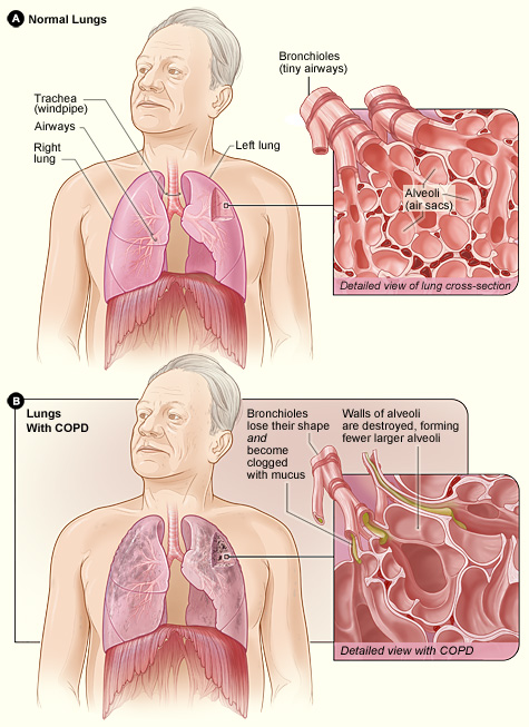
What smoke does to the airway. Smoke paralyzes and then destroys cilia, so the mucociliary escalator fails and smokers get more infections. The epithelium changes: goblet cells multiply, submucosal mucous glands enlarge, ciliated columnar cells are replaced by tougher stratified squamous cells (squamous metaplasia, a precursor of squamous cell cancer), and the basement membrane thickens. Smoke draws neutrophils and macrophages into the lungs, and they release proteases (elastase, matrix metalloproteinases) and oxidants that drive mucus hypersecretion and destroy alveolar walls.
Chronic bronchitis
Chronic bronchitis is defined clinically: a productive cough on most days for at least 3 months a year in 2 consecutive years, with other causes excluded. It is a disease of the airways: mucous-gland hypertrophy (the Reid index, gland thickness divided by wall thickness, rises above about 0.5; normal is below 0.4), goblet-cell hyperplasia, inflamed and narrowed small airways, and impaired or lost cilia, so mucus plugs the bronchioles. Poorly ventilated but perfused regions (low V̇/Q̇) cause hypoxemia and hypercapnia early, giving the "blue bloater" picture: cyanosis, a stocky build, polycythemia, and edema from right heart failure.

Cor pulmonale. Chronic alveolar hypoxia causes widespread hypoxic pulmonary vasoconstriction → pulmonary hypertension → the right ventricle hypertrophies and eventually fails → jugular venous distension, liver enlargement, ankle edema. COPD leads to right-sided, not left-sided, heart failure.
Emphysema
Emphysema is defined anatomically: permanent enlargement of the air spaces beyond the terminal bronchioles with destruction of their walls. Alveoli merge into fewer, larger sacs (bullae when larger than 1 cm).
- Protease–antiprotease imbalance. Neutrophil elastase digests the elastic fibers of the alveolar walls; α1-antitrypsin, made in the liver, normally blocks it. Smoking increases elastase (more neutrophils) and oxidizes α1-antitrypsin; inherited deficiency removes it.
- Patterns. Smoking causes centriacinar (centrilobular) emphysema, worst in the upper lobes, around the respiratory bronchioles. α1-antitrypsin deficiency causes panacinar emphysema, worst in the lower lobes, in young adults (often under 45), sometimes with liver disease.
- Physiology. Loss of elastic fibers raises compliance and lowers elastic recoil: the lungs fill easily but have little force to empty. The same fibers normally tether small airways open (radial traction), so without them the airways collapse during expiration, trapping air. RV, FRC and TLC rise; FEV₁ and the FEV₁/FVC ratio fall. Fewer alveolar walls mean less surface area and fewer capillaries: DLCO falls and alveolar dead space rises. Ruptured bullae are the commonest cause of secondary pneumothorax.
- "Pink puffer." Thin, breathless, breathing fast through pursed lips (which keeps back-pressure on the airways), leaning forward on the arms (tripod position) to use accessory muscles, with a barrel chest. Blood gases stay fairly normal until late because the patient hyperventilates and lost alveoli lose their capillaries too (V̇/Q̇ stays roughly matched). X-ray: hyperinflated, dark (hyperlucent) lungs and a flattened diaphragm.
| Feature | Chronic bronchitis ("blue bloater") | Emphysema ("pink puffer") |
|---|---|---|
| Definition | Clinical: productive cough ≥ 3 months/year for 2 years | Anatomic: destroyed, enlarged air spaces |
| Site | Bronchi and bronchioles | Acinus: respiratory bronchioles, alveoli |
| Main problem | Mucus, inflammation, narrowed airways | Lost elastic recoil, airway collapse |
| Cough and sputum | Prominent, productive | Little, late |
| Blood gases | Hypoxemia and hypercapnia early; cyanosis, polycythemia | Near normal until late |
| Compliance and DLCO | Compliance normal; DLCO near normal | Compliance high; DLCO low |
| Body habitus | Often overweight, edematous (cor pulmonale) | Thin, barrel chest, pursed lips |
Read the table row by row: the definitions, sites and DLCO are the facts tests ask; the "bloater/puffer" labels are textbook stereotypes, and real patients usually have both processes.
Diagnosis. Spirometry is the test: post-bronchodilator FEV₁/FVC < 0.70 confirms persistent obstruction. The GOLD system then grades severity by FEV₁ as a percent of predicted: GOLD 1 mild (≥ 80%), 2 moderate (50–79%), 3 severe (30–49%), 4 very severe (< 30%). Chest X-ray, blood gases, DLCO and an α1-antitrypsin level complete the workup.
Treatment and prevention. Stopping smoking is the only measure that slows the decline in FEV₁. Then: long-acting bronchodilators (LAMA such as tiotropium, LABA), inhaled corticosteroids for patients with frequent exacerbations, pulmonary rehabilitation (exercise training, breathing techniques), influenza and pneumococcal vaccines, antibiotics and oral steroids for exacerbations, long-term oxygen (at least 15 hours a day prolongs life when resting PaO₂ is 55 mmHg or below), lung volume reduction surgery or endobronchial valves, α1-antitrypsin replacement for the deficiency, and lung transplant. Diuretics such as furosemide do not treat COPD itself (only the edema of cor pulmonale). Patients often lose weight because the work of breathing burns calories and leaves them too tired to eat.
Pneumonia
Pneumonia is an infection of the lung parenchyma in which the alveoli fill with exudate (fluid, white cells, fibrin, microbes). It is a lower respiratory tract infection, and it can be caused by bacteria, viruses or fungi. It is most dangerous to infants, adults over 65, smokers and anyone with chronic disease or weak immunity.

Classified by setting: community-acquired (CAP); hospital-acquired (starting 48 hours or more after admission) and ventilator-associated, which involve tougher organisms such as Pseudomonas and MRSA; and aspiration pneumonia from inhaled stomach contents or mouth bacteria (stroke, seizures, alcohol), usually in the right lower lobe because the right main bronchus is more vertical.
| Group | Organisms and clues |
|---|---|
| Typical bacterial | Streptococcus pneumoniae (pneumococcus), the most common bacterial cause; lobar; rust-colored sputum. Haemophilus influenzae (COPD), Staphylococcus aureus (after influenza), Klebsiella (alcohol use; "currant-jelly" sputum), gram-negative bacilli |
| Atypical ("walking pneumonia") | Mycoplasma pneumoniae (young adults, dorms; no cell wall, so penicillins fail and macrolides are used), Chlamydophila pneumoniae, Legionella (contaminated water systems, cooling towers) |
| Viral | Influenza, RSV, SARS-CoV-2, adenovirus |
| Fungal | Pneumocystis jirovecii (HIV, immunosuppression), Histoplasma, Coccidioides |
How to read it: tests usually want S. pneumoniae as "most common bacterial cause" and accept that fungi can cause pneumonia.
Patterns. Lobar pneumonia consolidates an entire lobe (classically pneumococcal) and passes through congestion → red hepatization → gray hepatization → resolution. Bronchopneumonia is patchy, centered on bronchioles, in several lobes. Interstitial pneumonia (viral, atypical) inflames the alveolar walls more than the air spaces.
Physiology. Exudate removes alveoli from ventilation while blood keeps flowing past them: a shunt (V̇/Q̇ ≈ 0), so PaO₂ falls and responds only partly to oxygen. The inflamed respiratory membrane is also thicker, and the fluid-filled lung is less compliant. Rapid breathing usually keeps PaCO₂ normal or low.
Signs. Fever and chills, cough with purulent sputum (sometimes blood-streaked), pleuritic chest pain, dyspnea, tachypnea and tachycardia; older adults may show only confusion. Over the consolidation: crackles, bronchial breath sounds, increased tactile fremitus and dullness to percussion.
Diagnosis. Chest X-ray shows consolidation, often with air bronchograms (air-filled bronchi outlined by the white lung). Sputum Gram stain and culture, blood cultures, urine antigen tests (pneumococcus, Legionella), viral swabs and pulse oximetry follow; scores such as CURB-65 (confusion, urea, respiratory rate, blood pressure, age 65 or older; one point each) judge severity.
Treatment and prevention. Antibiotics for bacterial pneumonia (for example amoxicillin, a macrolide such as azithromycin, or doxycycline; broader drugs in hospital); antivirals such as oseltamivir for influenza; oxygen, fluids and fever control. Prevention: pneumococcal and influenza vaccines (also COVID-19 and RSV vaccines in eligible groups), hand hygiene, not smoking, and for hospital patients deep-breathing exercises, early mobilization and raising the head of the bed.
Sleep apnea
Sleep apnea is repeated pauses in breathing during sleep. An apnea is airflow stopping for 10 seconds or longer; a hypopnea is a partial reduction with a fall in oxygen saturation or an arousal.

| Type | Mechanism | Breathing effort during the pause | Risk factors and causes |
|---|---|---|---|
| Obstructive (OSA), by far the most common | Pharyngeal muscles relax in sleep; tongue and soft palate collapse the airway | Present: the chest and abdomen keep working against the blockage | Obesity (the strongest risk factor), thick neck, large tonsils and adenoids (the main cause in children), male sex, older age, alcohol and sedatives at night, small jaw, sleeping on the back |
| Central (CSA) | The brainstem stops sending signals to the breathing muscles | Absent | Heart failure (with Cheyne–Stokes breathing), stroke, opioids, high altitude |
| Mixed / complex | Mixed: an event starts central and ends obstructive. Complex (treatment-emergent): central events appear when OSA is treated with CPAP | Mixed | As above |
Consequences. During each pause the alveoli are not ventilated, so PaO₂ falls and PaCO₂ rises; the patient partly wakes (arousal), gasps and resumes breathing, often with a loud snort. Hundreds of arousals fragment sleep. Repeated hypoxia and sympathetic surges cause hypertension, atrial fibrillation, stroke and heart failure; hypoxic pulmonary vasoconstriction causes pulmonary hypertension; chronic hypoxemia stimulates erythropoietin, giving polycythemia.
Signs. Loud snoring, pauses or gasping witnessed by a bed partner, excessive daytime sleepiness, morning headaches, dry mouth on waking, poor concentration, nighttime urination. Obesity hypoventilation syndrome is different: an obese person who hypoventilates even while awake (daytime PaCO₂ above 45 mmHg); many also have OSA.
Diagnosis. Polysomnography (an overnight sleep study recording airflow, chest and abdominal effort, oxygen saturation, EEG and ECG), or a home sleep apnea test. Severity is the apnea–hypopnea index (AHI), events per hour of sleep: 5–15 mild, 15–30 moderate, 30 or more severe (in adults).
Treatment and prevention. CPAP (continuous positive airway pressure) is the most common and most effective treatment: air at a steady pressure through a mask acts as a pneumatic splint that holds the pharynx open. Others: weight loss, avoiding alcohol and sedatives, sleeping on the side, oral appliances that pull the jaw forward, surgery (tonsillectomy and adenoidectomy in children; uvulopalatopharyngoplasty), and hypoglossal nerve stimulation, which pushes the tongue forward during sleep. Central apnea is treated by treating the cause (heart failure, opioids) and with special ventilation modes (bilevel or adaptive servo-ventilation) or oxygen. Inhalers do not treat sleep apnea.
Cystic fibrosis
Cystic fibrosis (CF) is the most common lethal inherited disease in people of Northern European descent. It is autosomal recessive: a child must inherit a faulty CFTR gene (cystic fibrosis transmembrane conductance regulator) on chromosome 7 from each parent. More than 2,000 variants are known; the most common is F508del (ΔF508), a three-base deletion that removes phenylalanine 508, so the protein misfolds and is destroyed before reaching the cell membrane. In the US it affects about 1 in 2,500–3,500 white newborns.
What CFTR does. CFTR is a chloride channel in the apical membrane of epithelial cells, opened by cAMP (through protein kinase A) and ATP; it also carries bicarbonate and restrains the sodium channel ENaC. In the airway:
- Without CFTR, too little Cl⁻ is secreted into the airway and too much Na⁺ is absorbed.
- Water follows the salt back into the cells, so the airway surface liquid becomes dehydrated.
- The thin periciliary layer the cilia beat in collapses, and the mucus becomes thick and sticky.
- The mucociliary escalator fails; mucus plugs the airways and traps bacteria.
- Chronic infection and neutrophil inflammation follow (Staphylococcus aureus and Haemophilus influenzae in childhood, Pseudomonas aeruginosa in adolescents and adults, sometimes Burkholderia or Aspergillus), and the bronchi are permanently dilated and scarred (bronchiectasis). Respiratory failure is the usual cause of death.

Other organs. The disease was named for the cysts and fibrosis of the pancreas. Thick secretions block the pancreatic ducts, so digestive enzymes cannot reach the gut: pancreatic insufficiency, fat malabsorption with bulky, greasy stools (steatorrhea), poor weight gain and deficiency of the fat-soluble vitamins A, D, E and K; later, CF-related diabetes. About 1 in 6 newborns with CF has meconium ileus (the first stool blocks the intestine). Almost all men are infertile because the vas deferens is absent. Sweat is salty: in the sweat duct CFTR normally reabsorbs Cl⁻, so the defect leaves salt in the sweat (parents notice salty-tasting skin or salt crystals on clothes). Other findings: nasal polyps and sinusitis, digital clubbing, liver disease.

Diagnosis. US newborns are screened by measuring immunoreactive trypsinogen (IRT) in a heel-prick blood spot (high when the pancreatic ducts are blocked), often with a DNA panel. The confirmatory and most common diagnostic test is the sweat chloride test: pilocarpine iontophoresis stimulates sweating, and chloride ≥ 60 mmol/L is diagnostic (30–59 intermediate, below 30 unlikely). Genetic testing identifies the two CFTR variants. Spirometry shows obstruction; sputum cultures track infection.
Treatment. There is no cure, but survival has improved dramatically: children with CF in the 1950s rarely lived to school age, median survival reached the 30s by around 2000, and US registry estimates for babies born in the 2020s are now in the 60s (about 61–65 years), largely thanks to CFTR modulators.
- CFTR modulators fix the protein: correctors help it fold and reach the membrane, potentiators hold it open. Elexacaftor–tezacaftor–ivacaftor works for people with at least one F508del allele (about 90% of US patients); ivacaftor alone works for gating variants such as G551D.
- Airway clearance every day: chest physiotherapy with percussion and postural drainage, a high-frequency chest wall oscillation vest, breathing devices, exercise.
- Inhaled mucus thinners: hypertonic saline (draws water into the airway) and dornase alfa (DNase, which cuts the DNA released by dead neutrophils that thickens the sputum).
- Antibiotics for infections (inhaled tobramycin or aztreonam against Pseudomonas; azithromycin), bronchodilators.
- Pancreatic enzyme capsules with every meal, ADEK vitamins, extra salt and a high-calorie, high-fat diet.
- Lung transplant in end-stage disease.
Prevention means carrier screening and genetic counseling; the disease itself cannot be prevented by lifestyle.
Tuberculosis
Tuberculosis (TB) is caused by Mycobacterium tuberculosis, a slow-growing, aerobic, rod-shaped bacterium. Its waxy, mycolic-acid-rich cell wall resists ordinary stains, so it is identified with an acid-fast stain (Ziehl–Neelsen): the bacilli hold the red carbol-fuchsin after acid-alcohol washing. It spreads through the air in tiny droplet nuclei coughed out by someone with active lung disease; it is not spread by touch or shared dishes. About a quarter of the world's population carries the infection, and TB is among the leading infectious killers worldwide.
Primary infection. Inhaled bacilli reach the alveoli (usually in the mid-lung) and are eaten by alveolar macrophages, but they survive inside them by blocking phagosome–lysosome fusion. After a few weeks cell-mediated immunity (a type IV, delayed hypersensitivity) develops: T cells activate macrophages, which wall the bacteria off in granulomas of epithelioid macrophages, multinucleated Langhans giant cells and lymphocytes, with cheese-like caseous necrosis in the center (caseating granulomas). The first lesion is the Ghon focus; with the infected hilar lymph node it forms the Ghon complex, which may calcify (a calcified granuloma is a sign of old TB).
Latent versus active.
- Latent TB infection: bacteria survive inside granulomas but are held in check. No symptoms, normal chest X-ray, a positive skin or blood test, and not contagious. Without treatment about 5–10% develop active TB at some point in their lives.
- Active TB disease: bacteria multiply and cause symptoms; lung disease is contagious. Reactivation (secondary) TB happens when immunity weakens, and it favors the apices: the apical and posterior segments of the upper lobes and the superior segments of the lower lobes. The apex has the highest V̇/Q̇ and therefore the highest alveolar PO₂, which this aerobe likes, and poorer lymphatic clearance. Granulomas liquefy and erode into bronchi, leaving cavities. Spread through the blood gives miliary TB; outside the lungs TB affects lymph nodes (scrofula), spine (Pott disease), kidneys and meninges.
Signs. A cough lasting 3 weeks or more, hemoptysis (blood-tinged sputum), chest pain, fever, night sweats, weight loss and fatigue, sometimes swollen neck lymph nodes. Early TB can be silent and its symptoms mimic other diseases, which makes it hard to diagnose.
Risk factors. HIV (the strongest), diabetes, silicosis, malnutrition, immunosuppressive drugs, close contact with an active case, crowded settings (prisons, shelters, dormitories), birth in a high-burden country, healthcare work.
Diagnosis.
| Test | What it shows | Notes |
|---|---|---|
| Tuberculin skin test (Mantoux; PPD injected under the skin) | Infection (latent or active): induration read at 48–72 hours | Can be falsely positive after BCG vaccination |
| Interferon-γ release assay (IGRA, blood) | Infection (latent or active) | Not affected by BCG |
| Sputum acid-fast bacilli (AFB) smear | Active, infectious disease | Quick but not specific to M. tuberculosis |
| Sputum culture | Active disease; the gold standard | Takes weeks because the organism grows slowly; gives drug sensitivities |
| Nucleic acid amplification (NAAT) | M. tuberculosis DNA and rifampin resistance | Results in hours |
| Chest X-ray | Upper-lobe infiltrates and cavities, hilar nodes, calcified Ghon complex | Helps separate latent from active |
How to read it: the skin test and IGRA prove infection but cannot tell latent from active; the X-ray, smear and culture decide that.
Treatment. TB is curable with antibiotics, but only by several drugs for months, because the bacteria grow slowly, hide in granulomas and become resistant to single drugs. Active TB classically gets RIPE (rifampin, isoniazid, pyrazinamide, ethambutol) for 2 months, then rifampin and isoniazid for 4 more: 6 months, often as directly observed therapy. Side effects tests cite: isoniazid causes liver injury and peripheral neuropathy (prevented with vitamin B6), rifampin turns urine and tears orange, ethambutol affects vision. Latent TB is treated (for example 3 months of weekly isoniazid plus rifapentine, called 3HP, or 4 months of daily rifampin, called 4R) to prevent reactivation. MDR-TB resists at least isoniazid and rifampin and needs longer, harder regimens.
Prevention. Find and treat active cases, treat latent infection, isolate infectious patients in negative-pressure rooms with N95 respirators, and vaccinate infants with BCG (live, weakened Mycobacterium bovis) in high-burden countries, where it prevents severe childhood forms; it is not routinely used in the US.
Pulmonary edema
Pulmonary edema is excess fluid in the lung interstitium and then the alveoli. Fluid leaves capillaries when hydrostatic pressure pushes out harder than plasma oncotic pressure pulls in (the Starling forces), faster than the lymphatics can remove it. The pulmonary circuit normally runs at low pressure (about 25/8 mmHg), which keeps the alveoli dry.
- Cardiogenic (hydrostatic), the most common: left ventricular failure or mitral valve disease backs blood up into the pulmonary veins and capillaries. A hallmark of congestive heart failure.
- Non-cardiogenic (permeability): capillaries become leaky (ARDS, sepsis, inhaled toxins, near-drowning); also low plasma protein, lymphatic blockage, and high-altitude pulmonary edema (HAPE), where uneven hypoxic vasoconstriction overloads the capillaries that stay open.
Why it is debilitating. Fluid lengthens the diffusion distance, floods and collapses alveoli (less surface area, shunt), stiffens the lungs (less compliance), and stimulates juxtacapillary (J) receptors that drive rapid, shallow breathing. PaO₂ falls and DLCO falls.
Signs. Dyspnea, orthopnea (needs pillows to sleep), paroxysmal nocturnal dyspnea (PND, waking up breathless), a feeling of drowning, cough with pink, frothy sputum, crackles at the lung bases, a third heart sound (S3 gallop) from the failing ventricle, and in heart failure jugular venous distension and leg edema. Chest X-ray: enlarged heart, prominent upper-lobe vessels, Kerley B lines (short horizontal lines at the lung edges from fluid-filled septa), "bat-wing" opacities around the hila, pleural effusions; a high blood level of B-type natriuretic peptide (BNP), released by overstretched ventricles, supports a cardiac cause.
Treatment and prevention. Sit the patient upright, give oxygen (often CPAP), a loop diuretic such as furosemide to remove fluid, nitrates to dilate veins and reduce preload, and treat the cause (heart attack, arrhythmia, valve disease). HAPE is treated by descent and oxygen. Prevention: control blood pressure and heart disease, limit salt, take heart-failure drugs as prescribed, and ascend slowly at altitude.
Pleurisy and other pleural disorders
The pleural anatomy (visceral and parietal layers, potential space) is in Pleura.
Pleurisy (pleuritis) is inflammation of the pleura. Causes: viral infections (most common), pneumonia, tuberculosis, pulmonary embolism, autoimmune disease (lupus, rheumatoid arthritis), rib injury and cancer. The inflamed layers rub instead of gliding, so the patient has sharp, stabbing chest pain that worsens with a deep breath, cough or sneeze and eases when the breath is held; the pain comes from the parietal pleura, which has pain fibers. Breathing becomes shallow to avoid pain, and a pleural friction rub may be heard. Diagnosis rests on the history, the rub and imaging (X-ray or ultrasound) for an effusion. Treatment: treat the cause and give NSAIDs for pain and inflammation.
Pleural effusion is excess fluid in the pleural cavity. The lung beneath it is compressed; the chest is dull to percussion with reduced breath sounds.
| Type | Mechanism | Protein and lactate dehydrogenase (LDH) | Common causes |
|---|---|---|---|
| Transudate | Pressure imbalance pushes protein-poor fluid through intact vessels | Low | Heart failure (most common), cirrhosis, nephrotic syndrome |
| Exudate | Inflamed or leaky pleura releases protein-rich fluid | High | Pneumonia (parapneumonic), cancer, TB, pulmonary embolism |
Light's criteria call the fluid an exudate if any one is true: pleural/serum protein > 0.5, pleural/serum LDH > 0.6, or pleural LDH above two-thirds of the upper limit of normal for serum. Pus in the space is an empyema, blood a hemothorax, lymph a chylothorax. Large effusions are drained with a needle (thoracentesis) or chest tube.
Pneumothorax is air in the pleural cavity. The airtight seal that holds the lung against the chest wall is broken, intrapleural pressure rises from negative to atmospheric (or above), and the lung's elastic recoil collapses it.

- Primary spontaneous: a small apical bleb ruptures in a tall, thin young man, often a smoker.
- Secondary spontaneous: from underlying lung disease, most often emphysema (ruptured bullae); also CF, TB.
- Traumatic: a broken rib or a stab or gunshot wound tears the pleura; an open chest wound that draws air in with each breath is a sucking chest wound, covered in first aid with an occlusive dressing that lets air out but not in (a one-way valve).
- Tension pneumothorax: a one-way leak lets air in on each inspiration but not out, so intrapleural pressure becomes positive, the mediastinum and trachea shift away from the affected side, venous return falls and blood pressure drops. It is an emergency treated by immediate needle decompression and then a chest tube.
Signs: sudden one-sided sharp chest pain and breathlessness, absent breath sounds and hyperresonant percussion on that side. Treatment: observation and oxygen for a small one, needle aspiration or a chest tube (with an underwater seal or one-way valve) for larger ones; repeated ones may need pleurodesis (sealing the pleural layers together).
Atelectasis is collapse of part or all of a lung so that it holds no air. Types: resorption (an airway is blocked by mucus, a tumor or a foreign body and the trapped gas is absorbed), compression (by an effusion, pneumothorax or tumor), contraction (by scar) and surfactant loss (infant RDS, ARDS). It is common after surgery, when pain keeps breathing shallow, which is why patients use an incentive spirometer. A fetus's lungs are normally airless (fluid-filled) until the first breath.
Pulmonary fibrosis and pneumoconioses
Pulmonary fibrosis is scarring of the lung interstitium, the tissue between the alveoli and the capillaries. Fibroblasts lay down collagen, the alveolar walls thicken and stiffen, and eventually the lung becomes a network of cysts (honeycombing).
- Causes: idiopathic pulmonary fibrosis (IPF; no known cause; usually men over 50, often ex-smokers), mineral dusts (the pneumoconioses), healed infections (tuberculosis and severe pneumonia can leave scarred, fibrotic lung, as can ARDS), drugs (amiodarone, bleomycin, methotrexate, nitrofurantoin), radiation, autoimmune disease (scleroderma, rheumatoid arthritis), and organic dusts (hypersensitivity pneumonitis such as farmer's lung).
- Physiology: a restrictive pattern. Compliance falls, so the lungs are stiff and each breath costs more work; TLC, VC and FVC fall while the FEV₁/FVC ratio stays normal or high (stiff lungs recoil strongly). The thickened membrane slows diffusion, so DLCO falls and PaO₂ drops, especially in exercise. Patients breathe rapidly and shallowly, raising the dead-space fraction.
- Signs: progressive breathlessness on exertion, dry cough, fine "Velcro" crackles at the bases, clubbing.
- Diagnosis: restrictive spirometry with low DLCO; high-resolution CT shows basal, subpleural scarring and honeycombing; sometimes biopsy.
- Treatment: the antifibrotics pirfenidone and nintedanib slow (do not reverse) the scarring; oxygen, pulmonary rehabilitation, treating or removing the cause, and lung transplant. Prevention: avoid smoking and occupational dusts, monitor lung function on fibrosis-causing drugs.
Pneumoconioses are fibrotic lung diseases caused by inhaled mineral dust. Particles small enough to reach the alveoli (about 0.5–5 µm) are eaten by alveolar macrophages ("dust cells"); macrophages that cannot digest the dust release inflammatory and fibrogenic signals, die, and release the dust again, so the cycle repeats and scar builds up. The diseases take years to decades to appear and keep progressing after exposure ends.
| Disease | Dust and jobs | Where in the lung | Distinctive features |
|---|---|---|---|
| Silicosis | Crystalline silica (quartz): mining, quarrying, stone cutting, sandblasting, foundries, engineered-stone countertops | Upper lobes | Silicotic nodules; "eggshell" calcification of hilar lymph nodes; silica damages macrophages (disrupts phagolysosomes), so it raises the risk of TB; silica is also a lung carcinogen |
| Coal worker's pneumoconiosis ("black lung") | Coal dust: coal mining | Upper lobes | Coal macules and nodules; can progress to progressive massive fibrosis. Harmless carbon pigment in city dwellers and smokers is anthracosis |
| Asbestosis | Asbestos fibers: shipbuilding, insulation, roofing, brake linings, demolition | Lower lobes | Long, thin, durable fibers lodge in the lung; asbestos (ferruginous) bodies, golden-brown iron-coated dumbbell-shaped fibers; calcified pleural plaques; greatly raised risk of lung cancer (multiplied by smoking) and of mesothelioma, a pleural cancer that is otherwise rare |
How to read it: location separates them (silica and coal up, asbestos down), and the cancer and TB links are the most-tested extras. Another occupational lung disease tests sometimes add is berylliosis (aerospace and electronics work), which forms non-caseating granulomas like sarcoidosis.
Diagnosis and treatment. An occupational history plus restrictive spirometry, low DLCO and imaging. There is no cure: remove the exposure, stop smoking, give oxygen and pulmonary rehab, vaccinate, treat TB in silicosis, and consider transplant. Prevention is everything: wet cutting and dust suppression, ventilation, substituting safer materials, respirators, legal exposure limits (OSHA and MSHA in the US) and medical surveillance of exposed workers.
Lung cancer
Lung cancer is the leading cause of cancer death in the US for both men and women (about 1 in 5 cancer deaths). Cigarette smoking causes most of it (roughly 80–90% of lung cancer deaths); radon, a radioactive gas seeping from the ground into basements, is the second leading cause and the leading cause in people who never smoked. Other risks: secondhand smoke, asbestos (multiplied by smoking), silica, air pollution, arsenic and family history.
Lung cancers are grouped by how they behave and are treated:
| Type | Share | Origin and location | Key features |
|---|---|---|---|
| Non-small cell lung cancer (NSCLC) | About 85% | Slower growing; treated first with surgery when localized | |
| Adenocarcinoma | Most common type overall | Glandular cells, peripheral | Most common type in nonsmokers, women and young patients; driver mutations (EGFR, ALK, KRAS) guide targeted drugs |
| Squamous cell carcinoma | About a quarter | Bronchial epithelium after squamous metaplasia, central | Strongly linked to smoking; keratin pearls; can cavitate; may secrete PTH-related protein (hypercalcemia) |
| Large cell carcinoma | About 1 in 10 | Peripheral | Undifferentiated large cells; grows fast |
| Small cell lung cancer (SCLC) | About 10–15% | Neuroendocrine (Kulchitsky) cells, central near the hilum | Almost only in smokers; small "oat" cells with scant cytoplasm; very fast doubling, high growth fraction, early metastasis (often already spread at diagnosis); paraneoplastic SIADH (low blood sodium), ectopic ACTH (Cushing syndrome), Lambert–Eaton muscle weakness |
How to read it: NSCLC is the common, slower group treated surgically; SCLC is the rare, aggressive one treated with chemotherapy.
Signs.
- General: a persistent or changing cough, hemoptysis, chest pain, breathlessness, recurrent pneumonia behind a blocked bronchus, unexplained weight loss and fatigue.
- Local spread: hoarseness (tumor pressing the recurrent laryngeal nerve); a Pancoast tumor at the lung apex invades the brachial plexus (shoulder and arm pain) and the sympathetic chain, causing Horner syndrome (drooping eyelid, small pupil, no sweating on that side of the face); central tumors can compress the superior vena cava (SVC syndrome: swollen face and arms).
- Metastasis: brain, bone, liver and adrenal glands.
Diagnosis. Chest X-ray or CT finds a mass; tissue from bronchoscopy, needle biopsy or surgery confirms the type; tumor tests for EGFR, ALK, KRAS and other mutations and for PD-L1 guide drug choice; PET-CT and brain MRI stage it. NSCLC uses TNM staging from stage I to IV: T = size and local spread of the primary tumor, N = spread to lymph nodes, M = distant metastasis. SCLC is staged simply as limited (one side of the chest, fits in one radiation field) or extensive.
Treatment. NSCLC stage I–II: surgery (lobectomy) with or without chemotherapy; stage III: chemoradiation then immunotherapy; stage IV: targeted drugs for tumors with a driver mutation (EGFR or ALK inhibitors) or immunotherapy (PD-1/PD-L1 inhibitors), with or without chemotherapy. SCLC: chemotherapy (platinum plus etoposide) with radiation for limited disease, plus immunotherapy; it responds well at first but usually relapses, and surgery is rarely possible.
Prevention and screening. Not smoking or quitting (risk falls steadily after quitting), testing homes for radon, workplace protection. The US Preventive Services Task Force recommends yearly low-dose CT for adults aged 50–80 with a 20 pack-year smoking history who smoke now or quit within the past 15 years.
ARDS, infant RDS and pulmonary hypoplasia
Acute respiratory distress syndrome (ARDS) is diffuse damage to the alveolar–capillary membrane after a direct or indirect injury: sepsis (most common), pneumonia, aspiration, major trauma, pancreatitis, massive transfusion, burns, near-drowning. Inflammation makes the capillaries leaky, so protein-rich fluid floods the alveoli (non-cardiogenic edema) and inactivates surfactant; alveoli collapse, hyaline membranes of protein and dead cells line them, and later the lung may scar. The result is stiff, low-compliance lungs, a large shunt and hypoxemia that barely responds to oxygen. The Berlin definition requires: onset within 1 week of the insult; bilateral opacities on imaging; edema not fully explained by heart failure or fluid overload; and PaO₂/FiO₂ ≤ 300 mmHg (with PEEP ≥ 5 cmH₂O), graded mild (201–300), moderate (101–200) or severe (≤ 100).
Treatment supports the lung while the cause is treated: low tidal volume ventilation (about 6 mL/kg of predicted body weight) to avoid overstretching the few open alveoli, PEEP to hold alveoli open, prone positioning for severe cases, conservative fluids, and ECMO when all else fails.
Infant respiratory distress syndrome (IRDS, hyaline membrane disease) is caused by too little surfactant in a premature lung. The risk is highest before surfactant becomes adequate late in gestation (timeline in Lung development and aging). Without surfactant, surface tension collapses the alveoli at the end of each breath (atelectasis), compliance is low, and each breath must re-open them. Risk rises with lower gestational age, maternal diabetes (high fetal insulin delays surfactant), male sex, cesarean delivery without labor and birth asphyxia. Signs appear within minutes to hours of birth: tachypnea, expiratory grunting (the baby narrows the glottis to keep pressure in the alveoli), nasal flaring, chest retractions and cyanosis. The X-ray shows low-volume lungs with a ground-glass pattern and air bronchograms. Before birth, a lecithin/sphingomyelin ratio in amniotic fluid of 2 or more indicates mature lungs.
Treatment: CPAP, exogenous surfactant given down an endotracheal tube, mechanical ventilation and carefully limited oxygen (excess harms the eyes and lungs of premature babies). Prevention: antenatal corticosteroids (betamethasone) given to the mother when preterm birth is expected, which speed surfactant production and lung maturation, and delaying preterm delivery when possible.
Pulmonary hypoplasia is incomplete development of the lungs before birth: too few airways, alveoli and vessels. Fetal lungs need room to grow, amniotic fluid and fetal breathing movements.
- Too little room: congenital diaphragmatic hernia (CDH; abdominal organs herniate into the chest, most often through a left posterolateral defect, and compress the lung), large chest masses or effusions, a small rib cage (skeletal dysplasias).
- Too little fluid (oligohydramnios): kidney agenesis or blocked urine outflow (the Potter sequence: flat face, limb deformities and hypoplastic lungs) or early rupture of the membranes.
- Too little movement: neuromuscular disease that prevents fetal breathing.
At birth the baby has respiratory failure and pulmonary hypertension (few, muscular vessels). Diagnosis is often prenatal: ultrasound or MRI estimates lung size (the lung-to-head ratio in CDH). Management: delivery at a specialist center, gentle ventilation, inhaled nitric oxide, ECMO, then surgical repair of the hernia once the baby is stable; in selected severe CDH, fetoscopic tracheal occlusion (a balloon in the fetal trachea) traps lung fluid, which stretches the lung and stimulates its growth.
Respiratory drugs and therapies
Most respiratory drugs act on airway smooth muscle (bronchodilators, which exploit the β₂ and muscarinic receptors described in Airway resistance) or on inflammation (anti-inflammatories).
| Class | Examples | How it works | Used for |
|---|---|---|---|
| Short-acting β₂ agonist (SABA) | Albuterol (salbutamol), terbutaline, levalbuterol | Stimulates β₂ receptors → smooth muscle relaxes in minutes | Rescue in asthma and COPD; before exercise |
| Long-acting β₂ agonist (LABA) | Salmeterol, formoterol | Same, for 12 hours or more | Maintenance; in asthma only with an inhaled steroid |
| Anticholinergic (antimuscarinic) | Ipratropium (short), tiotropium (long) | Blocks vagal bronchoconstriction and mucus secretion | COPD maintenance; acute asthma add-on |
| Inhaled corticosteroid | Fluticasone, budesonide, beclomethasone | Suppresses airway inflammation | Asthma controller; COPD with exacerbations |
| Systemic corticosteroid | Prednisone, methylprednisolone | Same, stronger | Acute attacks and exacerbations |
| Leukotriene modifier | Montelukast (receptor blocker), zileuton | Blocks leukotriene-driven bronchoconstriction | Asthma, aspirin-sensitive asthma |
| Mast-cell stabilizer | Cromolyn | Prevents mast-cell degranulation | Allergic and exercise-induced asthma |
| Methylxanthine | Theophylline (aminophylline) | Relaxes bronchial smooth muscle (phosphodiesterase inhibition) | Add-on in asthma and COPD |
| Biologic | Omalizumab (anti-IgE), mepolizumab (anti-IL-5), dupilumab | Blocks a specific step of allergic inflammation | Severe asthma |
| Epinephrine | Epinephrine | α and β agonist: bronchodilation, less airway swelling | Anaphylaxis, life-threatening asthma |
| Mucoactive | Hypertonic saline, dornase alfa | Rehydrates mucus; cuts DNA in sputum | Cystic fibrosis |
| CFTR modulator | Elexacaftor–tezacaftor–ivacaftor, ivacaftor | Helps CFTR reach the membrane and open | Cystic fibrosis |
| Antifibrotic | Pirfenidone, nintedanib | Slow fibroblast activity | Idiopathic pulmonary fibrosis |
| Diuretic and vasodilator | Furosemide, nitrates | Remove fluid; reduce preload | Cardiogenic pulmonary edema |
| Anti-TB antibiotics | Rifampin, isoniazid, pyrazinamide, ethambutol | Kill or stop mycobacteria | Tuberculosis |
| Surfactant | Exogenous surfactant | Lowers alveolar surface tension | Infant RDS |
How to read it: decide whether the problem is a squeezed airway (bronchodilator), inflammation (steroid, leukotriene drug, biologic), mucus (CF drugs), fluid (diuretic), infection (antibiotic) or scar (antifibrotic).
Non-drug therapies tests mention: oxygen therapy (hypoxemia, carbon monoxide poisoning, acute severe asthma; titrated in CO₂ retainers; hyperbaric oxygen for CO poisoning), CPAP and BiPAP, mechanical ventilation, pulmonary rehabilitation, chest physiotherapy, chest tubes, incentive spirometry, and emergency airways (cricothyrotomy or tracheostomy when the upper airway is blocked; see Trachea and bronchial tree).
Breadth reference
These three tables are a lookup layer for the long tail of terms, names and numbers that tests pull in one question at a time. Type a word into a table's filter box ("carina", "Kohn", "mmHg", "not on 2027 list") to hide every row that does not match. Rows marked "not on 2027 list" are related conditions that appear as distractors; values are typical adult textbook figures, so use a question's own numbers when it gives them.
ReferenceRespiratory glossary A–Z· 449 rows
No rows match.
| Term | Meaning | Where it matters (exam angle or look-alike) |
|---|---|---|
| 2,3-BPG (2,3-bisphosphoglycerate, 2,3-DPG) | Negatively charged glycolysis by-product in red cells; binds the β-chain cavity of deoxy-Hb | Rises at altitude, in anemia and chronic hypoxia; shifts the O₂ curve right (lower affinity) |
| Abdominal thrusts | First aid for a choking conscious adult: fist above the navel, thrust in and up | Same as the Heimlich maneuver; alternate with 5 back blows; infants get back blows and chest thrusts |
| ABG (arterial blood gas) | Arterial sample giving pH, PaO₂, PaCO₂, HCO₃⁻ and saturation | Read pH → primary change → compensation; normal pH 7.35–7.45, PaCO₂ 35–45, HCO₃⁻ 22–26 |
| Accessory muscles of inspiration | Sternocleidomastoid, scalenes, pectoralis minor, serratus anterior (and others) | Used in forced inspiration and severe asthma/COPD; abdominal muscles are expiratory, not inspiratory accessories |
| Acetazolamide | Carbonic anhydrase inhibitor; causes mild metabolic acidosis that stimulates breathing | Prevents and treats acute mountain sickness; reduces altitude periodic breathing |
| Acidemia vs acidosis | Acidemia = blood pH below 7.35; acidosis = the process adding acid or removing base | Same pairing for alkalemia (pH above 7.45) and alkalosis |
| Acid-fast stain | Ziehl–Neelsen (carbol-fuchsin) stain that waxy mycolic-acid walls retain after acid-alcohol | Identifies mycobacteria (TB) as red "acid-fast bacilli (AFB)" in sputum |
| Acinus | Everything distal to one terminal bronchiole: respiratory bronchioles, alveolar ducts, sacs, alveoli | Unit of gas exchange; centriacinar vs panacinar emphysema |
| Acute bronchitis (not on 2027 list) | Short (1–3 week) inflammation of the bronchi, about 90% viral | No consolidation on X-ray (unlike pneumonia); antibiotics do not help a viral case |
| Acute mountain sickness (AMS) | Headache, nausea, fatigue, poor sleep within 6–12 h above about 2,500 m | Prevent by slow ascent and acetazolamide; treat by rest, descent, O₂ |
| Adam's apple | Common name of the laryngeal prominence of the thyroid cartilage | Not the cricoid; larger in males after puberty (testosterone) |
| Adenocarcinoma | Glandular non-small cell lung cancer, peripheral | Most common lung cancer overall and in nonsmokers, women and young patients; EGFR/ALK mutations |
| Adenoids | Pharyngeal tonsil on the posterior wall/roof of the nasopharynx | Enlarged adenoids and tonsils are the main cause of OSA in children |
| Agonal breathing | Irregular isolated gasps after cardiac arrest or severe brain hypoxia | Treat as no breathing: start CPR |
| AHI (apnea–hypopnea index) | Apneas + hypopneas per hour of sleep | Adults: under 5 normal, 5–15 mild, 15–30 moderate, 30 or more severe |
| Air bronchogram | Air-filled bronchi outlined against white, airless lung on X-ray | Seen in pneumonia consolidation and infant RDS |
| Alae (nasal) | Flared "wings" of the nose around each nostril | Shaped by the major and minor alar cartilages (hyaline); flare in respiratory distress |
| Albuterol (salbutamol) | Short-acting β₂ agonist (SABA) | Rescue inhaler for asthma and COPD; works in minutes; with terbutaline the classic "bronchodilator" answer |
| Allergic (extrinsic) asthma | Asthma triggered by allergens via IgE and mast cells | Childhood onset, with eczema/hay fever; pollen, dust mites, dander, mold; vs intrinsic (cold air, infection, exercise, aspirin) |
| Alpha-1 antitrypsin (α1-antitrypsin, AAT) | Liver-made protease inhibitor that blocks neutrophil elastase | Inherited deficiency → panacinar, lower-lobe emphysema in young adults (often with liver disease); replacement therapy exists |
| Alveolar dead space | Ventilated alveoli with no blood flow | Near zero in health; rises with pulmonary embolism, emphysema; anatomic + alveolar = physiologic dead space |
| Alveolar duct | Tube lined with alveoli between respiratory bronchiole and alveolar sacs | Respiratory zone; ends in an atrium |
| Alveolar gas equation | PAO₂ = FiO₂(PB − 47) − PaCO₂/0.8 | Gives about 100 mmHg at sea level; used for altitude questions |
| Alveolar macrophage | Phagocyte on the alveolar surface ("dust cell") | Respiratory-zone defense where there are no cilia; central to pneumoconiosis and TB |
| Alveolar sac | Grape-like cluster of alveoli at the end of an alveolar duct | Respiratory zone |
| Alveolar ventilation (V̇A) | (TV − dead space) × rate, about 4.2 L/min at rest | Better measure of effective ventilation than minute ventilation; deeper breathing raises it most |
| Alveolus | Thin-walled air sac (about 0.2 mm) wrapped in capillaries | Site of gas exchange; 300–500 million; simple squamous type I cells |
| Anaphylaxis (not on 2027 list) | Severe systemic allergic reaction with airway swelling and bronchospasm | Treat with epinephrine; may cause stridor and wheeze |
| Anatomic dead space | Volume of the conducting airways, about 150 mL (≈1 mL per lb body weight) | Nose to terminal bronchioles; alveoli are not part of it; a snorkel adds to it |
| Anemic hypoxia | Normal PaO₂ but too little working hemoglobin | Anemia, blood loss, CO poisoning; no rise in ventilation (carotid bodies sense PaO₂) |
| Anoxia | Total absence of oxygen in tissue | Stronger term than hypoxia |
| Antenatal corticosteroids | Betamethasone or dexamethasone given to the mother before expected preterm birth | Speed surfactant production; prevent infant RDS |
| Anthracosis | Harmless carbon-pigment deposits in city dwellers and smokers | Not the same as coal worker's pneumoconiosis |
| Anticholinergic (antimuscarinic) | Ipratropium (SAMA), tiotropium (LAMA) | Block vagal (ACh) bronchoconstriction and mucus; COPD maintenance |
| Aortic bodies | Peripheral chemoreceptors in the aortic arch | Signal via the vagus nerve (X); sense low PaO₂, high CO₂, H⁺ |
| Apex of the lung | Rounded top rising 2–3 cm above the medial clavicle | High V̇/Q̇ and PO₂; favored by reactivation TB; Pancoast tumors |
| Apnea | Pause in breathing; in adults airflow stopped 10 s or longer | Follows forced hyperventilation (low CO₂); sleep apnea; infants: 20 s or more |
| Apneustic breathing (apneusis) | Long gasping inspirations with a breath-hold at full inspiration | Upper/mid pons lesion (loss of pneumotaxic brake); classic "pons lesion" answer |
| Apneustic center | Lower-pons neurons that stimulate the DRG and prolong inspiration | Normally checked by the pneumotaxic center and vagal input |
| ARDS (acute respiratory distress syndrome) | Diffuse alveolar damage with leaky capillaries within 1 week of an insult | Berlin: bilateral opacities, not cardiac, PaO₂/FiO₂ ≤ 300; Division C National only |
| Aryepiglottic folds | Mucosal folds from the epiglottis edges to the arytenoids | Lateral boundaries of the laryngeal inlet; contain cuneiform cartilages |
| Arytenoid cartilages | Paired pyramidal (mostly hyaline) cartilages on top of the cricoid | Anchor vocal folds; pivot to open/close them; with corniculates "move the vocal cords" |
| Asbestos (ferruginous) bodies | Golden-brown, iron-coated, dumbbell-shaped asbestos fibers | Hallmark of asbestosis ("iron-containing inclusion bodies") |
| Asbestosis | Fibrosis from inhaled asbestos fibers | Lower lobes; pleural plaques; shipyards, insulation; raises mesothelioma and lung cancer (× smoking) |
| Aspergillosis (not on 2027 list) | Lung infection or allergy caused by the mold Aspergillus | Seen in CF and immunosuppression; a fungal distractor |
| Aspiration | Inhaling food, fluid or stomach contents into the airway | Usually right lower lobe (right main bronchus more vertical); causes aspiration pneumonia |
| Asthma | Chronic airway inflammation with hyperresponsiveness and reversible obstruction | Regional; "primarily an inflammatory process"; wheeze, cough, chest tightness |
| ATA / atm | Atmospheres absolute: 1 atm = 760 mmHg = 101.3 kPa | Each 10 m of seawater adds 1 atm; hyperbaric O₂ at 2–3 ATA |
| Atelectasis | Collapse of part or all of a lung so it holds no air | Resorption, compression, contraction, surfactant loss; normal state of the lung before the first breath |
| Atrium (alveolar) | Space at the end of an alveolar duct opening into alveolar sacs | Differs from the duct mainly in shape |
| Atypical pneumonia | "Walking pneumonia" from Mycoplasma, Chlamydophila, Legionella | Macrolides (e.g. azithromycin); Mycoplasma lacks a cell wall so penicillins fail |
| Auditory (Eustachian) tube | Tube from the middle ear to the nasopharynx | Equalizes ear pressure (opens on yawning/swallowing); ringed by tubal tonsils |
| Auscultation | Listening to the chest with a stethoscope | Breath sounds: vesicular, bronchovesicular, bronchial; adventitious: crackles, wheeze, rhonchi, rub |
| Baroreceptors | Pressure sensors in the carotid sinus and aortic arch | Falling blood pressure raises ventilation; rising pressure lowers it |
| Barrel chest | Increased front-to-back chest diameter | Air trapping in emphysema |
| Basal cells | Stem cells at the base of pseudostratified airway epithelium | Part of the respiratory epithelium (with ciliated and goblet cells) |
| BCG vaccine | Live attenuated Mycobacterium bovis (bacille Calmette–Guérin) | Prevents severe childhood TB in high-burden countries; can cause false-positive skin test |
| Berylliosis (not on 2027 list) | Lung disease from beryllium (aerospace, electronics) | Non-caseating granulomas like sarcoidosis |
| Bicarbonate (HCO₃⁻) | Main form of CO₂ in blood (about 70%) | Made in red cells by carbonic anhydrase, carried in plasma; normal 22–26 mmol/L |
| BiPAP (BPAP, bilevel positive airway pressure) | Noninvasive ventilation with separate inspiratory and expiratory pressures | COPD exacerbations, obesity hypoventilation; also used for OSA |
| Bleb / bulla | Air space in or under the pleura (bulla larger than 1 cm) | Rupture causes spontaneous pneumothorax; bullae in emphysema |
| Body plethysmography | Patient sits in a sealed box; Boyle's law gives thoracic gas volume | Measures FRC, RV and TLC, which spirometry cannot |
| Bradypnea | Slow rate, below 12 breaths/min in adults | Opioids, sedatives, head injury, hypothermia, deep sleep |
| Bronchial arteries | Small branches mostly from the thoracic aorta | Feed lung tissue with oxygenated blood (nutritive circulation); part drain into pulmonary veins |
| Bronchial breath sounds | Loud, hollow, tubular sounds normally heard only over the trachea | Heard over consolidated lung in lobar pneumonia |
| Bronchiectasis (not on 2027 list) | Permanent dilation and scarring of bronchi | Complication of CF and repeated infection; copious sputum, clubbing |
| Bronchioles | Airways under about 1 mm with smooth muscle and no cartilage | First branches without cartilage; narrow in asthma; club cells |
| Bronchiolitis (not on 2027 list) | Viral (mostly RSV) infection of bronchioles in children under 2 | Wheeze, fast breathing; supportive care |
| Bronchoalveolar lavage (BAL) | Washing a lung segment through a bronchoscope and collecting fluid | Samples cells, microbes, elastase |
| Bronchoconstriction | Narrowing of airways by smooth muscle contraction | Vagus/ACh, histamine, leukotrienes, irritants, low local PCO₂ |
| Bronchodilation | Widening of airways by smooth muscle relaxation | Sympathetic/epinephrine on β₂, albuterol, high local PCO₂ |
| Bronchodilator response | Rise in FEV₁ after a bronchodilator | Classic cutoff: at least 12% and 200 mL (newer 2022 ATS/ERS: more than 10% of predicted); suggests asthma |
| Bronchopulmonary dysplasia (BPD, not on 2027 list) | Chronic lung disease of premature babies after ventilation and O₂ | Complication of infant RDS treatment |
| Bronchopulmonary segment | Lung region supplied by one segmental (tertiary) bronchus and artery branch | Right 10, left 8–10; can be removed surgically on its own |
| Bronchoscopy | Viewing the airways with a flexible or rigid scope | Biopsy of central tumors, BAL, foreign-body removal |
| Bronchovesicular sounds | Intermediate sounds near the main bronchi | May be heard over consolidation in pneumonia |
| Bucket-handle motion | Lower ribs swing up and out during inspiration | Widens the chest side to side (transverse diameter) |
| Capnography (ETCO₂) | Measurement of exhaled CO₂ at the end of each breath | Normal end-tidal CO₂ about 35–45 mmHg; confirms tube placement |
| Carbaminohemoglobin (HbCO₂) | CO₂ bound to amino groups of globin | About 20–23% of CO₂; favored when Hb is deoxygenated; not on the heme |
| Carbonic anhydrase | Zinc enzyme speeding CO₂ + H₂O ⇌ H₂CO₃ | Abundant in red cells; works both directions; inhibited by acetazolamide |
| Carbon monoxide (CO) poisoning | CO binds heme about 200–250× more tightly than O₂ | Cherry-red skin, normal pulse-ox reading, left shift; treat 100% or hyperbaric O₂ |
| Carboxyhemoglobin (HbCO) | CO bound to the heme iron | Blocks O₂ binding; normal under about 3% in nonsmokers |
| Cardiac notch | Indentation in the left lung's anterior border for the heart | Left lung only; why the left lung is smaller |
| Carina | Ridge of the last tracheal cartilage where the trachea splits | About T4–T5 (sternal angle); most sensitive cough trigger |
| Carotid bodies | Peripheral chemoreceptors at the carotid bifurcation | Sense PaO₂ below about 60 mmHg, plus CO₂ and H⁺; signal via glossopharyngeal nerve (IX) |
| Caseous necrosis | Cheese-like central necrosis in a granuloma | Hallmark of TB (caseating granulomas) |
| Central chemoreceptors | Medullary sensors bathed in CSF | Respond to H⁺ formed from CO₂; main drive in health; not to O₂ |
| Central sleep apnea (CSA) | Brainstem stops sending breathing signals during sleep | No breathing effort during pauses; heart failure (Cheyne–Stokes), opioids, altitude |
| Centriacinar (centrilobular) emphysema | Destruction centered on respiratory bronchioles, upper lobes | Smoking type; vs panacinar (AAT deficiency, lower lobes) |
| CFTR | Cystic fibrosis transmembrane conductance regulator: cAMP-regulated Cl⁻ channel | Gene on chromosome 7; F508del most common; defect causes CF |
| Chest tube (tube thoracostomy) | Drain placed into the pleural space | Pneumothorax, hemothorax, empyema; underwater seal or one-way valve |
| Chloride shift (Hamburger shift) | HCO₃⁻ leaves red cells in exchange for Cl⁻ via band 3 | Keeps charge balanced; reverses in the lungs; venous RBCs hold more Cl⁻ |
| Choanae | Posterior (internal) nares connecting nasal cavity to nasopharynx | Choanal atresia = congenital blockage, newborn distress relieved by crying |
| Chronic bronchitis | Productive cough most days ≥ 3 months/year for 2 consecutive years | Clinical definition; "blue bloater"; excess mucus, Reid index above 0.5 |
| Chylothorax | Lymph (chyle) in the pleural cavity | Milky effusion; thoracic duct injury |
| Cilia | Motile hair-like projections on airway epithelial cells | Beat mucus toward the pharynx (mucociliary escalator); absent in alveoli; smoke paralyzes them |
| Closed quotient | Fraction of each vocal-fold vibration cycle that the glottis is closed | High in pressed voice, low in breathy voice |
| Clubbing | Bulbous fingertips with loss of the nail-bed angle | CF, fibrosis, lung cancer, bronchiectasis; not typical of uncomplicated COPD or asthma |
| Club cells | Dome-shaped secretory cells of bronchioles (formerly Clara cells) | Detoxify, secrete protective proteins, act as stem cells; bronchiolar counterpart of type II cells |
| cmH₂O | Pressure unit used for ventilators and lung pressures | 1 mmHg ≈ 1.36 cmH₂O; −5 cmH₂O ≈ −4 mmHg |
| Coal worker's pneumoconiosis | Fibrosis from coal dust ("black lung") | Upper lobes; coal macules; progressive massive fibrosis |
| Compliance | Change in lung volume per change in pressure (ΔV/ΔP) | ↑ emphysema, aging; ↓ fibrosis, edema, RDS; surfactant raises it |
| Conchae (turbinates) | Three scroll-shaped shelves on the lateral nasal wall | Increase surface area and turbulence to warm, humidify and filter; inferior concha has erectile tissue |
| Conducting zone | Nose to terminal bronchioles | No gas exchange; warms, cleans, humidifies; = anatomic dead space |
| Congenital diaphragmatic hernia (CDH) | Abdominal organs herniate into the chest through a diaphragm defect | Usually left posterolateral (Bochdalek); compresses lung → pulmonary hypoplasia |
| Consolidation | Lung airspaces filled with fluid or exudate so tissue is solid | Pneumonia; dull percussion, bronchial breath sounds, increased fremitus |
| Conus elasticus (cricothyroid membrane) | Elastic membrane from the cricoid up to the vocal ligaments | Its median part is the site of emergency cricothyrotomy |
| COPD (chronic obstructive pulmonary disease) | Persistent, largely irreversible airflow limitation | Chronic bronchitis + emphysema; post-bronchodilator FEV₁/FVC < 0.70; smoking main cause |
| Corniculate cartilages | Paired small elastic cartilages on the arytenoid tips | Move with the arytenoids |
| Cor pulmonale | Right ventricular enlargement/failure from lung disease | Hypoxic vasoconstriction → pulmonary hypertension; COPD strains the right heart |
| Corticosteroid, inhaled (ICS) | Fluticasone, budesonide, beclomethasone | Asthma controller backbone; rinse mouth to prevent thrush |
| Coryza | Common cold: acute viral rhinitis | Upper respiratory infection; distractor in emphysema questions |
| Costal breathing | Breathing mainly by rib movement | Relied on when the diaphragm or phrenic nerves fail |
| Cough | Deep breath, glottis closes, pressure builds, glottis bursts open | Clears lower airway; trachealis contracts; carina and larynx most sensitive |
| Cough suppressant vs expectorant | Antitussive (dextromethorphan, codeine) suppresses cough; expectorant (guaifenesin) thins and loosens mucus | Neither is a bronchodilator (theophylline relaxes airway muscle) |
| CPAP (continuous positive airway pressure) | Steady positive pressure delivered by mask | Pneumatic splint for OSA (most common treatment); also infant RDS and pulmonary edema |
| Crackles (rales) | Brief popping sounds, mostly inspiratory | Fluid or reopening alveoli: pneumonia, edema (bases), fibrosis ("Velcro") |
| Cribriform plate | Perforated part of the ethmoid bone forming the nasal roof | Olfactory nerve fibers pass through it |
| Cricoid cartilage | Signet-ring hyaline cartilage below the thyroid | Only complete cartilage ring in the airway |
| Cricothyroid muscle | Intrinsic laryngeal muscle that tilts the thyroid cartilage | Tenses the vocal folds → higher pitch |
| Cricothyrotomy | Emergency opening through the cricothyroid membrane | Faster than tracheostomy when the upper airway is blocked |
| Croup (laryngotracheobronchitis, not on 2027 list) | Viral (parainfluenza) swelling below the vocal folds in young children | Barking cough, inspiratory stridor, "steeple sign"; dexamethasone |
| Cuneiform cartilages | Paired small elastic rods in the aryepiglottic folds | Stiffen the folds |
| CURB-65 | Pneumonia severity score: Confusion, Urea, Respiratory rate ≥30, low BP, age ≥65 | 1 point each; higher scores → hospital or ICU care |
| Cyanide poisoning | Cyanide blocks mitochondrial cytochrome oxidase | Histotoxic hypoxia: O₂ delivered but not used |
| Cyanosis | Bluish skin and mucosa when more than about 5 g/dL Hb is deoxygenated | Central (lips) vs peripheral (nail beds); absent in severe anemia and CO poisoning |
| Cystic fibrosis (CF) | Autosomal recessive CFTR chloride-channel defect | Thick dehydrated mucus, salty sweat, Pseudomonas, pancreatic insufficiency; Regional |
| Dead space (physiologic) | Anatomic plus alveolar dead space | Normally ≈ anatomic; measured by the Bohr equation |
| Decompression sickness (the bends) | N₂ dissolved at depth forms bubbles on fast ascent | Joint pain, numbness, paralysis; Henry's law; recompression in a hyperbaric chamber |
| Defensins | Antimicrobial peptides in airway mucus | Chemical defense with lysozyme, lactoferrin, IgA, cathelicidin |
| Deoxyhemoglobin (reduced Hb, HHb) | Hemoglobin with no O₂ on the heme | Binds H⁺ and CO₂ better (Haldane effect); darker red |
| Diaphragm | Dome-shaped skeletal muscle between thorax and abdomen | Main inspiratory muscle (about 2/3–3/4 of quiet volume change); contracts and moves down; phrenic C3–C5 |
| Diaphragmatic openings | Caval (T8, IVC), esophageal hiatus (T10), aortic hiatus (T12) | "I 8 10 Eggs At 12"; there is no spinal opening |
| Diffusion | Passive movement of gas down its partial-pressure gradient | The only way O₂ and CO₂ cross the respiratory membrane (simple diffusion, no active transport) |
| DLCO (diffusing capacity for carbon monoxide) | Uptake of a small amount of inhaled CO per minute per mmHg | Low in emphysema, fibrosis, edema; normal in asthma; high with alveolar hemorrhage |
| Dornase alfa | Inhaled DNase that cuts neutrophil DNA in sputum | Thins CF mucus |
| DPPC (dipalmitoylphosphatidylcholine) | Main phospholipid of surfactant (a lecithin) | Lowers surface tension; measured in the L/S ratio |
| DRG (dorsal respiratory group) | Mainly inspiratory neurons in the dorsal medulla (nucleus of the solitary tract) | Integrates chemoreceptor and stretch input; drives the diaphragm via phrenic nerves |
| Dry powder inhaler (DPI) | Breath-actuated inhaler needing a fast, strong inhalation | One of three main inhaler types (with MDI and soft mist) |
| Dullness to percussion | Thud instead of resonance over the chest | Fluid (effusion) or solid lung (consolidation) |
| Dust cells | Alveolar macrophages | Engulf dust; key cell in pneumoconiosis |
| Dynamic lung volumes | Measures that depend on volume and speed: FVC, FEV₁, PEF, MVV | Reveal obstruction; static volumes (TV, IRV, ERV, VC) do not |
| Dyspnea | Feeling of difficult, labored breathing; shortness of breath | Symptom of many disorders; "labored, gasping" |
| ECMO (extracorporeal membrane oxygenation) | External circuit that oxygenates blood like a heart–lung machine | Last resort in ARDS, CDH, pulmonary hypoplasia |
| EGFR / ALK | Driver mutations tested in non-small cell lung cancer | Guide targeted drugs (EGFR or ALK inhibitors), mostly in adenocarcinoma |
| Egophony | "E" heard as "A" through the stethoscope | Consolidation or top of an effusion |
| Elastase | Neutrophil protease that digests elastic fibers | Excess over α1-antitrypsin destroys alveolar walls in emphysema |
| Elastic cartilage | Flexible cartilage with elastic fibers | Epiglottis, corniculate, cuneiform; not in the trachea |
| Elastic recoil | Inward pull of lungs toward smaller volume (elastin + surface tension) | Drives passive expiration; lost in emphysema; chest wall recoils outward |
| Elastin | Elastic protein of the lung extracellular matrix | Maintains parenchyma and holds small airways open (radial traction) |
| Emphysema | Permanent enlargement of air spaces beyond terminal bronchioles with wall destruction | Obstructive; ↑ compliance, ↓ DLCO, barrel chest, "pink puffer" |
| Empyema | Pus in the pleural cavity | Complication of pneumonia; needs drainage |
| Endotracheal intubation | Tube through mouth or nose between the vocal folds into the trachea | No incision; used for mechanical ventilation |
| Epiglottis | Spoon-shaped elastic cartilage flap anchored to the thyroid cartilage | Tips back over the laryngeal inlet in swallowing; open (not closed) at rest |
| Epiglottitis (not on 2027 list) | Bacterial swelling of the epiglottis (classically H. influenzae type b) | Drooling, tripod position, stridor, "thumb sign"; airway emergency |
| Epinephrine | α and β agonist hormone/drug | Dilates bronchioles (β₂), lowers resistance; anaphylaxis and life-threatening asthma |
| EPO (erythropoietin) | Kidney hormone that raises red cell production | Rises with hypoxia at altitude and in chronic lung disease → polycythemia |
| Epworth Sleepiness Scale | Questionnaire scoring daytime sleepiness (0–24) | Above 10 suggests excessive sleepiness, e.g. OSA |
| ERV (expiratory reserve volume) | Extra air forcibly exhaled after a normal expiration | About 1,100–1,200 mL; ERV = FRC − RV; falls lying down and in obesity |
| Eupnea | Normal quiet breathing, about 12–20/min | A 19/min resting adult is eupneic; healthy sleep is eupnea (slightly slower) |
| Exercise-induced bronchoconstriction (EIB) | Airway narrowing from fast breathing of cool, dry air | Diagnosed by FEV₁ fall of 10% or more after exercise; prevent with SABA 15 min before |
| Expiration | Air flowing out of the lungs | Quiet: passive recoil; forced: internal intercostals and abdominals |
| External intercostals | Muscles between ribs that lift the ribs | Contract in inspiration (with the diaphragm) |
| External nares | Nostrils | Entrance to the nasal vestibule |
| External respiration | Gas exchange between alveolar air and pulmonary capillary blood | Not ventilation; "pulmonary gas exchange"; also called hematosis |
| Exudate | Protein-rich effusion (high protein and LDH) | Pneumonia, cancer, TB, PE; Light's criteria |
| F508del (ΔF508) | Three-base CFTR deletion removing phenylalanine 508 | Most common CF variant; protein misfolds and is degraded |
| False vocal cords | Vestibular folds above the true cords | Make no sound; help close the airway |
| Fauces | Opening from the mouth into the oropharynx | Palatine tonsils lie in its lateral walls (between the arches) |
| FEF₂₅–₇₅ | Mean forced expiratory flow over the middle half of the FVC | Reflects small airways; falls early in obstruction |
| FeNO (fractional exhaled nitric oxide) | NO concentration in exhaled breath | High (above about 50 ppb in adults) in eosinophilic (Th2) asthma |
| Fetal hemoglobin (HbF) | α₂γ₂ hemoglobin with weak 2,3-BPG binding | Left-shifted (P₅₀ about 19 mmHg); pulls O₂ from maternal blood |
| FEV₁ | Volume forcibly exhaled in the first second | Always less than FVC; falls in obstruction (and restriction) |
| FEV₁/FVC ratio | Fraction of FVC exhaled in 1 s | Below 0.70 = obstruction; normal or high in restriction |
| FiO₂ | Fraction of inspired oxygen (0.21 in room air) | Used in P/F ratio and alveolar gas equation |
| Fissures (lung) | Grooves dividing lobes: oblique (both lungs) and horizontal (right only) | Horizontal separates right upper and middle lobes |
| Flail chest | Segment of chest wall broken in several places moving paradoxically | Moves inward on inspiration |
| Flow–volume loop | Plot of airflow against volume in forced breathing | Obstructive: scooped; restrictive: small normal shape; OSA may show "sawtooth" flutter |
| Forced breathing | Breathing that recruits accessory and expiratory muscles | Contrast quiet (tidal) breathing; the two main breathing patterns |
| FRC (functional residual capacity) | Air left after a normal quiet expiration = ERV + RV (about 2,300–2,400 mL) | Lung inward recoil balances chest outward recoil; no muscle effort; not measurable by spirometry |
| Friction (airway) | Air rubbing on airway walls | Main nonelastic source of airway resistance |
| FVC (forced vital capacity) | Total air forcibly exhaled after maximal inspiration | Higher in swimmers, singers, wind players; low in restriction |
| Glossopharyngeal nerve (IX) | Cranial nerve carrying carotid body signals | Carotid bodies → IX; aortic bodies → vagus (X) |
| Glottis | Vocal folds plus the rima glottidis between them | Not the epiglottis; closes in swallowing, cough, Valsalva |
| Goblet cells | Mucus-secreting (mucin) columnar cells | Conducting airways; multiply in chronic bronchitis and asthma; absent from terminal bronchioles |
| GOLD grades | COPD severity by post-bronchodilator FEV₁ % predicted | 1 ≥ 80%, 2 50–79%, 3 30–49%, 4 < 30% |
| Granuloma | Organized collection of macrophages (epithelioid, giant cells) and lymphocytes | Caseating in TB; non-caseating in sarcoidosis, berylliosis |
| HACE (high-altitude cerebral edema) | Brain swelling at altitude | Confusion, ataxia; descent, O₂, dexamethasone |
| Haemophilus influenzae | Gram-negative bacterium | Pneumonia in COPD; early CF infections; type b causes epiglottitis |
| HAPE (high-altitude pulmonary edema) | Lung edema at altitude from uneven hypoxic vasoconstriction | Usually 2–4 days after ascent; pink frothy sputum; descent and O₂; nifedipine prevents |
| Hard palate | Bony roof of the mouth (maxillae and palatine bones) | Separates nasal from oral cavity |
| HbS (sickle hemoglobin) | Mutant β-chain hemoglobin | Right-shifted curve (lower affinity); sickles when deoxygenated |
| Heliox | Helium–oxygen mixture | Lower density reduces turbulent resistance in upper-airway obstruction |
| Hematosis | Oxygenation of venous blood in the lungs | Synonym for external respiration |
| Hemoglobin (Hb) | Four globin chains (α₂β₂ in adults), each with an Fe²⁺ heme | Carries up to 4 O₂ (8 O atoms); 1.34 mL O₂ per gram; buffers H⁺ |
| Hemoptysis | Coughing up blood | TB, lung cancer, bronchiectasis, pneumonia; pink froth in edema |
| Hemothorax | Blood in the pleural cavity | Trauma; chest tube |
| Hiccup (singultus) | Involuntary diaphragm spasm with sudden glottis closure | No useful purpose; irritation of the diaphragm or phrenic nerve |
| Hilum (hilus) | Indentation on the lung's medial surface | Where bronchus, pulmonary vessels, nerves and lymphatics enter (root of the lung) |
| Histotoxic hypoxia | Cells cannot use delivered O₂ | Cyanide blocks cytochrome oxidase |
| Honeycombing | Cystic, scarred end-stage lung on CT | Pulmonary fibrosis (IPF) |
| Hyaline cartilage | Glassy cartilage rich in type II collagen | Tracheal C-rings, bronchial plates, thyroid, cricoid, most of arytenoids, nose |
| Hyaline membranes | Eosinophilic protein-and-dead-cell lining of alveoli | Result of ARDS and infant RDS ("hyaline membrane disease"), not their cause |
| Hyoid bone | U-shaped bone above the larynx | Suspends the larynx by the thyrohyoid membrane but is not a laryngeal cartilage |
| Hyperbaric oxygen | 100% O₂ at 2–3 atm in a chamber | CO poisoning, decompression sickness, gas gangrene |
| Hypercapnia | PaCO₂ above about 45 mmHg | Hypoventilation; respiratory acidosis |
| Hyperinflation | Over-expanded lungs from trapped air | Emphysema and acute severe asthma: dark lungs, flat diaphragm |
| Hyperpnea | Deeper (often faster) breathing matching metabolic need; PaCO₂ normal | Exercise; not hyperventilation |
| Hyperresonance | Booming percussion note | Air: pneumothorax, emphysema |
| Hypersensitivity pneumonitis (not on 2027 list) | Immune lung reaction to organic dusts (farmer's lung, bird fancier's lung) | Can lead to fibrosis |
| Hyperventilation | Ventilation above metabolic need; PaCO₂ falls | Respiratory alkalosis; dizziness, tingling (low ionized Ca²⁺); anxiety most common cause |
| Hypocapnia | PaCO₂ below about 35 mmHg | Hyperventilation; cerebral vasoconstriction → light-headedness |
| Hypoglossal nerve stimulation | Implant that pushes the tongue forward during sleep | OSA treatment when CPAP fails |
| Hypopnea | Abnormally shallow breaths (reduced tidal volume) | In sleep scoring: ≥30% airflow fall for ≥10 s with desaturation or arousal |
| Hypoventilation | Ventilation below metabolic need; PaCO₂ rises | Respiratory acidosis; opioids, obesity, neuromuscular weakness; only hypoxemia cause that raises PaCO₂ much |
| Hypoxemia | Low O₂ in arterial blood (low PaO₂ or saturation) | Leads to hypoxia; vs hypoxia (tissue) |
| Hypoxia | Too little O₂ reaching or used by tissues | Four types: hypoxemic, anemic, ischemic (stagnant), histotoxic |
| Hypoxic pulmonary vasoconstriction | Pulmonary arterioles constrict where alveolar PO₂ is low | Opposite of systemic vessels; diverts blood; at altitude → pulmonary hypertension, HAPE |
| IC (inspiratory capacity) | TV + IRV, max air inhaled from resting expiratory level | About 3,500 mL |
| Idiopathic pulmonary fibrosis (IPF) | Fibrosis of unknown cause, usually men over 50 | Restrictive, Velcro crackles, clubbing; pirfenidone, nintedanib |
| IgE | Antibody class bound to mast cells | Allergic asthma (type I hypersensitivity); omalizumab blocks it |
| IGRA (interferon-γ release assay) | Blood test for TB infection (QuantiFERON, T-SPOT) | Not affected by BCG; cannot tell latent from active |
| Immunotherapy (PD-1/PD-L1 inhibitors) | Drugs releasing the brakes on T cells | Advanced NSCLC and SCLC; PD-L1 testing guides use |
| Incentive spirometer | Device prompting slow deep breaths | Prevents postoperative atelectasis |
| Induration | Raised, hard area of a skin test | Measured (not redness) for the TB skin test at 48–72 h |
| Infant RDS (IRDS, NRDS, hyaline membrane disease) | Surfactant deficiency in premature lungs | Grunting, flaring, retractions; ground-glass X-ray; surfactant and CPAP; Division C National only |
| Inferior meatus | Groove under the inferior concha | Receives the nasolacrimal duct |
| Influenza | Seasonal RNA virus infection | Viral pneumonia; risk of secondary S. aureus pneumonia; yearly vaccine |
| Inspiration | Air flowing into the lungs | Active: diaphragm and external intercostals contract |
| Intercostal muscles | Muscles between the ribs | External: inspiration; internal (interosseous): forced expiration |
| Internal respiration | Gas exchange between systemic capillary blood and tissue cells | O₂ unloaded, CO₂ loaded |
| Interstitial lung disease (ILD) | Diseases of the tissue between alveoli and capillaries | Fibrosis, pneumoconioses, sarcoidosis; restrictive, low DLCO |
| Intrapleural pressure | Pressure in the pleural space, about −4 mmHg at rest | Always below alveolar pressure; becomes positive in forced expiration and tension pneumothorax |
| Intrapulmonary (intra-alveolar) pressure | Pressure inside the alveoli | Equals atmospheric between breaths; ≈ −1 inspiration, +1 expiration; very high in cough |
| Intrinsic (non-allergic) asthma | Asthma not driven by allergens | Adult onset; cold air, exercise, infection, aspirin/NSAIDs, stress |
| Intrinsic vs extrinsic laryngeal muscles | Intrinsic move the cords; extrinsic move the whole larynx | Extrinsic muscles raise the larynx in swallowing so the epiglottis covers it |
| Irritant receptors | Sensors in airway epithelium | Dust, smoke → cough, bronchoconstriction (vagus); sneeze from nose (trigeminal) |
| IRV (inspiratory reserve volume) | Extra air forcibly inhaled after a normal inspiration | Largest of the four volumes (about 3,000 mL) |
| Ischemic (stagnant) hypoxia | Normal blood O₂ but too little blood flow | Heart failure, shock, blocked artery |
| Isoniazid | First-line TB antibiotic | Liver injury, peripheral neuropathy (give vitamin B6); RIPE |
| J receptors (juxtacapillary) | Sensors in alveolar walls near capillaries | Edema/congestion → rapid shallow breathing and breathlessness |
| Kaposi sarcoma | Vascular tumor associated with HIV/HHV-8 | Not a type of lung cancer (distractor) |
| Kartagener syndrome (not on 2027 list) | Primary ciliary dyskinesia with situs inversus | Immotile cilia → sinusitis, bronchiectasis, male infertility |
| Klebsiella pneumoniae | Gram-negative cause of pneumonia | Alcohol use; "currant-jelly" sputum |
| kPa | Kilopascal pressure unit | 1 kPa ≈ 7.5 mmHg; 13.3 kPa ≈ 100 mmHg |
| Lamina propria | Loose areolar connective tissue under the epithelium | Part of the mucosa |
| Lamina reticularis | Layer under the airway basement membrane | Thickens in asthma remodeling |
| Laryngectomy | Surgical removal of the larynx | Breathes through a stoma; cannot speak normally |
| Laryngitis | Inflammation of the larynx/vocal cords | Hoarseness, sore throat, dry cough |
| Laryngomalacia (not on 2027 list) | Floppy supraglottic tissue in infants | Most common cause of infant inspiratory stridor |
| Laryngopharynx (hypopharynx) | Lowest pharynx, hyoid to esophagus | Air and food; nonkeratinized stratified squamous; contains piriform recesses |
| Larynx | Voice box, about C3–C6, nine cartilages | Air only; first structure of the lower tract (A&P convention); voice, airway protection; not for food |
| LDCT (low-dose CT) | Low-radiation chest CT | Yearly lung cancer screening: age 50–80, ≥20 pack-years, quit within 15 years |
| Lecithin/sphingomyelin (L/S) ratio | Amniotic-fluid ratio of surfactant phospholipid to a reference lipid | 2 or more indicates mature fetal lungs |
| Legionella | Bacterium from contaminated water systems | Atypical pneumonia (Legionnaires' disease); urine antigen test |
| Leukotriene modifier | Montelukast, zafirlukast (receptor blockers), zileuton (synthesis inhibitor) | Asthma only (incl. aspirin-sensitive); not for emphysema |
| Leukotrienes | Lipid mediators from mast cells and eosinophils | Most potent bronchoconstrictors in asthma |
| Lingual tonsil | Lymphoid tissue at the base of the tongue | Oropharynx; part of Waldeyer's ring |
| Lingula | Tongue-like flap of the left superior lobe | Counterpart of the right middle lobe |
| Lobes | Right lung 3 (superior, middle, inferior); left lung 2 | Five lobes total; five lobar (secondary) bronchi |
| Lung bud | Outgrowth of the foregut that forms the lungs | Appears about the end of week 3 to week 4 of development |
| Lung cancer | Malignant tumor of the lung | Leading cause of US cancer death (~1 in 5); smoking ~80–90%; radon second |
| Lung volume reduction surgery | Removing the worst emphysematous lung | Lets the rest expand; endobronchial valves are a non-surgical version |
| Macrolide | Antibiotic class (azithromycin, clarithromycin, erythromycin) | Atypical pneumonia; azithromycin in CF |
| Mast cell | Tissue cell with IgE receptors and histamine granules | Degranulates in allergic asthma; cromolyn stabilizes |
| MDI (metered-dose inhaler) | Pressurized canister delivering a measured puff | Best with a spacer |
| MDR-TB / XDR-TB | TB resistant to isoniazid + rifampin (MDR) or more drugs (XDR) | Longer second-line regimens (e.g. bedaquiline) |
| Meatus (nasal) | Groove under each concha (superior, middle, inferior) | Sinuses drain into meatuses |
| Meconium ileus | Newborn bowel blocked by thick first stool | About 1 in 6 newborns with CF |
| Mediastinum | Central chest compartment between the pleural sacs | Heart, great vessels, trachea, esophagus, thymus, nerves |
| Medulla oblongata | Brainstem region with the DRG and VRG | Sets basic rhythm; most common "breathing center" answer |
| Mesothelioma | Cancer of the pleura (mesothelium) | Strongly tied to asbestos; otherwise rare; latency decades |
| Metaplasia (squamous) | Ciliated columnar epithelium replaced by stratified squamous | Smoking; precursor of squamous cell carcinoma |
| Methacholine challenge | Inhaled bronchoconstrictor in rising doses | Airway hyperresponsiveness confirms asthma |
| Methemoglobin | Hemoglobin with iron oxidized to Fe³⁺ | Cannot bind O₂ |
| Methylxanthine | Theophylline, aminophylline, caffeine | Relaxes bronchial smooth muscle (PDE inhibition); caffeine for apnea of prematurity |
| Miliary TB | TB spread through the blood as millet-seed-sized lesions | Disseminated disease; immunosuppressed |
| Minute ventilation (V̇E) | TV × rate, about 6 L/min at rest | Also "pulmonary ventilation"; up to 100+ L/min in exercise |
| Mucociliary escalator | Mucus layer moved by cilia toward the pharynx | Swallowed or coughed out; fails in CF, smoking, cold air |
| Mycobacterium tuberculosis | Slow-growing aerobic acid-fast rod | Causes TB; airborne droplet nuclei |
| Mycoplasma pneumoniae | Wall-less bacterium | Walking pneumonia in young adults; penicillins fail |
| Myoglobin | One-chain, one-heme muscle O₂ store | Hyperbolic curve far left; releases O₂ only at very low PO₂ |
| NAAT (nucleic acid amplification test) | PCR test for M. tuberculosis DNA | Results in hours; detects rifampin resistance |
| Nasal cannula | Prongs in the nostrils delivering O₂ | Low flow; each L/min adds roughly 3–4% to FiO₂ |
| Nasal cavity | Air space behind the nose, divided by the septum | Filters, warms, humidifies; olfaction |
| Nasal flaring | Widening of the nostrils on inspiration | Infant respiratory distress |
| Nasal septum | Midline wall: septal cartilage, vomer, perpendicular plate of the ethmoid | Divides the nasal cavity in two |
| Nasal vestibule | Skin-lined entry just inside the nostrils | Stratified squamous with vibrissae, sebaceous and sweat glands |
| Nasolacrimal duct | Drains tears into the inferior meatus | Why crying causes a runny nose |
| Nasopharynx | Pharynx above the soft palate | Air only; pseudostratified ciliated; adenoids; auditory tube openings |
| Nebulizer | Turns liquid medicine into a mist breathed over minutes | Small children, severe attacks |
| Negative-pressure breathing | Lowering lung pressure below atmospheric to draw air in | Humans; frogs and ventilators use positive pressure |
| Neutrophils | Granulocytes recruited by smoke and infection | Release elastase and proteases in COPD; neutrophil DNA thickens CF sputum |
| Nitric oxide (inhaled) | Selective pulmonary vasodilator gas | Pulmonary hypertension of newborns; does not change Hb saturation curve |
| Nitrogen narcosis | Anesthetic-like effect of high N₂ partial pressure at depth | Henry's law; impaired judgment |
| Non-rebreather mask | Mask with reservoir bag and one-way valves | Delivers high FiO₂ (about 60–90%) |
| Nonrespiratory air movements | Cough, sneeze, hiccup, yawn, sigh, laugh, cry, Valsalva | Sneeze is always reflexive; coughing can be voluntary |
| Non-small cell lung cancer (NSCLC) | About 85% of lung cancers: adenocarcinoma, squamous, large cell | Slower; surgery when localized; TNM staging |
| Obesity hypoventilation syndrome | Obese person who hypoventilates awake (daytime PaCO₂ > 45) | Often with OSA; "Pickwickian syndrome" |
| Oblique fissure | Fissure in both lungs | Separates upper and lower lobes (right: also middle) |
| Obstructive disease | Airflow limited, especially expiration | FEV₁/FVC < 0.70, high TLC/RV: asthma, COPD, CF |
| Obstructive sleep apnea (OSA) | Pharynx collapses in sleep while effort continues | Most common type; obesity, snoring, daytime sleepiness, hypertension, polycythemia; CPAP |
| Olfactory epithelium | Pseudostratified sensory epithelium in the nasal roof | Olfactory neurons (non-motile cilia), Bowman's glands |
| Oligohydramnios | Too little amniotic fluid | Kidney agenesis or membrane rupture → pulmonary hypoplasia (Potter sequence) |
| Opioids | Morphine, fentanyl, heroin | Depress medullary CO₂ response → bradypnea, apnea; naloxone reverses |
| Oropharynx | Pharynx from soft palate to hyoid (back of the mouth) | Air and food; stratified squamous; palatine and lingual tonsils |
| Orthopnea | Dyspnea lying flat, relieved sitting up | Left heart failure, pulmonary edema |
| Ostia | Small openings of the paranasal sinuses into the nasal cavity | Blocked ostia cause sinusitis |
| Oxygen therapy | Supplemental O₂ by cannula, mask or ventilator | COPD target saturation 88–92%; long-term O₂ ≥15 h/day prolongs life if PaO₂ ≤ 55 |
| Oxygen toxicity | Lung and brain damage from very high PO₂ | Deep diving, prolonged high FiO₂; pure O₂ is toxic over time |
| Oxyhemoglobin (HbO₂) | O₂ bound to heme iron | Forms in pulmonary capillaries; bright red |
| P₅₀ | PO₂ at which Hb is 50% saturated (about 26–27 mmHg) | Higher = right shift; HbF about 19 |
| Palatine tonsils | Tonsils in the lateral walls of the oropharynx/fauces | "The" tonsils |
| Panacinar emphysema | Uniform destruction of whole acini, lower lobes | α1-antitrypsin deficiency |
| Paradoxical breathing | Abdomen (or chest segment) moves inward on inspiration | Diaphragm paralysis/fatigue, flail chest |
| Paranasal sinuses | Frontal, ethmoid, sphenoid, maxillary air spaces | Lighten skull, resonance, mucus, warm/humidify; none in the nasal bone |
| Paraneoplastic syndrome | Remote effects of tumor products | SCLC: SIADH, ectopic ACTH, Lambert–Eaton; squamous: PTHrP hypercalcemia |
| Parenchyma | Functional lung tissue: alveoli and respiratory passages | vs stroma/interstitium |
| Parietal pleura | Pleura lining chest wall, diaphragm, mediastinum | Has pain fibers (pleurisy pain) |
| Peak expiratory flow (PEF, PEFR) | Fastest flow at the start of a forced breath, L/min | Peak flow meter; asthma zones green 80–100%, yellow 50–80%, red < 50% of personal best |
| PEEP (positive end-expiratory pressure) | Pressure held in the airway at end-expiration | Keeps alveoli open in ARDS (≥5 cmH₂O in Berlin definition) |
| Percussion | Tapping the chest to judge underlying air/fluid | Resonant (normal), dull (fluid/solid), hyperresonant (air) |
| Peripheral chemoreceptors | Carotid and aortic bodies | Only O₂ sensors; fire strongly below PaO₂ ≈ 60 mmHg |
| Pertussis (whooping cough, not on 2027 list) | Bordetella pertussis infection | Paroxysmal cough with inspiratory "whoop"; Tdap/DTaP vaccine |
| Pharyngeal tonsil | Adenoids in the nasopharynx | Part of Waldeyer's ring |
| Pharynx | Muscular throat tube about 13 cm, nasal cavity to larynx/esophagus | Shared air and food path; skeletal constrictor muscles; upper tract |
| Phrenic nerve | Nerve to the diaphragm from C3–C5 | "C3, 4, 5 keep the diaphragm alive"; damage forces costal breathing |
| Pilocarpine iontophoresis | Drug driven into skin by current to make it sweat | Collects sweat for the chloride test (CF) |
| Piriform recess (sinus) | Pocket on each side of the laryngeal inlet in the laryngopharynx | Fish bones and swallowed objects lodge here |
| Platypnea | Breathlessness when upright, relieved lying down | Opposite of orthopnea |
| Pleura | Double serous membrane around each lung | Visceral on lung, parietal on wall; reduces friction, adhesion, compartmentalization |
| Pleural effusion | Excess fluid in the pleural cavity | Dull percussion, reduced sounds; transudate vs exudate |
| Pleural fluid | Thin serous film (about 10–20 mL per side) | Lubricates; surface tension makes lungs follow the chest wall |
| Pleural friction rub | Grating sound of inflamed pleura | Pleurisy |
| Pleurisy (pleuritis) | Inflammation of the pleura | Sharp pain on inspiration; State & National |
| Pleurodesis | Sealing the pleural layers together | Prevents recurrent pneumothorax or effusion |
| PM2.5 / PM10 | Particulate matter under 2.5 / 10 µm | Fine particles reach the alveoli; air pollution and COPD/asthma |
| Pneumoconiosis | Lung fibrosis from mineral dust | 2027: silicosis, coal worker's lung, asbestosis |
| Pneumocystis jirovecii | Fungus causing pneumonia | HIV and immunosuppression |
| Pneumonectomy | Removal of a whole lung | A person can live with one lung |
| Pneumonia | Infection filling alveoli with exudate | Lower-tract; bacterial, viral or fungal; consolidation; shunt |
| Pneumotaxic center (pontine respiratory group) | Upper-pons center that switches off inspiration | Regulates rate; smooths inspiration–expiration transition; damage → apneustic breathing |
| Pneumothorax | Air in the pleural cavity | Intrapleural pressure → atmospheric; lung collapses; hyperresonance, absent sounds |
| Polycythemia | Raised red cell mass/hematocrit | Chronic hypoxemia: altitude, COPD ("blue bloater"), OSA |
| Polysomnography | Overnight sleep study (airflow, effort, SpO₂, EEG, ECG) | Diagnoses sleep apnea; effort present = obstructive |
| Pons | Brainstem region above the medulla | Pneumotaxic and apneustic centers; fine-tunes rhythm |
| Posterior cricoarytenoid | Intrinsic laryngeal muscle | Only abductor (opener) of the vocal folds |
| PPD (purified protein derivative) | Tuberculin antigen injected in the Mantoux skin test | Positive = infection, not necessarily disease |
| Pseudomonas aeruginosa | Gram-negative bacterium | Chronic infection in CF adolescents/adults; ventilator pneumonia |
| Pseudostratified ciliated columnar epithelium | Respiratory epithelium with goblet and basal cells | Nasal cavity, nasopharynx, trachea, bronchi |
| Pulmonary arteries | Carry deoxygenated blood from the right ventricle to the lungs | Only arteries with deoxygenated blood (adult) |
| Pulmonary circulation | Low-pressure, low-resistance, high-compliance circuit (about 25/8 mmHg) | Gas exchange circuit; vs bronchial circulation |
| Pulmonary edema | Fluid in lung interstitium and alveoli | Usually left heart failure; pink frothy sputum, orthopnea, S3, crackles |
| Pulmonary embolism (not on 2027 list) | Clot blocking a pulmonary artery | Dead space (V̇/Q̇ → ∞); sudden dyspnea, pleuritic pain |
| Pulmonary fibrosis | Interstitial scarring | Restrictive; low compliance and DLCO; dry cough, clubbing |
| Pulmonary hypertension | Mean pulmonary artery pressure above 20 mmHg (older cutoff 25) | Hypoxic vasoconstriction, COPD, altitude, hypoplasia; leads to cor pulmonale |
| Pulmonary hypoplasia | Incomplete fetal lung development | CDH, oligohydramnios, absent fetal breathing; Division C National only |
| Pulmonary veins | Usually four; carry oxygenated blood to the left atrium | Only veins with oxygenated blood; highest blood PO₂ |
| Pulse oximetry (SpO₂) | Red/infrared light estimate of Hb saturation | Normal 95–100%; falsely normal in CO poisoning |
| Pulsus paradoxus | Systolic pressure fall over 10 mmHg on inspiration | Severe asthma, cardiac tamponade |
| Pursed-lip breathing | Exhaling through narrowed lips | Back-pressure holds floppy airways open in emphysema |
| Radon | Radioactive gas from soil | Second leading cause of lung cancer; leading in never-smokers |
| Residual volume (RV) | Air left after a maximal expiration (about 1,200 mL) | Cannot be measured by spirometry; prevents alveolar collapse; rises with age and obstruction |
| Resistance (airway) | Opposition to airflow; R ∝ 1/r⁴ | Halve radius → 16×; bronchiole diameter is the variable part; medium bronchi carry most in health |
| Respiratory acidosis | Low pH from high PaCO₂ | Hypoventilation; kidneys retain HCO₃⁻ |
| Respiratory alkalosis | High pH from low PaCO₂ | Hyperventilation (anxiety, altitude); kidneys excrete HCO₃⁻ |
| Respiratory bronchiole | First airway with alveoli in its wall | Start of the respiratory zone (about generation 17) |
| Respiratory membrane | Alveolar epithelium + fused basement membranes + capillary endothelium (about 0.5 µm) | Thickened by edema/fibrosis; no ciliated mucosa in it |
| Respiratory zone | Respiratory bronchioles, alveolar ducts, sacs, alveoli | Gas exchange |
| Restrictive disease | Lungs or chest cannot expand fully | Low TLC/VC/FVC, ratio normal or high: fibrosis, pneumoconiosis, obesity, neuromuscular |
| Retractions | Skin pulled in between/below ribs or above sternum on inspiration | Respiratory distress, especially infants |
| Rhinitis | Inflammation of the nasal mucosa | Runny nose, congestion, sneezing (allergic or viral) |
| Rhonchi | Low-pitched snoring/rattling sounds that may clear with cough | Mucus in larger airways |
| Rifampin | First-line TB antibiotic | Turns urine and tears orange; RIPE; MDR-TB = resistant to isoniazid + rifampin |
| Rima glottidis | Opening between the vocal folds | Part of the glottis; closed by adductors (lateral cricoarytenoids, arytenoids) |
| RIPE | Rifampin, isoniazid, pyrazinamide, ethambutol | Active TB: 2 months RIPE then 4 months RI (6 months) |
| RQ (respiratory quotient) | CO₂ produced ÷ O₂ consumed | Carbohydrate 1.0, fat 0.7, protein 0.8, mixed 0.8 |
| RSV (respiratory syncytial virus) | Common respiratory virus | Bronchiolitis in infants; pneumonia in older adults; vaccines and infant antibody now exist |
| SABA vs LABA | Short-acting (rescue, minutes, 4–6 h) vs long-acting (12 h+) β₂ agonists | LABA never alone in asthma (always with ICS) |
| Sarcoidosis (not on 2027 list) | Multisystem non-caseating granulomas | Bilateral hilar lymphadenopathy; distractor for TB |
| Serous membrane | Thin membrane secreting watery fluid | Pleura is serous (simple squamous mesothelium) |
| Shunt | Perfused but unventilated alveoli (V̇/Q̇ → 0) | Pneumonia, atelectasis, ARDS; hypoxemia responds poorly to O₂ |
| SIADH | Inappropriate antidiuretic hormone secretion → low blood sodium | Paraneoplastic in small cell lung cancer |
| Silicosis | Fibrosis from crystalline silica | Upper lobes; eggshell node calcification; raises TB risk (damages macrophage phagolysosomes); quarry, sandblasting |
| Simple squamous epithelium | Single layer of flat cells | Type I alveolar cells, capillary endothelium, mesothelium |
| Sinusitis | Inflammation of the paranasal sinuses | Blocked ostia; common in CF and Kartagener |
| Sleep apnea | Repeated breathing pauses in sleep | Regional; obstructive, central, mixed |
| Small airways | Airways under 2 mm diameter | "Quiet zone": contribute little resistance in health; disease there is missed early |
| Small cell lung cancer (SCLC) | Neuroendocrine ("oat cell") cancer, central | Smokers; fast, early metastasis, high growth fraction; chemo + radiation; limited vs extensive |
| Sneeze | Forced expiration through the nose (uvula drops) | Clears the upper airway; trigeminal afferents; reflexive |
| Snorkel | Tube extending the conducting zone | Adds dead space (V = πr²L) |
| Soft palate | Muscular posterior roof of the mouth ending in the uvula | Rises to close the nasopharynx during swallowing |
| Spirometer / spirometry | Device/test measuring volumes and flows breathed | Gives TV, IRV, ERV, VC, FVC, FEV₁; not RV, FRC, TLC |
| Sputum | Mucus coughed up from the lower airways | Rusty (pneumococcus), currant-jelly (Klebsiella), pink frothy (edema), purulent |
| Squamous cell carcinoma | Non-small cell cancer from metaplastic bronchial epithelium | Central, smokers, keratin pearls, cavitation, PTHrP hypercalcemia |
| Status asthmaticus | Severe asthma attack unresponsive to standard rescue therapy | Emergency; rising PaCO₂ is ominous |
| Streptococcus pneumoniae | Gram-positive diplococcus (pneumococcus) | Most common bacterial pneumonia (community-acquired); lobar; rusty sputum |
| Stridor | Loud high-pitched inspiratory sound | Upper airway (larynx/trachea) obstruction: foreign body, croup, epiglottitis |
| Subcutaneous emphysema | Air trapped under the skin | Crackling (crepitus) after barotrauma or pneumothorax |
| Surface tension | Cohesive pull of water molecules at the air–liquid interface | Collapses alveoli; major part of lung recoil; reduced by surfactant |
| Surfactant | Phospholipid–protein mix (mostly DPPC; SP-A, B, C, D) from type II cells | ↓ surface tension, ↑ compliance, prevents collapse; deep breaths release it |
| SVC syndrome | Compression of the superior vena cava | Swollen face and arms; central lung cancer (often SCLC) |
| Sweat chloride test | Measures Cl⁻ in sweat after pilocarpine | ≥ 60 mmol/L diagnostic of CF |
| Tachypnea | Rapid rate, above 20/min in adults | Fever, pneumonia, anxiety, restriction |
| Tactile fremitus | Vibration felt on the chest while the patient speaks | ↑ consolidation; ↓ effusion, pneumothorax |
| Tension pneumothorax | One-way leak making intrapleural pressure positive | Trachea shifts away; hypotension; needle decompression |
| Terminal bronchiole | Last purely conducting airway (about generation 16) | Anatomically lower tract but functionally conducting zone; no cartilage |
| Th2 cells | Helper T cells driving allergy | IL-4 (IgE switch), IL-5 (eosinophils), IL-13 (mucus) in asthma |
| Theophylline (aminophylline) | Methylxanthine bronchodilator | Relaxes bronchial smooth muscle; narrow therapeutic range |
| Thoracentesis | Needle drainage of pleural fluid | Diagnostic or therapeutic for effusion |
| Thyrohyoid membrane | Membrane from the thyroid cartilage up to the hyoid | Suspends the larynx from the hyoid |
| Thyroid cartilage | Largest laryngeal cartilage (hyaline) | Laryngeal prominence (Adam's apple) |
| Tidal volume (TV, VT) | Air moved per quiet breath, about 500 mL | About 10% of TLC; rises first in exercise |
| TLC (total lung capacity) | Maximum air the lungs can hold = VC + RV | About 5,800–6,000 mL; does not change with exercise or training |
| TNM staging | Tumor size, Node spread, Metastasis | NSCLC stages I–IV; SCLC uses limited vs extensive |
| Tonsils | Lymphoid tissue around the pharynx | Pharyngeal, tubal, palatine, lingual = Waldeyer's ring |
| Trachea | Windpipe from larynx (C6) to carina (T4–T5) | 16–20 hyaline C-rings; anterior to esophagus; 10–12 cm long |
| Trachealis | Smooth muscle bridging the open back of the C-rings | Contracts in coughing to narrow the lumen and speed airflow |
| Tracheostomy / tracheotomy | Surgical opening into the trachea through the neck (tracheotomy = the incision) | Bypasses upper airway blockage; long-term ventilation |
| Transient tachypnea of the newborn (not on 2027 list) | Fast breathing from delayed clearance of fetal lung fluid | Often after cesarean; resolves in 1–3 days; vs infant RDS |
| Transpulmonary pressure | Alveolar minus intrapleural pressure (about +4 mmHg) | Holds lungs open; rises in inspiration |
| Transudate | Protein-poor effusion (low protein and LDH) | Heart failure (most common), cirrhosis, nephrotic syndrome |
| Transversus thoracis | Inner chest-wall muscle | Expiratory; does not contract in inhalation |
| Trepopnea | Breathlessness lying on one side, relieved on the other | Not "relieved by sitting" (that is orthopnea) |
| Tripod position | Leaning forward on braced arms | Recruits accessory muscles: COPD, epiglottitis |
| True vocal cords (vocal folds) | Lower pair of laryngeal folds with vocal ligaments | Vibrate for sound; stratified squamous, pearly white |
| Tuberculin skin test (TST, Mantoux) | Intradermal PPD read for induration at 48–72 h | Positive ≥5, ≥10 or ≥15 mm by risk group; false positive after BCG |
| Tuberculosis (TB) | Infection by M. tuberculosis | Airborne; granulomas, Ghon complex; apical reactivation, cavities; State & National |
| Type I alveolar cells | Flat cells covering about 95% of the alveolar surface | Gas exchange; most numerous by area (fewer by count than type II) |
| Type II alveolar cells | Cuboidal cells covering about 5% of the surface | Secrete surfactant; replace type I cells; dysfunctional in infant RDS |
| Upper vs lower respiratory tract | A&P convention: upper = nose, sinuses, pharynx; lower = larynx downward | Clinical texts may put the larynx in the upper tract; bronchi are never upper |
| Uvula | Tag hanging from the soft palate | With the soft palate, blocks the nasopharynx in swallowing |
| Vagus nerve (X) | Parasympathetic and sensory nerve to the lungs | Bronchoconstriction, mucus; carries stretch, irritant, J-receptor and aortic body signals |
| Vallecula | Pocket between the base of the tongue and the epiglottis | Landmark for intubation; food can pool there |
| Ventilator-associated pneumonia (VAP) | Pneumonia 48 h or more after intubation | Tougher organisms (Pseudomonas, MRSA) |
| Venturi mask | Mask mixing O₂ and air through a fixed jet | Precise FiO₂ (about 24–60%); useful in CO₂ retainers |
| Vesicular breath sounds | Soft, low-pitched normal sounds over most of the lungs | Normal; replaced by bronchial sounds over consolidation |
| Vestibular folds | False vocal cords | No phonation; close airway, support epiglottis function |
| Vibrissae | Coarse hairs in the nasal vestibule | Filter large particles |
| Visceral pleura | Pleura fused to the lung surface | Innermost layer; first layer a surgeon meets on the lung; no pain fibers |
| Vital capacity (VC) | IRV + TV + ERV, max air exhaled after max inhalation | "Total exchangeable air"; about 4,600–4,800 mL; falls with age |
| Vocal ligament | Elastic core of each true vocal fold | Runs from thyroid cartilage (front) to the arytenoid vocal process (back) |
| Voluntary control (cerebral cortex) | Motor cortex override via corticospinal tracts | Speech, singing, breath-holding; cerebellum does not control breathing |
| VO₂max | Maximum rate of O₂ uptake and use | Limited by heart and muscle, not lungs; rises with training |
| Vomer | Bone forming the lower back of the nasal septum | Below the perpendicular plate of the ethmoid |
| V/Q ratio (V̇/Q̇) | Ventilation ÷ perfusion, about 0.8 overall | High at apex, low at base; 0 = shunt, ∞ = dead space |
| VRG (ventral respiratory group) | Ventrolateral medullary neurons incl. the pre-Bötzinger complex | Rhythm generator; forced expiration and deep breathing; damage → respiratory arrest |
| Wheeze | Continuous high-pitched musical sound, mostly expiratory | Narrowed small airways: asthma, COPD; one-sided suggests tumor or foreign body |
| Yawn | Very deep inspiration with jaws wide open | Opens auditory tubes; ventilates alveoli |
ReferenceEponyms and named signs· 71 rows
No rows match.
| Name | What it is | Exam fact |
|---|---|---|
| Adam's apple | Laryngeal prominence of the thyroid cartilage | Not the cricoid; more prominent in males after puberty |
| Armstrong limit | Altitude (about 18–19 km) where pressure falls to 47 mmHg | Body water boils at 37 °C; no breathing possible without a pressure suit |
| Biot's breathing | Clusters of quick, shallow, equal breaths separated by apnea | Medulla/pons damage, opioids; severe form = ataxic breathing; not "clinically equivalent" to Cheyne–Stokes |
| Bochdalek hernia | Posterolateral congenital diaphragmatic hernia, usually left | Commonest CDH; compresses the lung → pulmonary hypoplasia (Morgagni = anterior, rarer) |
| Bohr dead-space equation | VD = VT × (PaCO₂ − PECO₂)/PaCO₂ | Measures physiologic dead space from exhaled CO₂ |
| Bohr effect | ↑ CO₂ and H⁺ (↓ pH) lower Hb's O₂ affinity | Right shift that unloads O₂ in active tissue; partner of the Haldane effect |
| Bowman's glands | Olfactory glands in the olfactory mucosa | Found in olfactory, not respiratory, mucosa |
| Boyle's law | P₁V₁ = P₂V₂ at constant temperature | Basis of ventilation; divers must exhale on ascent; body plethysmography |
| Bötzinger / pre-Bötzinger complex | Medullary neuron groups in the ventral respiratory group | Pre-Bötzinger = rhythm generator (pacemaker); Bötzinger neurons are mainly expiratory |
| Calmette–Guérin (BCG) | Live attenuated M. bovis vaccine | Prevents severe childhood TB; causes false-positive Mantoux; IGRA unaffected |
| Charcot–Leyden crystals | Needle-shaped crystals from broken-down eosinophils | Asthma sputum |
| Charles's law | V₁/T₁ = V₂/T₂ at constant pressure (kelvin) | Inhaled air expands as it is warmed to 37 °C |
| Cheyne–Stokes breathing | Crescendo–decrescendo breaths then apnea | Heart failure, stroke, brain injury, drug overdose, altitude; central sleep apnea |
| Chvostek and Trousseau signs | Facial twitch on tapping / carpal spasm with a BP cuff | Low ionized calcium, e.g. from hyperventilation alkalosis (tingling, spasms) |
| Clara cells | Old eponym for club cells of the bronchioles | Renamed because of Max Clara's Nazi-era research; bronchiolar counterpart of type II cells |
| Curschmann spirals | Coiled mucus casts of small airways | Asthma sputum |
| Cushing's triad | Hypertension, bradycardia and irregular breathing | Raised intracranial pressure; not a feature of sickle cell |
| Dalton's law | Total pressure = sum of partial pressures | PO₂ = 0.21 × PB; explains low PO₂ at altitude |
| Eustachian tube | Auditory tube from middle ear to nasopharynx | Opens on yawning/swallowing to equalize pressure |
| Fick principle | V̇O₂ = cardiac output × (CaO₂ − CvO₂) | Rest ≈ 250 mL O₂/min; links extraction to cardiac output |
| Fick's law of diffusion | Gas transfer ∝ area × ΔP × solubility / thickness | Emphysema ↓ area; fibrosis and edema ↑ thickness; diaphragm force not a factor |
| Gay-Lussac's law | P ∝ T at constant volume | Distractor in Boyle's/Dalton's questions |
| Ghon focus / Ghon complex | Primary TB lesion (focus) plus infected hilar lymph node (complex) | May calcify: a calcified granuloma means old TB |
| Goodpasture syndrome (not on 2027 list) | Anti-basement-membrane antibodies attack lungs and kidneys | Pulmonary hemorrhage (high DLCO) + glomerulonephritis |
| Graham's law | Diffusion rate ∝ 1/√(molar mass) | O₂ diffuses slightly faster than CO₂ in gas, but CO₂'s solubility wins in tissue |
| Haldane effect | Deoxygenated Hb carries more CO₂ and H⁺ | O₂ loading in the lungs releases CO₂; "higher saturation carries more CO₂" is backwards |
| Hamburger shift | The chloride shift: HCO₃⁻ out of red cells, Cl⁻ in | Band 3 exchanger; reverses in the lungs |
| Hamman's sign | Crunching sound with each heartbeat | Air in the mediastinum (pneumomediastinum), e.g. after barotrauma |
| Heimlich maneuver | Abdominal thrusts for choking | Pushes diaphragm up, forcing out residual air; alternate with 5 back blows |
| Henderson–Hasselbalch equation | pH = 6.1 + log[HCO₃⁻/(0.03 × PCO₂)] | Ratio 20:1 gives pH 7.40 |
| Henry's law | Dissolved gas ∝ partial pressure (× solubility) | Bends, CO₂ in soda, hyperbaric O₂ |
| Hering–Breuer reflex | Airway stretch receptors → vagus → stop inspiration | Prevents overinflation; acts at large tidal volumes in adults; cutting vagi gives slower, deeper breaths |
| Hoover's sign | Lower ribs move inward on inspiration | Flattened diaphragm from hyperinflation in COPD |
| Horner syndrome | Ptosis, miosis, anhidrosis on one side of the face | Pancoast tumor invading the sympathetic chain |
| Hutchinson (John) | Invented the spirometer (1840s) and coined "vital capacity" | History question on spirometry |
| Kartagener syndrome | Primary ciliary dyskinesia with situs inversus | Sinusitis, bronchiectasis, male infertility (immotile cilia) |
| Kerley B lines | Short horizontal lines at lung bases on X-ray | Interstitial edema (pulmonary edema, heart failure) |
| Koch, Robert | Discovered M. tuberculosis (1882) | World TB Day is 24 March |
| Kohn, pores of | Openings between neighboring alveoli | Collateral ventilation around blocked airways (canals of Lambert link bronchioles to alveoli) |
| Kulchitsky cells | Neuroendocrine cells of airway epithelium | Origin of small cell lung cancer |
| Kussmaul breathing | Deep, labored, regular breaths | Metabolic acidosis (DKA, kidney failure) |
| Kussmaul's sign | Jugular venous pressure rises on inspiration | Constrictive pericarditis; look-alike of Kussmaul breathing |
| Laennec, René | Invented the stethoscope (1816) | Auscultation history |
| Lambert–Eaton syndrome | Antibodies to presynaptic calcium channels → proximal weakness | Paraneoplastic in small cell lung cancer; improves with repeated use (unlike myasthenia) |
| Langhans giant cells | Multinucleated macrophages with peripheral horseshoe nuclei | In TB granulomas |
| Laplace's law | P = 2T/r for a sphere | Small alveoli need more pressure; surfactant lowers T most in small alveoli |
| Le Chatelier's principle | An equilibrium shifts to undo a change | Bicarbonate buffer: removing CO₂ shifts away from carbonic acid (pH up) |
| Light's criteria | Exudate if pleural/serum protein > 0.5, pleural/serum LDH > 0.6, or pleural LDH > 2/3 upper normal serum LDH | Separates exudate from transudate (any one criterion) |
| Mallampati score | Grades view of the throat with mouth open (I–IV) | High class = crowded airway: hard intubation, OSA risk |
| Mantoux test | Tuberculin skin test with intradermal PPD | Read induration at 48–72 h; infection not disease |
| Müller maneuver | Forced inspiration against a closed glottis | Opposite of Valsalva; mimics OSA effort |
| Morgagni hernia | Anterior (retrosternal) diaphragmatic hernia | Rarer than Bochdalek |
| Ondine's curse | Congenital central hypoventilation syndrome (PHOX2B gene) | Breathing stops in sleep because automatic control fails |
| Pancoast tumor | Lung-apex (superior sulcus) cancer | Shoulder/arm pain (brachial plexus), Horner syndrome |
| Pickwickian syndrome | Old name for obesity hypoventilation syndrome | Obesity + daytime hypercapnia (PaCO₂ > 45) |
| Poiseuille's law | Resistance ∝ 1/r⁴ | Halving radius raises resistance 16× |
| Pott disease | Tuberculosis of the spine | Extrapulmonary TB |
| Potter sequence | Oligohydramnios (e.g. kidney agenesis) → flat face, limb deformities, pulmonary hypoplasia | Too little amniotic fluid stops lung growth |
| Ranke complex | Healed, calcified Ghon complex | Sign of old primary TB |
| Reid index | Mucous gland thickness ÷ bronchial wall thickness | Above about 0.5 in chronic bronchitis (normal below 0.4) |
| Simon focus | Apical TB seeding from primary infection | Site of later reactivation TB |
| Starling forces | Hydrostatic vs oncotic pressures across capillaries | High pulmonary capillary pressure (LV failure) → pulmonary edema |
| Tdap / Bordetella (pertussis) | Bordetella pertussis causes whooping cough | Paroxysmal cough with inspiratory "whoop"; vaccine prevents |
| Valsalva maneuver | Forced expiration against a closed glottis | Raises abdominal/thoracic pressure for defecation, urination, childbirth |
| Virchow's triad | Stasis, endothelial injury, hypercoagulability | Risk factors for clots that cause pulmonary embolism |
| Waldeyer's ring | Pharyngeal, tubal, palatine and lingual tonsils | Lymphoid ring guarding airway and gut entrance |
| Weibel model | Symmetric airway tree, trachea = generation 0 to alveolar sacs = 23 | Gens 0–16 conducting, 17–19 respiratory bronchioles, 20–23 ducts/sacs; limitation: real airways branch asymmetrically |
| Westermark sign / Hampton hump | X-ray signs of pulmonary embolism (oligemia / wedge infarct) | PE distractors |
| West zones | Apex-to-base zones of lung perfusion set by alveolar vs vascular pressures | Explains low apical and high basal blood flow (V̇/Q̇ high at apex) |
| Winter's formula | Expected PaCO₂ = 1.5 × HCO₃⁻ + 8 ± 2 | Checks respiratory compensation in metabolic acidosis |
| Ziehl–Neelsen stain | Acid-fast stain (carbol-fuchsin, acid-alcohol, methylene blue) | Mycobacteria stain red (AFB) |
ReferenceNumbers to know· 139 rows
No rows match.
| Quantity | Value | Typical question |
|---|---|---|
| Tidal volume (TV) | About 500 mL (≈ 6–8 mL/kg) | "Air per quiet breath"; about 10% of TLC |
| Inspiratory reserve volume (IRV) | About 3,000 mL (Marieb male 3,100, female 1,900) | Largest of the four volumes |
| Expiratory reserve volume (ERV) | About 1,100–1,200 mL (female ~700) | ERV = VC − IC = FRC − RV |
| Residual volume (RV) | About 1,200 mL (female ~1,100) | "Air left after maximal expiration"; not by spirometry |
| Inspiratory capacity (IC) | About 3,500 mL | TV + IRV |
| Functional residual capacity (FRC) | About 2,300–2,400 mL | ERV + RV; resting balance point |
| Vital capacity (VC) | About 4,600–4,800 mL (female ~3,100) | IRV + TV + ERV |
| Total lung capacity (TLC) | About 5,800–6,000 mL (female ~4,200) | VC + RV; unchanged by exercise |
| Anatomic dead space | About 150 mL (≈ 1 mL per lb body weight) | Subtract per breath in alveolar ventilation |
| Physiologic dead space fraction (VD/VT) | About 0.2–0.35 at rest | Rises with PE, emphysema, rapid shallow breathing |
| Minute ventilation | About 6 L/min at rest (500 mL × 12) | Up to 100+ L/min in heavy exercise |
| Alveolar ventilation | About 4.2 L/min at rest ((500 − 150) × 12) | Deeper breaths raise it more than faster breaths |
| Respiratory rate, adult | 12–20 breaths/min (OpenStax and MedlinePlus give 12–18) | Above 20 tachypnea; below 12 bradypnea |
| Respiratory rate, infants and children | Newborns/infants about 30–60; 10-year-olds about 18–30 | Rate depends mostly on age |
| Breaths per day | About 20,000 (12–20/min × 1,440 min ≈ 17,000–29,000) | Daily-breath estimation questions |
| Air breathed per day | About 8,600 L at rest (6 L/min × 1,440); popular figure 10,000–11,000 L | Estimation questions |
| Water lost by breathing | About 300–400 mL/day | Insensible loss; air saturated with water at 47 mmHg |
| Quiet breath timing | Inspiration about 2 s, expiration about 3 s | Inspiratory neurons fire in a ramp, then stop |
| Diaphragm share of quiet breathing | About 2/3–3/4 of volume change | Phrenic nerves C3–C5 |
| Diaphragm excursion | About 1–1.5 cm in quiet breathing, up to about 10 cm in deep breathing | Moves down when it contracts |
| Atmospheric pressure, sea level | 760 mmHg = 760 torr = 1 atm = 101.3 kPa | 1 torr = 1 mmHg (mercury rises 1 mm) |
| Intrapulmonary (alveolar) pressure | 0 between breaths; about −1 inspiration; +1 expiration (mmHg relative) | Equals atmospheric at end of each phase |
| Intrapleural pressure | About −4 mmHg (−5 cmH₂O) at rest; −6 mmHg (−7.5 to −8 cmH₂O) end-inspiration | Always below alveolar; positive only in forced expiration/tension pneumothorax |
| Transpulmonary pressure | About +4 mmHg at rest | P_alv − P_ip; holds lungs open |
| Cough pressure | Alveolar pressure can exceed 100 mmHg | Glottis closes, then bursts open |
| Unit conversions | 1 mmHg ≈ 1.36 cmH₂O ≈ 0.133 kPa; 1 kPa ≈ 7.5 mmHg | Tests mixing cmH₂O or kPa |
| Lung compliance | About 200 mL/cmH₂O (lungs alone); about 100–110 with chest wall | C = ΔV/ΔP worked examples |
| Airway resistance rule | R ∝ 1/r⁴ | Halve radius → 16× resistance; 16× radius → 1/65,536 |
| Surface tension | Water about 70 dyn/cm; alveolar fluid with surfactant about 5–30 dyn/cm | Why surfactant matters |
| Work of breathing | About 3–5% of total body energy at rest | Rises steeply in fibrosis, asthma, COPD |
| Airway generations | About 23 branchings (Weibel 0–23) | Terminal bronchioles ≈ gen 16; gas exchange begins ≈ gen 17 |
| Number of alveoli | About 300–500 million (adult) | Not 30 million per lung; fixed after childhood |
| Alveolar diameter | About 0.2 mm (200–300 µm) | Laplace and surface tension |
| Alveolar surface area | About 70 m² (estimates 50–100 m²) | "Tennis court" comparison (really about 1/4 of a doubles court, ~260 m²) |
| Respiratory membrane thickness | About 0.5 µm (0.2–0.6; tests accept "0.5–1 µm") | Edema and fibrosis thicken it |
| Type I vs type II cell coverage | Type I about 95% of surface; type II about 5% | Type II more numerous by count |
| Airway length (popular figure) | About 1,500 miles (2,400 km) of airways | Trivia multiple choice |
| Trachea | 10–12 cm long, 2–2.5 cm wide, 16–20 C-rings, C6 to T4–T5 | Carina at the sternal angle |
| Main bronchi | Right ≈ 2.5 cm, steep (~25° from vertical); left ≈ 5 cm, ~45° | Why aspiration goes right |
| Larynx | About 5 cm, C3–C6, 9 cartilages (3 unpaired, 3 paired) | "How many cartilages" |
| Pharynx | About 13 cm long | Shared air/food path |
| Lobes and segments | Right 3 lobes, 10 segments; left 2 lobes, 8–10 segments; 5 lobar bronchi | "How many secondary bronchi" = 5 |
| Right lung share | About 55% of total lung volume | Right is larger and heavier, but shorter |
| Pleural fluid | About 10–20 mL per side (≈ 0.1–0.3 mL/kg) | Potential space |
| Lung borders | Lung base at ribs 6 (midclavicular), 8 (midaxillary), 10 (back); pleura 2 ribs lower (8, 10, 12) | Thoracentesis below the lung edge |
| Diaphragm openings | Caval T8, esophageal T10, aortic T12 | No spinal opening |
| Cilia | Beat about 10–20 times/s; mucus moves about 1 cm/min | Mucociliary escalator |
| Particle deposition | Above ~10 µm trapped in nose; ~2–10 µm in bronchi; ~0.5–5 µm reach alveoli | Pneumoconiosis size questions; PM2.5 |
| Air composition (dry) | N₂ 78%, O₂ 20.9%, Ar 0.93%, CO₂ ~0.04% | Rank N₂ > O₂ > Ar > CO₂ |
| PO₂ of dry air | 0.21 × 760 ≈ 159–160 mmHg | Dalton's law |
| Humidified tracheal PO₂ | 0.21 × (760 − 47) ≈ 150 mmHg | Water vapor added first |
| Water vapor pressure at 37 °C | 47 mmHg | Subtract in alveolar gas equation |
| Alveolar PO₂ / PCO₂ | About 100–104 / 40 mmHg | Alveolar CO₂ much higher than air (0.3) |
| Arterial PO₂ / PCO₂ | PaO₂ about 95–100 (normal 75–100) / PaCO₂ 35–45 (40) | A–a difference 5–10 mmHg in young adults |
| Venous PO₂ / PCO₂ | About 40 / 45–46 mmHg | Gradient O₂ ≈ 60 mmHg vs CO₂ ≈ 6 mmHg |
| Expired air | O₂ about 15–16%, CO₂ about 3.6–4% | Dead-space air dilutes alveolar air |
| Tissue PO₂ | 40 mmHg or less (often 20–40) | Internal respiration gradient |
| Arterial pH, HCO₃⁻ | pH 7.35–7.45; HCO₃⁻ 22–26 mmol/L | ABG interpretation |
| Henderson–Hasselbalch constants | pK 6.1; CO₂ solubility 0.03 mmol/L/mmHg; normal ratio 20:1 | pH = 7.40 at HCO₃⁻ 24, PCO₂ 40 |
| Compensation rules | Respiratory acidosis: HCO₃⁻ ↑ ~1 per 10 mmHg PaCO₂ (acute), ~3.5–4 (chronic); Winter's PaCO₂ = 1.5×HCO₃⁻ + 8 ± 2 | Mixed-disorder questions (enrichment) |
| CSF pH | About 7.32 (less buffered than blood) | Central chemoreceptors sense CSF H⁺ |
| CO₂ response | Each 1 mmHg rise in PaCO₂ raises ventilation about 2–5 L/min (typically ~3) | CO₂ is the strongest stimulus |
| Peripheral chemoreceptor threshold | PaO₂ below about 60 mmHg | O₂ drive; altitude |
| Hering–Breuer threshold (adults) | Tidal volume above about 1.5 L | Mostly active in exercise; stronger in infants |
| O₂ carried | About 98.5% on Hb, 1.5% dissolved (some texts 97–98% / 2–3%) | "% dissolved" questions |
| Hb O₂ capacity | 1.34 mL O₂ per g Hb (1.34–1.39); 4 O₂ per Hb | Oxygen content formula |
| Dissolved O₂ | 0.003 mL O₂/dL per mmHg | Only 0.3 mL/dL at PaO₂ 100 |
| Arterial O₂ content | About 20 mL O₂/dL | CaO₂ = 1.34 × Hb × SaO₂ + 0.003 × PaO₂ |
| Venous O₂ content | About 15 mL O₂/dL at rest | Tissues take 5 mL/dL (≈25%) |
| O₂ consumption / CO₂ production | About 250 / 200 mL/min at rest | RQ 0.8 |
| O₂ delivery | About 1,000 mL O₂/min (CO 5 L/min × 20 mL/dL) | Fick principle examples |
| Hemoglobin concentration | Men about 13.5–17.5 g/dL; women about 12–15.5 g/dL | Rises at altitude (EPO) |
| Hb molecules per red cell | About 250–280 million (270 million often quoted) | Trivia |
| Saturation anchor points | PO₂ 100 → 97–98%; 60 → 90%; 40 → 75%; 27 → 50% | Sketch or read the curve |
| SpO₂ | Normal 95–100%; COPD target 88–92% | Avoid over-oxygenating CO₂ retainers |
| P₅₀ values | HbA 26–27 mmHg; HbF about 19; myoglobin about 1–3 | Left vs right shift |
| Fetal arterial PO₂ | About 20–30 mmHg | Why HbF must be left-shifted |
| 2,3-BPG in red cells | About 4–5 mmol/L; rises within hours–days at altitude | Right shift |
| Hill coefficient of Hb | About 2.7–3 (myoglobin 1) | Cooperativity makes the curve sigmoidal |
| CO₂ transport | Bicarbonate ~70% (texts 60–70%); carbamino 20–23%; dissolved 7–10% | Rank forms |
| CO affinity for Hb | About 200–250× that of O₂ | CO poisoning |
| HbCO half-life | About 4–6 h on room air; ~1–1.5 h on 100% O₂; shorter with hyperbaric O₂ | Treatment questions |
| Cyanosis threshold | More than about 5 g/dL deoxygenated Hb | Absent in severe anemia |
| Brain hypoxia | Some brain cells start dying in under 5 minutes without O₂ (MedlinePlus) | First aid urgency |
| Transit time in pulmonary capillary | About 0.75 s; O₂ equilibrates in ~0.25 s | Diffusion reserve lost in fibrosis during exercise |
| V/Q ratio | About 0.8 overall; about 3 at apex, about 0.6 at base | Apex favored by TB |
| Pulmonary artery pressure | About 25/8 (mean ~15) mmHg | Pulmonary hypertension: mean above 20 (older 25) |
| Pulmonary capillary pressure vs oncotic | About 7 mmHg hydrostatic vs about 28 mmHg plasma oncotic (Guyton) | Edema when hydrostatic pressure rises far above normal |
| Diffusion ratio CO₂:O₂ | CO₂ about 20× more soluble, diffuses ~20× faster in tissue | Why diffusion block lowers PaO₂ first |
| Respiratory quotient | Carbohydrate 1.0; fat 0.7; protein 0.8; mixed 0.8 | RER > 1 in heavy exercise |
| VO₂ units | Rest about 3.5 mL/kg/min (1 MET); untrained VO₂max ≈ 35–45, elite endurance 70–85 mL/kg/min | Training questions |
| Exercise saturation of venous blood | Falls from 75% to about 25% | Wider a–v O₂ difference |
| Altitude bands | High 1,500–3,500 m; very high 3,500–5,500 m; extreme above 5,500 m (some schemes split off 1,500–2,500 m as intermediate) | Classifying a hike |
| Barometric pressure at altitude | Denver ~630, Mexico City ~580, 3,000 m ~525, Everest summit ~250 mmHg | O₂ fraction stays 21% |
| Everest summit | 8,849 m; inspired PO₂ about 43 mmHg; about one-third atm | PO₂ ≈ 0.07 atm |
| Airliner cabin | Pressurized to about 1,800–2,400 m (6,000–8,000 ft) | COPD patients need O₂ to fly |
| AMS / HAPE timing | AMS 6–12 h above ~2,500 m, resolves in 1–3 days; HAPE after 2–4 days | Altitude illness vignettes |
| Acclimatization timeline | Ventilation ↑ minutes–hours; kidney HCO₃⁻ excretion 1–3 days; 2,3-BPG hours–days; red cells weeks | "What happens first" |
| Diving pressure | Each 10 m (33 ft) of seawater adds 1 atm | At 10 m a breath-hold doubles in volume on surfacing |
| Oxygen toxicity limits | CNS risk above PO₂ about 1.4–1.6 atm; lung injury with prolonged high FiO₂ | Divers; "pure O₂ is toxic" |
| Hyperbaric O₂ | 100% O₂ at 2–3 atm; PaO₂ near 2,000 mmHg; dissolved O₂ ≈ 6 mL/dL | CO poisoning, bends |
| Lung development | Lung bud ~week 4; pseudoglandular wk 5–17; canalicular 16–26; saccular 24–38; alveolar 36 wk to ~8 yr | Order the stages |
| Surfactant timeline | Starts weeks 20–24; viability 22–24; adequate 34–36 | Infant RDS risk |
| L/S ratio | 2 or more = mature lungs | Amniocentesis |
| Respiratory system development begins | About 3.5–4 weeks of gestation | Lung bud from foregut |
| Apnea definitions | Adult ≥10 s; infant ≥20 s (or shorter with bradycardia/desaturation) | Sleep apnea and apnea of prematurity |
| AHI cutoffs (adults) | < 5 normal; 5–15 mild; 15–30 moderate; ≥ 30 severe | AHI = events ÷ hours slept |
| Hypopnea scoring | Airflow ↓ ≥30% for ≥10 s with ≥3% desaturation or arousal (AASM) | Sleep study definitions |
| FEV₁/FVC | Normal about 0.75–0.85 in young adults; < 0.70 obstruction | Classify spirometry |
| Bronchodilator reversibility | FEV₁ ↑ ≥12% and ≥200 mL (classic); 2022 ATS/ERS: >10% of predicted | Asthma vs COPD |
| Exercise challenge | FEV₁ fall ≥10% after 6–8 min hard exercise | EIB diagnosis |
| Peak-flow zones | Green 80–100%, yellow 50–80%, red < 50% of personal best | Asthma action plan |
| GOLD grades | 1 ≥80%, 2 50–79%, 3 30–49%, 4 <30% FEV₁ predicted | COPD severity |
| Chronic bronchitis definition | Productive cough ≥3 months/year for 2 consecutive years | Definition matching |
| Long-term O₂ in COPD | ≥15 h/day prolongs life when resting PaO₂ ≤ 55 mmHg | Treatment |
| Reid index | Normal < 0.4; chronic bronchitis > 0.5 | Histology |
| Sweat chloride | ≥60 mmol/L CF; 30–59 intermediate; < 30 unlikely | CF diagnosis |
| CF genetics | 1 in 2,500–3,500 white newborns; carriers ~1 in 25–30; two carriers → 25% per child | Punnett/Hardy–Weinberg |
| CF meconium ileus | About 1 in 6 newborns with CF | Presentation |
| TB skin test cutoffs | ≥5 mm (HIV, recent contacts, immunosuppressed); ≥10 mm (recent immigrants from high-burden countries, injection drug users, residents/staff of high-risk congregate settings, children under 4); ≥15 mm (no risk); read at 48–72 h | Interpreting induration |
| TB burden | About 1/4 of the world infected; 5–10% of latent infections ever become active | Latent vs active |
| TB cough threshold | Cough of 3 weeks or more | Screening question |
| TB treatment | RIPE 2 months + RI 4 months = 6 months; latent: 3HP (12 weekly doses) or 4 months rifampin | Regimen questions |
| Pneumonia timing | Hospital-acquired ≥48 h after admission; ventilator-associated ≥48 h after intubation | Classify pneumonia |
| CURB-65 | 1 point each; 0–1 outpatient, 2 consider hospital, ≥3 severe | Severity scoring |
| ARDS (Berlin) | Onset ≤1 week; P/F ≤300 mild 201–300, moderate 101–200, severe ≤100 (PEEP ≥5) | Grade from PaO₂/FiO₂ |
| Normal P/F ratio | About 400–500 (100/0.21 ≈ 476) | Sanity check |
| Ventilator tidal volume (ARDS) | 6 mL/kg predicted body weight; plateau ≤30 cmH₂O | "Which setting" questions |
| Nasal cannula FiO₂ | Roughly 24% at 1 L/min, +3–4% per L/min (to ~44% at 6 L/min) | O₂ therapy estimates |
| Lung cancer facts | ~1 in 5 US cancer deaths; smoking ~80–90%; NSCLC ~85%, SCLC ~10–15%; adenocarcinoma ~40% | Epidemiology multiple choice |
| Lung cancer screening | Yearly LDCT, age 50–80, ≥20 pack-years, current or quit ≤15 years (USPSTF 2021) | Eligibility with pack-years |
| Radon action level | 4 pCi/L (EPA) | Test homes; second cause of lung cancer |
| Pleural effusion on X-ray | About 200 mL blunts the costophrenic angle on a PA film (~50–75 mL on lateral) | Imaging trivia |
| Thoracentesis volume | Usually no more than about 1–1.5 L at a time | Avoid re-expansion pulmonary edema |
| Needle decompression site | Adults (ATLS 10): 4th–5th intercostal space just anterior to the midaxillary line; classic/children: 2nd space midclavicular line | Tension pneumothorax |
| CPR and choking | 5 back blows + 5 abdominal thrusts; compressions 100–120/min, 5–6 cm deep, 30:2 | First aid |
| Pulse oximeter wavelengths | About 660 nm (red) and 940 nm (infrared) | How SpO₂ works |
| End-tidal CO₂ | About 35–45 mmHg | Capnography |
| Lung function peak | Early to mid 20s, then slow decline | Aging questions |
Common pitfalls
FRC is not ERV
Functional residual capacity is the air left after a quiet exhalation: ERV + RV (about 2,300 mL). The expiratory reserve volume is only the extra air you can still push out (about 1,100 mL). Some bank keys mix them up; use the definitions. Likewise, the air left after a maximal expiration is RV, not the air left after a passive one.
Tidal volume, minute ventilation and alveolar ventilation are different numbers
Shallow, fast breathing can keep minute ventilation constant while alveolar ventilation collapses, because each breath loses the same 150 mL to dead space.
The conducting zone does not exchange gas
Terminal bronchioles only conduct. Respiratory bronchioles are the first airways with alveoli.
Emphysema and fibrosis move compliance in opposite directions
Emphysema raises compliance (lost elastic tissue); fibrosis lowers it. Both lower DLCO, for different reasons: less area versus a thicker membrane.
Chronic bronchitis and emphysema are not the same disease
Chronic bronchitis is an airway disease defined by symptoms (productive cough for 3 months a year, 2 years running; excess mucus from enlarged glands and goblet cells). Emphysema is an alveolar disease defined by anatomy (destroyed, enlarged air spaces). "Inflammation of the bronchial tubes with persistent cough" is bronchitis, not emphysema.
COPD strains the right heart, not the left
Hypoxic pulmonary vasoconstriction raises pulmonary artery pressure, which loads the right ventricle (cor pulmonale). Left heart failure causes pulmonary edema, the reverse direction of cause and effect.
Asthma is reversible; COPD is mostly not
Both are obstructive. A large improvement in FEV₁ after a bronchodilator points to asthma. Bronchodilators still relieve symptoms in emphysema; they simply do not restore normal function.
Tuberculosis is bacterial, and pneumonia is a lower-tract infection
M. tuberculosis is an acid-fast bacterium treated with antibiotics. Pneumonia infects the alveoli (lower respiratory tract) and can be bacterial, viral or fungal.
Cystic fibrosis is genetic and recessive
Autosomal recessive, CFTR on chromosome 7; two carrier parents have a 25% chance per child. It is not caused by smoking or infection and has no cure, though modulators treat the defect. The faulty protein is an ion channel in epithelial cells; goblet cells make the mucus, but the mucus is thick because it is dehydrated.
Small cell is the aggressive lung cancer, not the common one
About 85% of lung cancers are non-small cell, and adenocarcinoma is the most common single type (and the most common in nonsmokers). Small cell grows and spreads fastest. Lung cancer causes about 1 in 5 US cancer deaths, not a third.
Steroids before preterm birth help surfactant
Antenatal corticosteroids accelerate surfactant production. Insulin (maternal diabetes) delays it. Infant RDS is a surfactant deficiency, not a "lack of hyaline membrane": hyaline membranes are the result.
Pneumothorax makes intrapleural pressure less negative
Air entering the pleural space raises intrapleural pressure from about −4 mmHg toward 0 (atmospheric); in a tension pneumothorax it becomes positive. The lung collapses from its own recoil.
Carbon dioxide, not oxygen, normally drives breathing
Central chemoreceptors respond to CO₂ through H⁺ in the CSF. Oxygen matters strongly only below a PaO₂ of about 60 mmHg, which is why altitude triggers it.
Bohr versus Haldane
Bohr: CO₂ and H⁺ change hemoglobin's grip on O₂ (more CO₂ → less O₂ affinity, so CO₂ and H⁺ push O₂ off). Haldane: O₂ changes hemoglobin's grip on CO₂ (less O₂ → more CO₂ and H⁺ carried). A statement that "higher O₂ saturation lets blood carry more CO₂" is the Haldane effect backwards. Work out the direction rather than recalling the name.
Altitude lowers pressure, not the percentage of oxygen
Air on a mountain is still 21% O₂. The inspired PO₂ falls because barometric pressure falls.
Hyperpnea is not hyperventilation, and eupnea is not hyperpnea
In hyperpnea (exercise) ventilation rises to match a higher CO₂ output, so PaCO₂ stays normal. In hyperventilation ventilation exceeds CO₂ output, so PaCO₂ falls. Normal quiet breathing is eupnea; a 19-breath/min resting adult is still eupneic. Cheyne–Stokes waxes and wanes smoothly; Biot's has abrupt clusters of equal breaths.
Check the level for your division
Lung cancer and treatments are State-level for Division B but National-only for Division C; ARDS, infant RDS and pulmonary hypoplasia appear only in Division C.
How tournaments test this
The Scio.ly test archive holds 1,787 respiratory questions from public tests in the 2017-18 season (990) and the 2022-23 season (797); 953 pass the trust filter for practice quizzes. The practice bank holds 2,921 respiratory questions: about 350 exported from these archive tests, about 960 from a 2025 import of tests the archive also holds, and about 1,530 from older tournament tests the archive does not have. In an earlier classification of the bank, 2,854 of its questions fell inside the 2027 rules. A question can carry more than one subtopic tag.
- Division and format. Division C makes up 68% of the archive (1,222 questions), Division B 29% (524), and the remaining 41 questions (2%) come from tests filed under both divisions. Archived questions are 49% multiple choice, 43% free response, 4% true/false and 3% select-all.
- Topic weights in the bank. Disorders together are about a sixth of the bank; the table gives each main topic's share of in-scope questions.
| Topic | Share | Topic | Share |
|---|---|---|---|
| Mechanics of ventilation | 9.7% | COPD and emphysema | 4.3% |
| Upper airway | 9.7% | Cystic fibrosis | 2.5% |
| Lung volumes | 7.4% | Asthma | 2.4% |
| Gas exchange | 6.7% | Sleep apnea | 1.9% |
| Dissociation curve | 6.4% | Pneumonia | 1.2% |
| Lower airway | 6.2% | Edema and pleural disorders | 1.2% |
| Breathing patterns | 5.4% | TB; lung cancer | 0.8% each |
| Histology | 5.1% | Fibrosis and pneumoconioses | 0.7% |
| Control of breathing | 4.9% | Treatment | 0.6% |
| National-only disorders | 0.2% |
- Task shapes. "Which of the following" appears in about 10% of questions, explain-or-why in 6%, case scenarios in 4%, figure identification in 3.3%, graph or table interpretation in 2.5% and calculations in 2%. Disorder questions are mostly definition matching ("alveolar walls break down…" → emphysema) and short case vignettes.
- Recurring terms. "Carbon dioxide" (155 archived questions), "gas exchange" (141), "tidal volume" (112), "vital capacity" (79), "partial pressure" (73), "cystic fibrosis" (66) and "dissociation curve" (58).
- Answers that keep coming back. Alveoli (40), emphysema (36), diaphragm (23), asthma (22), larynx (22), vital capacity (20), pneumonia (17), trachea (17), medulla oblongata (14), cystic fibrosis, surfactant, tidal volume and residual volume (13 each), sleep apnea (12) and visceral pleura (12).
- Classic disorder vignettes. Smoker with productive cough or barrel chest (COPD, emphysema); child with salty sweat, greasy stools and Pseudomonas (CF); obese snorer with daytime sleepiness and hypertension (obstructive sleep apnea); wheezing child or wheeze after exercise or allergen (asthma); fever, productive cough and consolidation (pneumonia); night sweats, weight loss, hemoptysis and upper-lobe cavity (TB); quarry worker (silicosis) or shipyard worker (asbestosis); orthopnea, S3 and pink frothy sputum (pulmonary edema); smoker with hemoptysis and weight loss, or with Cushing syndrome or low sodium (lung cancer, small cell); premature baby grunting (infant RDS).
- What to expect in 2027. A labeled figure of the airway or a micrograph to identify; a spirogram or flow–volume loop to read and classify; a case vignette to diagnose; a two-step calculation (alveolar ventilation, dead space, alveolar O₂ at altitude, O₂ content, pack-years, AHI); and a curve-shift question. Exercise, altitude and breathing patterns are Regional topics in both divisions, so expect them at every level.
Test yourself
These questions come from real tournament tests in the Scio.ly question bank; the explanations are ours. Click an answer to check it. Questions whose key looked wrong or ambiguous, or that fall outside the 2027 rules, were left out.
- 1
Which of the following is NOT a mechanism used by the lungs to protect the body from particulate matter?
Practice more Anatomy and Physiology questions →
- 1
How many major phases are there in pulmonary ventilation?
Practice more Anatomy and Physiology questions →
- 1
How does a decrease in metabolism with an unchanged alveolar ventilation rate affect the partial pressures of O₂ and CO₂ in the alveoli?
Practice more Anatomy and Physiology questions →
- 1
Where are pulmonary stretch receptors located in the body?
Practice more Anatomy and Physiology questions →
- 1
A 35 year old patient is a heavy smoker and has chronic cough, fatigue and breathlessness on exertion. What disease is he most likely suffering from?
Practice more Anatomy and Physiology questions →
References and further reading
- OpenStax, Anatomy and Physiology 2e, chapter 22 (The Respiratory System): https://openstax.org/books/anatomy-and-physiology-2e/pages/22-introduction
- National Heart, Lung, and Blood Institute, lung health topics (asthma, COPD, cystic fibrosis, sleep apnea, ARDS, respiratory distress syndrome): https://www.nhlbi.nih.gov/health-topics
- Global Initiative for Chronic Obstructive Lung Disease (GOLD), spirometric criteria and severity grades for COPD: https://goldcopd.org
- World Health Organization, COPD fact sheet: https://www.who.int/news-room/fact-sheets/detail/chronic-obstructive-pulmonary-disease-(copd)
- Global Initiative for Asthma (GINA), asthma management strategy: https://ginasthma.org
- CDC, tuberculosis (testing, latent and active TB, treatment regimens) and pneumonia: https://www.cdc.gov/tb/ and https://www.cdc.gov/pneumonia/
- NIOSH, coal workers' pneumoconiosis, silicosis and asbestosis: https://www.cdc.gov/niosh/
- National Cancer Institute, small cell and non-small cell lung cancer treatment summaries: https://www.cancer.gov/types/lung
- American Cancer Society, key statistics for lung cancer: https://www.cancer.org/cancer/types/lung-cancer/about/key-statistics.html
- US Preventive Services Task Force, lung cancer screening recommendation (2021): https://www.uspreventiveservicestaskforce.org
- Cystic Fibrosis Foundation, diagnosis and sweat test: https://www.cff.org
- MedlinePlus Genetics, cystic fibrosis: https://medlineplus.gov/genetics/condition/cystic-fibrosis/
- MedlinePlus, pulmonary hypoplasia, altitude illness and spirometry: https://medlineplus.gov
- StatPearls (NCBI Bookshelf), reviews of ARDS, pleural effusion, pneumothorax and obstructive sleep apnea: https://www.ncbi.nlm.nih.gov/books/NBK430685/
- SciOly Wiki, Anatomy and Physiology (another summary of the event): https://scioly.org/wiki/index.php/Anatomy_and_Physiology
- Practice more: the archived Anatomy & Physiology tests in the Scio.ly test archive.
Condensed facts
A condensed sheet for the one 8.5 × 11 page (both sides) the rules allow. That one page must also hold the digestive and immune systems, so this section is more than you can fit: pick the rows you are weakest on. Typical adult values; textbooks differ slightly, so use a question's own numbers when it gives them.
Condensed facts: airway path and anatomy
| Laryngeal cartilage | No. | Type | Note |
|---|---|---|---|
| Thyroid | 1 | Hyaline | Largest, Adam's apple |
| Cricoid | 1 | Hyaline | Only complete ring |
| Epiglottis | 1 | Elastic | Covers inlet in swallowing |
| Arytenoid | 2 | Hyaline | Move vocal folds |
| Corniculate | 2 | Elastic | On arytenoids |
| Cuneiform | 2 | Elastic | In aryepiglottic folds |
Hyoid is a bone, not a laryngeal cartilage. Pitch = vocal fold tension (cricothyroid); loudness = airflow force. Posterior cricoarytenoid is the only abductor. True folds = stratified squamous; false (vestibular) folds make no sound.
| Region | Epithelium | Cartilage | Smooth muscle | Glands / goblet |
|---|---|---|---|---|
| Vestibule | Stratified squamous, hairs | Hyaline (nose) | None | Sebaceous, sweat |
| Nasal cavity, nasopharynx | Pseudostratified ciliated columnar | None | None | Many |
| Oro-, laryngopharynx | Nonkeratinized stratified squamous | None | Skeletal | Mucous |
| Trachea, bronchi | Pseudostratified ciliated columnar | Rings → plates | Trachealis → spiral layer | Many → fewer |
| Bronchioles | Simple columnar → cuboidal, club cells | None | Thick (relative) | No glands; few goblet cells, none in terminal bronchioles |
| Alveoli | Simple squamous type I (95% area) + cuboidal type II | None | None | None; macrophages |
Type II cells: surfactant (DPPC), replace type I. Club cells: protect, detoxify. Kulchitsky cells → small cell cancer. Respiratory membrane ≈ 0.5 µm; ~300–500 million alveoli, ~70 m².
Condensed facts: mechanics, volumes and ventilation
Volumes (Guyton values):
| Volume / capacity | Value (mL) | Formula |
|---|---|---|
| TV | 500 | — |
| IRV | 3,000 | — |
| ERV | 1,100 | — |
| RV | 1,200 | Not by spirometry |
| IC | 3,500 | TV + IRV |
| FRC | 2,300 | ERV + RV (not by spirometry) |
| VC | 4,600 | IRV + TV + ERV |
| TLC | 5,800 | VC + RV |
| Formula | Use |
|---|---|
| V̇_E = TV × f | Minute ventilation (≈ 6 L/min) |
| V̇_A = (TV − V_D) × f | Alveolar ventilation (≈ 4.2 L/min) |
| V_D = V_T × (PaCO₂ − PECO₂)/PaCO₂ | Bohr dead space |
| FEV₁/FVC < 0.70 | Obstruction; reversible if FEV₁ ↑ ≥ 12% and 200 mL |
| Pack-years = packs/day × years | Smoking dose; screen at 50–80 y, ≥ 20 pack-years |
| AHI = (apneas + hypopneas)/hours | 5–15 mild, 15–30 moderate, ≥ 30 severe |
| P/F = PaO₂/FiO₂ | ARDS ≤ 300 (≤ 200 moderate, ≤ 100 severe) |
| FRC = V₁(C₁ − C₂)/C₂ | Helium dilution |
Obstructive: ratio ↓, TLC/RV ↑, scooped loop. Restrictive: ratio normal/↑, TLC ↓, small loop. DLCO ↓ in emphysema, fibrosis, edema; normal in asthma.
Condensed facts: gas laws and gas exchange
| Law | Statement | Respiratory use |
|---|---|---|
| Boyle | P × V constant (T fixed) | Ventilation; divers must exhale on ascent |
| Dalton | P_gas = fraction × P_total | PO₂ air = 0.21 × 760 ≈ 160 mmHg |
| Henry | Dissolved gas ∝ partial pressure × solubility | CO₂ 20× more soluble than O₂; hyperbaric O₂; decompression |
| Charles | V ∝ T (P fixed) | Air warmed in airways expands |
| Graham | Diffusion rate ∝ 1/√(molar mass) | Smaller gas diffuses faster |
| Fick | Flow ∝ A × ΔP × solubility / thickness | Emphysema ↓A; fibrosis/edema ↑ thickness |
Alveolar gas: P_AO₂ = FiO₂(P_B − 47) − PaCO₂/0.8 ≈ 100 mmHg at sea level.
| Site | PO₂ (mmHg) | PCO₂ (mmHg) |
|---|---|---|
| Dry air | 160 | 0.3 |
| Alveolar | 100 | 40 |
| Arterial | 95–100 | 40 |
| Venous / tissue | 40 | 46 |
V̇/Q̇ ≈ 0.8 overall; apex high (TB likes it), base low. Shunt (V̇/Q̇ → 0): pneumonia, atelectasis, ARDS; poor O₂ response. Dead space (V̇/Q̇ → ∞): pulmonary embolism. Hypoxic pulmonary vasoconstriction diverts blood from poorly ventilated alveoli. Blood transit 0.75 s, equilibrates in 0.25 s.
Condensed facts: oxygen and carbon dioxide transport
| Right shift (unload O₂, P₅₀ ↑) | Left shift (load O₂, P₅₀ ↓) |
|---|---|
| ↑ CO₂, ↑ H⁺ (↓ pH), ↑ temperature, ↑ 2,3-BPG | ↓ CO₂, ↑ pH, ↓ temperature, ↓ 2,3-BPG |
| Exercise, altitude (BPG), fever, anemia (BPG) | Fetal Hb, CO, myoglobin (far left), hypothermia |
Condensed facts: breathing patterns and control
| Pattern | Description | Cause |
|---|---|---|
| Eupnea | Normal quiet, 12–20/min | Rest |
| Tachypnea / bradypnea | Fast / slow rate | Fever, pneumonia / opioids, brain injury |
| Hyperpnea | ↑ depth/rate matching need; PaCO₂ normal | Exercise |
| Hyperventilation | Exceeds need; PaCO₂ ↓, alkalosis; tingling, dizziness | Anxiety, altitude |
| Hypoventilation | Below need; PaCO₂ ↑, acidosis | Opioids, COPD, obesity |
| Dyspnea | Labored breathing, breathlessness | Many disorders |
| Orthopnea | Dyspnea lying flat | Pulmonary edema, heart failure |
| Apnea | Pause in breathing | Sleep apnea, after hyperventilation |
| Cheyne–Stokes | Crescendo–decrescendo depth, then apnea | Heart failure, stroke, central apnea |
| Kussmaul | Deep, labored, regular | Metabolic acidosis (DKA) |
| Biot's | Clusters of equal quick breaths, then apneas | Medulla or pons damage |
| Ataxic | Completely irregular rate and depth | Medullary damage; near arrest |
| Apneustic | Prolonged inspiratory gasps | Pons lesion |
| Center or receptor | Location | Role |
|---|---|---|
| VRG (pre-Bötzinger complex) | Medulla | Rhythm generator; forced breathing |
| DRG | Medulla | Integrates sensory input; inspiration |
| Pneumotaxic (pontine respiratory group) | Upper pons | Ends inspiration, so regulates rate; overactivity → fast, shallow breaths (higher dead-space fraction) |
| Apneustic center | Lower pons | Prolongs inspiration |
| Central chemoreceptors | Medulla surface (CSF) | H⁺ from CO₂; main drive |
| Peripheral chemoreceptors | Carotid bodies (IX), aortic bodies (X) | PaO₂ < 60 mmHg, also CO₂, H⁺ |
| Stretch receptors | Airway smooth muscle (vagus) | Hering–Breuer reflex stops overinflation |
| Irritant receptors | Airway epithelium (vagus; IX in pharynx) | Cough, sneeze, bronchoconstriction |
| J receptors | Alveolar walls near capillaries | Rapid shallow breathing in edema |
Condensed facts: acid–base, exercise and altitude
Henderson–Hasselbalch: pH = 6.1 + log[HCO₃⁻ / (0.03 × PCO₂)]. Normal pH 7.35–7.45, PaCO₂ 35–45 mmHg, HCO₃⁻ 22–26 mmol/L. ABG: pH → which value explains it → has the other moved the same way (partial) and is pH back in range (full)?
| Disorder | pH | Primary change | Compensation | Examples |
|---|---|---|---|---|
| Respiratory acidosis | ↓ | PaCO₂ ↑ | Kidneys retain HCO₃⁻ (days) | COPD, opioids, severe sleep apnea, late asthma |
| Respiratory alkalosis | ↑ | PaCO₂ ↓ | Kidneys excrete HCO₃⁻ | Anxiety, altitude, early asthma/pneumonia/edema |
| Metabolic acidosis | ↓ | HCO₃⁻ ↓ | Hyperventilation (Kussmaul), PaCO₂ ↓ | DKA, lactic acidosis, kidney failure |
| Metabolic alkalosis | ↑ | HCO₃⁻ ↑ | Hypoventilation, PaCO₂ ↑ | Vomiting, diuretics |
| Exercise | High altitude |
|---|---|
| Ventilation ↑ at once (neural), then with CO₂; TV first, then rate; up to > 100 L/min | P_B ↓ (O₂ still 21%); PIO₂ ↓; peripheral chemoreceptors fire below PaO₂ 60 |
| PaO₂, PaCO₂ stay normal (hyperpnea) until the ventilatory threshold | Hyperventilation → respiratory alkalosis → kidneys excrete HCO₃⁻ (1–3 days) |
| Right shift (heat, acid, CO₂); a–v O₂ difference widens | 2,3-BPG ↑ (right shift); EPO → polycythemia (weeks) |
| More capillaries open; V̇/Q̇ improves at the apex | Hypoxic pulmonary vasoconstriction → pulmonary hypertension, HAPE; also HACE, AMS, periodic breathing |
| Training: stronger muscles, ↑ VO₂max; lung volumes barely change | Prevent: slow ascent, acetazolamide; treat: descent, O₂, dexamethasone (HACE) |
Condensed facts: disorders
Levels: R = Regional (both divisions); SN = State & National (both); B-SN/C-N = State & National in B, National only in C; C-N = Division C National only. For every disorder, diagnosis is C-N in Division C, and treatment and prevention are B-SN/C-N.
| Disorder | Level | Key mechanism | Hallmark signs | Diagnosis (C-N) | Treatment (B-SN/C-N) |
|---|---|---|---|---|---|
| Asthma | R | Th2/IgE, mast cells → histamine, leukotrienes; bronchoconstriction + edema + mucus; reversible | Expiratory wheeze, cough at night, triggers; childhood | FEV₁ ↑ ≥ 12% after SABA; methacholine; Charcot–Leyden crystals, Curschmann spirals | SABA rescue; inhaled steroid controller; LABA, montelukast, anti-IgE; epinephrine if life-threatening; peak-flow zones green 80–100%, yellow 50–80%, red < 50% |
| Chronic bronchitis | R | Gland hypertrophy, goblet hyperplasia, lost cilia, squamous metaplasia | Productive cough ≥ 3 mo/yr × 2 yr; "blue bloater", cyanosis, cor pulmonale | FEV₁/FVC < 0.70 | Quit smoking, LAMA/LABA, rehab, vaccines, O₂ (88–92%) |
| Emphysema | R | Elastase > α1-antitrypsin; walls destroyed; ↑ compliance, ↓ recoil, air trapping; centriacinar (smoking, upper) vs panacinar (AAT, lower) | "Pink puffer", barrel chest, pursed lips, tripod | Obstructive, ↑ TLC/RV, ↓ DLCO, flat diaphragm | As COPD; AAT replacement; volume reduction; transplant |
| Pneumonia | R | Alveoli fill with exudate → shunt; S. pneumoniae most common; atypical Mycoplasma; Pneumocystis in HIV | Fever, productive (rusty) cough, pleuritic pain, crackles, bronchial sounds, dullness | CXR consolidation; sputum culture | Antibiotics (macrolide for atypical), antivirals, O₂; pneumococcal + flu vaccines |
| Sleep apnea | R | OSA: pharynx collapses (obesity, tonsils, male); CSA: no brainstem signal (heart failure, opioids) | Snoring, witnessed pauses, daytime sleepiness, hypertension, polycythemia | Polysomnography; AHI ≥ 5 | CPAP (pneumatic splint), weight loss, oral appliance, surgery, hypoglossal stimulation |
| Cystic fibrosis | R | Autosomal recessive CFTR Cl⁻ channel, chr 7, F508del; dehydrated mucus, failed clearance | Salty sweat, Pseudomonas, bronchiectasis, steatorrhea, meconium ileus, ADEK deficiency, male infertility, clubbing | Sweat Cl⁻ ≥ 60 mmol/L (pilocarpine); IRT newborn screen; genetics | Modulators (elexacaftor/tezacaftor/ivacaftor), airway clearance, saline, dornase, antibiotics, enzymes |
| Tuberculosis | SN | M. tuberculosis, acid-fast aerobe, airborne; caseating granulomas, Ghon complex; reactivation at apices, cavities | Cough ≥ 3 wk, hemoptysis, night sweats, fever, weight loss | TST/IGRA (infection); AFB smear, culture, NAAT, CXR (active) | RIPE 2 mo + RI 4 mo; latent: 3 mo isoniazid + rifapentine or 4 mo rifampin; BCG |
| Pulmonary edema | SN | ↑ capillary hydrostatic pressure (LV failure) or leaky capillaries (ARDS, HAPE) | Orthopnea, paroxysmal nocturnal dyspnea, pink frothy sputum, basal crackles, S3 | CXR (Kerley B, bat-wing), BNP | Upright, O₂, furosemide, nitrates, treat cause |
| Pleurisy | SN | Inflamed pleura (viral, pneumonia, lupus, PE) | Sharp pain on inspiration, friction rub | Exam, imaging for effusion | NSAIDs, treat cause |
| Pulmonary fibrosis | SN | Interstitial scar; ↓ compliance, thick membrane; IPF, drugs, radiation, autoimmune | Dry cough, Velcro crackles, clubbing | Restrictive, ↓ DLCO, honeycombing on CT | Pirfenidone, nintedanib, O₂, transplant |
| Silicosis | SN | Silica (quarry, sandblasting); upper lobes; eggshell nodes | Slow dyspnea | Restrictive + history | Remove exposure; ↑ TB and cancer risk |
| Coal worker's lung | SN | Coal dust; upper lobes; progressive massive fibrosis | "Black lung" | Restrictive + history | Remove exposure, supportive |
| Asbestosis | SN | Asbestos (shipyards, insulation); lower lobes; ferruginous bodies, pleural plaques | Dyspnea decades later | Restrictive + history | Supportive; ↑ mesothelioma, lung cancer (× smoking) |
| Small cell lung cancer | B-SN/C-N | Kulchitsky cells, central, smokers; fast, early metastasis | SIADH, ectopic ACTH (Cushing), Lambert–Eaton | CT, biopsy; limited vs extensive | Chemo (platinum + etoposide) + radiation + immunotherapy |
| Non-small cell lung cancer | B-SN/C-N | ~85%: adenocarcinoma (peripheral, nonsmokers, most common), squamous (central, PTHrP hypercalcemia), large cell | Cough, hemoptysis, weight loss; Pancoast → Horner | CT, biopsy, EGFR/ALK/PD-L1, TNM | Surgery if early; chemoradiation; targeted drugs; immunotherapy; LDCT screening |
| ARDS | C-N | Sepsis, pneumonia, aspiration, trauma → leaky capillaries, hyaline membranes, shunt | Severe dyspnea within 1 wk, refractory hypoxemia | Berlin: bilateral opacities, not cardiac, P/F ≤ 300 | Low TV 6 mL/kg, PEEP, prone, treat cause |
| Infant RDS | C-N | Surfactant deficiency in prematurity (before ~34–36 wk), maternal diabetes; alveolar collapse | Grunting, flaring, retractions, cyanosis | Ground-glass CXR, air bronchograms; L/S < 2 | CPAP, exogenous surfactant; antenatal betamethasone |
| Pulmonary hypoplasia | C-N | Small lungs: CDH, oligohydramnios (Potter, renal agenesis, early membrane rupture), no fetal breathing | Respiratory failure, pulmonary hypertension at birth | Prenatal ultrasound/MRI, lung-to-head ratio | Ventilation, inhaled nitric oxide, ECMO, hernia repair, fetal tracheal occlusion |
| Pneumothorax (related) | — | Air in pleural space; intrapleural → 0 or positive (tension) | Sudden pain, absent sounds, hyperresonance, tracheal shift (tension) | Exam, CXR | Needle decompression, chest tube |